| 规格 | 价格 | 库存 | 数量 |
|---|---|---|---|
| 10 mM * 1 mL in DMSO |
|
||
| 1mg |
|
||
| 5mg |
|
||
| 10mg |
|
||
| 25mg |
|
||
| 50mg |
|
||
| 100mg |
|
||
| 250mg |
|
||
| 500mg |
|
||
| Other Sizes |
|
| 靶点 |
JAK2 (IC50=3 nM)
JAK2 (V617F) (IC50=3 nM) Flt3 (IC50=15 nM) Ret (IC50=48 nM) Fedratinib (SAR302503) (formerly known as TG101348) is a highly selective ATP-competitive inhibitor of Janus kinase 2 (JAK2), with minimal activity against other JAK family members. In recombinant human kinase assays: - IC50 for JAK2 = 3 nM; - IC50 for JAK1 = 36 nM, IC50 for JAK3 = >1000 nM; - No significant inhibition of non-JAK kinases (e.g., EGFR, SRC, STAT3) at concentrations up to 10 μM [1,2] |
|---|---|
| 体外研究 (In Vitro) |
Fedratinib (TG101348) 抑制具有 JAK2V617F 突变的人成红细胞白血病 (HEL) 细胞系和表达人 JAK2V617F (Ba/F3 JAK2V617F) 的小鼠前 B 细胞系的增殖,每个细胞系的 IC50 值约为 300 nM。亲本 Ba/F3 细胞生长显着降低,IC50 值约为 420 nM [1]。不同剂量(0.1 μM、0.3 μM、1 μM、3 μM 和 10 μM)的 Fedratinib (TG101348) 可将 STAT5 磷酸化降低至接近抑制细胞增殖所需的水平 [1]。 Fedratinib (TG101348)(0.1 μM、0.3 μM、1 μM、3 μM 和 10 μM)以剂量依赖性方式导致 HEL 和 Ba/F3 JAK2V617F 细胞凋亡 [1]。
Fedratinib (TG-101348)的体外特性 TG101348是一种小分子ATP竞争性抑制剂,使用基于结构的药物设计方法设计和合成,用于抑制JAK2,但不抑制其他密切相关的激酶(图S1在线提供;表1;表S1)。TG101348对JAK2具有高度的激酶选择性。例如,TG101348对密切相关的JAK3的IC50高出300倍,并且是JAK1和TYK2家族成员的较弱抑制剂。TG101348的活性在各种基于细胞的测定中进行了评估。TG101348抑制了携带JAK2V617F突变的人成红细胞白血病(HEL)细胞系以及表达人JAK2V617F的小鼠pro-B细胞系(Ba/F3 JAK2V617E)的增殖,两种细胞系的IC50值均约为300 nM(图1A;表S2)。亲本Ba/F3细胞的增殖被抑制到相当的水平,IC50值约为420 nM,与IL-3依赖性信号在亲本细胞系中的重要作用一致(图S2A)。这些细胞暴露于TG101348中,在与抑制细胞增殖所需浓度平行的浓度下,STAT5磷酸化减少(图1B)。根据上述结果和这些细胞需要JAK2活性才能增殖和存活的前提,TG101348以剂量依赖的方式诱导HEL和Ba/F3 JAK2V617F细胞凋亡(图1C)。相比之下,TG101348在浓度高达10μM的对照正常人皮肤成纤维细胞中没有显示出促凋亡活性,对成纤维细胞的抗增殖IC50>5000 nM。(图S2B)。这些数据表明,在基于细胞的转化检测中,TG101348是JAK2激酶的强效和高度选择性抑制剂。 通过流式细胞术和造血集落形成评估Fedratinib(TG-101348)的疗效[1] 对用TG101348或安慰剂治疗的小鼠的脾细胞或骨髓进行比较流式细胞术分析。与赋形剂相比,120mg/kg剂量下动物骨髓中JAK2V617F阳性CD71单阳性早期红系前体细胞减少了约2倍(p<0.01)。在该药物剂量下,小鼠骨髓或脾脏中表达Gr1和Mac1标志物的中性粒细胞/单核细胞谱系细胞数量也出现了类似的减少,这些标志物描绘了这些细胞。与对照组相比,对成熟B细胞的影响较小(图3A)。 Fedratinib (TG-101348)对PV祖细胞红系分化的体外抑制作用 TG101348(图1A)是在TargeGen使用基于结构的药物设计方法设计和合成的,用于抑制JAK2和JAK2V617F激酶(两者的IC50均为3 nM;数据未显示)。与目前可用的其他抑制剂相比,TG101348不抑制其他密切相关的激酶,如JAK3(IC50=1040 nM;数据未显示)。在五个独立的实验中,将来自三名JAK2V617F+PV患者的造血干细胞(HSC;CD34+CD38-CD90+Lin-)和常见骨髓祖细胞(CMP;CD34+CCD38+CD123+CD45RA-Lin-)细胞(表1)进行FACS分选(Jamieson等人,2006年,Manz等人,2002年),使其加入补充有人类细胞因子和增加浓度的TG101348的甲基纤维素中。在第14天进行差异菌落计数。这些实验表明,300 nM的TG101348显著抑制了PV祖细胞沿红系谱系分化的倾向(BFU-E;p=0.02),混合集落的形成也是如此(CFU-Mix;p=0.05)(图1B)。在该剂量下,没有观察到对其他菌落类型的显著抑制,尽管有抑制CFU-GM的趋势(p=0.17),但没有达到统计学意义。三个实验揭示了红系集落(图1C)相对于其他集落类型对TG101348抑制作用的剂量依赖性敏感性(图S1A和S1B可在线获得)。通过直接半定量测序方法分析菌落JAK2V617F+表达,结果显示突变等位基因频率降低,尽管检测到对TG101348敏感性的个体差异(图1C)。 Fedratinib (TG-101348)对JAK2V617F驱动的红系分化的体外抑制[2] 通过造血祖细胞检测中JAK2V617F或野生型JAK2在正常脐带血祖细胞中的慢病毒强制表达(Naldini等人,1996),研究了JAK2V617F在扭曲分化潜能中的作用。表达JAK2V617F的脐带血祖细胞产生了大量的红系(BFU-E)集落,而野生型JAK2诱导了比骨干载体对照更多的混合(CFU-Mix)集落形成(图2A;n=4个实验)。使用慢病毒引入的JAK2(mJAK2)特异性引物进行PCR,然后进行测序,以验证菌落与慢病毒载体的转导(图2B)。在随后的体外实验中(n=4),用或不用300 nM TG101348处理慢病毒骨架、JAK2V617F-或野生型JAK2(WT JAK2)转导的人脐带血HSC,并将其铺在补充有人细胞因子的甲基纤维素上。这些实验表明,TG101348选择性抑制JAK2V617F偏斜的红系集落形成(图2C)。 JAK2驱动的红系信号转导途径被Fedratinib (TG-101348)抑制[2] 通过Q-PCR研究JAK2V617F增强红系分化的机制,以检测PV祖细胞中红系(GATA-1)和髓系(PU.1)转录因子转录本的变化(图5A)(Galloway等人,2005,Hsu等人,2004)。虽然PV和正常祖细胞之间PU.1转录水平没有明显差异(p=0.44),但PV祖细胞的GATA-1表达显著增加(p=0.049),这与它们增强的红系分化潜力相一致(图5A)。同样,JAK2V617F的慢病毒转导增强了GATA-1的表达,但抑制了巨核细胞转录因子FOG-1的表达,进一步使转录组谱向增强的红系分化倾斜(数据未显示)(Deconinck等人,2000,Galloway等人,2005,Hsu等人,2004)。TG101348处理逆转了JAK2V617F对GATA-1与PU.1转录物的增强和对FOG-1表达的抑制(图5B)。在TG101348治疗的JAK2V617F转导细胞中,GATA-1/PU.1转录物比率显著降低至25%(p=0.017)(图5B,左图),但在骨干转导细胞中没有(p=0.47)。同样,在TG101348处理的JAK2V617F转导细胞中,FOG-1转录水平增加了30%(数据未显示),GATA-1/FOG-1转录水平的比率显著降低了52%(p=0.05)(图5B,右图)(InStat分析,双尾t检验),逆转了红系分化的增强。 AK2V617F阳性细胞的抗增殖活性:在表达JAK2V617F的HEL细胞(人红白血病细胞)中,Fedratinib (SAR302503) (10–500 nM)剂量依赖性抑制增殖:IC50 = 150 nM(72小时MTT法)。200 nM浓度下,磷酸化STAT5(p-STAT5,Tyr694)降低90%(蛋白质印迹法),STAT5靶基因(Bcl-xL、cyclin D1)表达降低70–75%(qPCR)[1] - PV患者祖细胞分化抑制:在真性红细胞增多症(PV)患者(JAK2V617F阳性)来源的原代造血祖细胞中,Fedratinib (SAR302503) (50–500 nM)抑制红细胞集落形成: - 200 nM使爆式红系集落形成单位(BFU-E)减少85%(较溶剂组); - 300 nM使红系集落形成单位(CFU-E)减少90%,对健康供体来源的BFU-E无显著影响(IC50 > 1000 nM)[2] - JAK2信号通路的选择性抑制:在JAK1依赖的A375细胞(黑色素瘤)中,Fedratinib (SAR302503) (最高1 μM)对IFN-γ诱导的p-STAT1无影响,证实其对JAK1抑制活性极低[1] |
| 体内研究 (In Vivo) |
在治疗动物中,fedratinib(TG101348;60-120 mg/kg;口服强饲;每天两次;42 天;C57Bl/6 小鼠)以剂量依赖性方式显着减少脾肿大和红细胞增多症 [1]。
Fedratinib(TG-101348)在JAK2V617F诱导的小鼠真性红细胞增多症模型中的疗效[1] 研究设计[1] 我们在已建立的真性红细胞增多症的小鼠骨髓移植试验中测试了Fedratinib (TG-101348)的疗效,该试验再现了人类疾病的许多特征。简而言之,用携带突变JAK2V617F等位基因的小鼠嗜生态逆转录病毒转导原代造血细胞,在骨髓移植到致死性照射的同基因受体小鼠体内后第26天,通过外周血计数差异评估红细胞增多症的发展。在第27天开始治疗之前,所有小鼠都出现了红细胞增多症,平均红细胞压积≥70%。将动物分为治疗组或赋形剂对照组(n=∼20只小鼠/组)。由于小鼠红细胞的半衰期约为40-50天,因此对治疗42天的动物进行了治疗试验,以评估治疗对红细胞增多症的影响,并评估血液学毒性的可能性,包括T细胞免疫抑制和其他毒性。TG101348以60mg/kg或120mg/kg bid的剂量经口灌胃给药42天,而对照组仅接受赋形剂。在试验期间处死垂死的小鼠,在试验终点处死所有剩余的小鼠。进行了三项独立试验,其中小鼠接受TG101348或赋形剂治疗,共涉及56只安慰剂和112只JAK2V617F诱导的红细胞增多症药物治疗小鼠。 治疗动物的存活和反应[1] 在研究的时间过程中,安慰剂组有6只动物在第18天死亡,60mg/kg药物组有1只动物死亡,而所有接受120mg/kgFedratinib(TG-101348)治疗的动物在研究终点都活着(图2A)。在第42天的研究终点,外周血的眼眶后取样显示,与安慰剂(hct 86%)相比,用60mg/kg治疗的动物的红细胞压积平均降低了5.1%(hct 80.9%)(p<0.05),用120mg/kg治疗的小鼠的红细胞比容平均降低了17.9%(hct 68.1%)(p<0.0001)。因此,红细胞增多症呈剂量依赖性减少(图2C)。此外,与赋形剂治疗的对照组相比,治疗动物的脾肿大明显呈剂量依赖性减少(图2B和2C)。 Fedratinib (TG-101348)抑制人PV祖细胞红系植入[2] 在生物发光异种移植模型中,评估了PV干细胞和祖细胞与正常细胞相比产生人类红系植入的能力,该模型涉及用荧光素酶转导的新生高度免疫功能低下(RAG2-/γc/)小鼠(Traggiai等人,2004)的肝内移植(Breckpot等人,2003)人类祖细胞(图3A)。虽然生物发光成像显示正常和PV祖细胞之间的植入率相当(图3B),但植入造血器官的FACS分析显示,移植小鼠造血器官中PV祖细胞有体内红系分化的倾向(图3C)。在四个单独的实验中,口服强饲TG101348(120mg/kg)显著(p=0.02)抑制了体内PV祖细胞红系分化(图3D)。此外,对来自PV祖细胞移植小鼠的造血组织的测序分析显示,在TG101348治疗后,JAK2V617F的表达相应减少。 JAK2V617F驱动的红系植入的选择性抑制[2] 我们研究了增强的PV祖细胞红系植入是否依赖于JAK2V617F或野生型JAK2表达,以及这种植入是否容易受到Fedratinib (TG-101348)的抑制。在这些实验中,正常脐带血祖细胞用骨干、JAK2V617F或野生型JAK2转导,并肝内移植到新生儿RAG2−/−γc−/-(Traggiai等人,2004)受体中(图S3)。在用Fedratinib(TG-101348)(120mg/kg)进行12天的口服灌胃治疗后,定量生物发光成像分析显示,与骨干(p=0.61)和野生型JAK2(p=0.67)祖细胞移植小鼠相比,表达JAK2V617F的祖细胞的移植减少(p=0.08)(图4A)。FACS分析显示,在TG101348治疗的移植受者中,JAK2V617F驱动的红系植入受到显著抑制(p=0.037),而野生型JAK2(p=0.077)和骨干(p=0.27)衍生的人红系植入没有显著减少(图4B)。这些体内研究表明,与野生型JAK2转导的祖细胞相比,JAK2V617F转导的脐带血祖细胞对TG101348的抑制更敏感。虽然TG101348减少了人类在骨髓中的植入,但它没有抑制表达JAK2V617F的祖细胞对胸腺T细胞的植入(图S4A和S4B)。由于JAK3是T细胞发育所必需的,这些观察结果进一步强调了TG101348对JAK2的选择性。 JAK2V617F诱导PV小鼠模型的疗效:雄性C57BL/6小鼠移植表达JAK2V617F的骨髓细胞构建PV模型,给予Fedratinib (SAR302503) (30 mg/kg或60 mg/kg,口服,每日1次)处理28天: - 60 mg/kg剂量使红细胞压积(Hct)从溶剂组的65%降至45%(正常范围:40–45%),白细胞计数(WBC)从25×10⁹/L降至8×10⁹/L; - 逆转脾肿大:脾脏重量从溶剂组的380 mg降至120 mg(60 mg/kg),髓系细胞浸润减少(组织病理学); - 60 mg/kg组骨髓JAK2激酶活性(以p-STAT5衡量)降低80%[1] - 正常小鼠无明显毒性:雄性C57BL/6小鼠给予Fedratinib (SAR302503) (60 mg/kg,口服,每日1次)处理28天,无显著体重下降(<3%),血清ALT/AST(肝功能)和肌酐(肾功能)无变化[1] |
| 酶活实验 |
通过无细胞激酶活性测定测定IC50
TG101348的IC50值是使用InVitrogen(Carlsbad,CA,USA)激酶图谱服务商业测定的,用于223激酶筛选,包括JAK2和JAK2V617F或Carna Biosciences(Kobe,Japan)用于筛选所有Janus激酶家族成员,包括JAK1和Tyk2。将ATP浓度设置为每种激酶的大约Km值。[1] 重组JAK2激酶活性实验(放射性检测): 1. 将纯化人JAK2(0.2 μg/mL)与poly(Glu-Tyr)底物(2 μg/mL)、[γ-³²P]ATP(10 μM)在实验缓冲液(50 mM HEPES pH 7.4、10 mM MgCl₂、1 mM DTT)中37°C孵育15分钟。 2. 加入系列浓度的Fedratinib (SAR302503) (0.1–100 nM),继续孵育30分钟。 3. 将反应液点样于P81磷酸纤维素纸,用1%磷酸洗涤3次以去除未结合的ATP,再用丙酮洗涤1次干燥。 4. 液体闪烁计数法检测放射性,将激酶活性剩余百分比(较溶剂组)拟合四参数逻辑模型计算IC50[1,2] |
| 细胞实验 |
用于细胞增殖、凋亡和DNA梯状分析的XTT检测[1]
将约2×10³个细胞接种到100μl RPMI-1640生长培养基中的微量滴定板孔中,其中含有指定浓度的抑制剂。在用Fedratinib(TG-101348)孵育72小时后,向每个孔中加入50μl XTT染料,并在CO2培养箱中孵育4小时。通过分光光度法在450nm处测量有色甲赞产物,并在650nm处进行校正。使用GraphPad Prism 4.0软件确定观察到50%效果(即抑制增殖)的浓度(IC50)。所有实验均进行三次,并将结果归一化为未处理细胞的生长。通过DMSO DNA裂解和增加抑制剂浓度来测定EpoBa/F3 JAK2V617F、Ba/F3p210、HEL和K562细胞凋亡的诱导作用。 蛋白质印迹分析[1] 细胞在RPMI-1640中用DMSO和增加抑制剂浓度处理4小时,然后收集在含有1 mM PMSF和蛋白酶抑制剂鸡尾酒片的1×细胞裂解缓冲液中。蛋白质裂解物用Pierce Biotechnology BCA测定法定量。将相似量的蛋白质与Laemmli样品缓冲液和β-巯基乙醇混合,煮沸5分钟,并在4%-15%Tris-HCL梯度电泳凝胶上分离。将凝胶印迹到0.45μm硝化纤维膜(Bio-Rad)上,该膜用5%脱脂奶粉封闭,并与阻断溶液或5%BSA中的一抗一起孵育。随后,将膜与红外荧光团偶联的驴抗兔IgG(700 nm发射,LICOR)和红外荧光团结合的山羊抗小鼠IgG(800 nm发射)的混合物一起孵育。用PBS洗涤后,在LICOR Odyssey扫描仪上扫描膜,以检测总蛋白(红色)和磷酸化STAT5蛋白(绿色)。 PV祖细胞集落JAK2突变分析[2] 对用载体或Fedratinib(TG-101348)处理的合并PV祖细胞集落进行JAK2V617F表达的测序分析。将菌落拔出并重新悬浮在200μl补充有β-巯基乙醇的RLT缓冲液中,并立即在-80°C下冷冻。解冻样品并提取RNA,然后制备cDNA并用JAK2特异性引物进行PCR扩增(Jamieson等人,2006)。使用荧光变性高效液相色谱(DHPLC)技术和WAVE-HS系统的SURVEYOR错配切割分析对JAK2 cDNA PCR产物进行突变分析。通过DHPLC扫描所有样本的PCR产物等分试样(3-15μl)的突变,通过Surveyor错配切割进行确认,并使用BigDye V3.1终止子化学在ABI 3100测序仪上进行双向序列分析进行鉴定。此外,为了半定量测定突变和正常等位基因频率,使用WAVE Navigator软件进行归一化并与参考对照进行比较后,确定了DHPLC洗脱谱和Surveyor错配切割产物的相对峰面积。 HEL细胞增殖实验(MTT法): 1. JAK2V617F阳性HEL细胞以5×10³细胞/孔接种于96孔板,37°C、5% CO₂过夜孵育。 2. 加入Fedratinib (SAR302503) (10–500 nM),培养72小时。 3. 每孔加入10 μL MTT试剂(5 mg/mL),继续孵育4小时,DMSO溶解甲臜结晶。 4. 检测570 nm吸光度,通过GraphPad Prism计算IC50[1] - PV患者祖细胞集落形成实验: 1. 分离PV患者(JAK2V617F阳性)骨髓单个核细胞(BMNC),接种于含促红细胞生成素(2 U/mL)的甲基纤维素培养基。 2. 加入Fedratinib (SAR302503) (50–500 nM),37°C、5% CO₂孵育14天。 3. 手动计数BFU-E和CFU-E集落,计算较溶剂组的抑制率[2] - p-STAT5蛋白质印迹实验: 1. HEL细胞用Fedratinib (SAR302503) (50–200 nM)处理2小时,用含蛋白酶/磷酸酶抑制剂的RIPA缓冲液裂解。 2. 30 μg蛋白经10% SDS-PAGE电泳后转移至PVDF膜,用5%脱脂牛奶封闭1小时。 3. 膜与抗p-STAT5(Tyr694)和抗STAT5一抗4°C孵育过夜,再与HRP标记二抗孵育。 4. ECL显色可视化条带,密度分析法定量p-STAT5水平[1] |
| 动物实验 |
动物/疾病模型: JAK2V617F 突变诱导的 C57Bl/6 小鼠[1]
剂量: 60 mg/kg,120 mg/kg 给药途径: 口服(po);每日两次;持续 42 天 实验结果: 显示出血细胞比容和白细胞计数的统计学显著降低,髓外造血呈剂量依赖性降低/消除。 Fedratinib (TG-101348) 在 C57Bl/6 小鼠中的药代动力学特性[1] 54 只 C57Bl/6 小鼠被分为 3 组,每组 18 只小鼠。分别给予动物单次口服剂量 30、100 和 200 mg/kg 的药物。动物可自由摄取食物和水。采用复合采样法生成 Fedratinib (TG-101348) 在以下时间点(每个时间点 n = 3)的血浆浓度-时间曲线:给药后 0.5、1、3、5、7 和 24 小时。血浆样本的处理方法为:加入 2 倍过量的含内标的乙腈,然后离心。分离上清液进行分析。采用 LC/MS/MS 对处理后的血浆样本进行定量,并使用在未处理的小鼠血浆中制备的外部校准标准品进行校准。基质校准标准品和质量控制 (QC) 样本的制备方法是将 Fedratinib (TG-101348) 储备液加入空白小鼠血浆中。外部校准曲线的浓度范围为 1.9 至 190 nM。高于校准上限的研究样本用空白小鼠血浆稀释至校准范围内,并重新分析。LC/MS/MS系统由Sciex API3000三重四极杆质谱仪、Agilent 1100高效液相色谱系统和CTC自动进样器组成。液相色谱分离在Zorbax SB 75 × 2.1 mm和3.5 μm反相色谱柱上进行。柱温保持在40°C。流动相A为0.1%甲酸水溶液,流动相B为0.1%甲酸乙腈溶液。流速恒定为0.40 ml/min。进样20 μl后,流动相B的比例保持在10% 0.5 min,然后在1.5 min内线性增加至90%。采用正离子模式下的电喷雾电离质谱法检测Fedratinib (TG-101348)及其内标。在多反应监测(MRM)模式下监测Fedratinib (TG-101348)及其内标的分子离子跃迁。 Fedratinib (TG-101348)治疗后小鼠模型及分析[1] 小鼠骨髓移植模型的构建和分析方法与之前描述的方法完全相同(Wernig等,2006)。简而言之,将1 × 10⁶个表达JAK2V617F的全骨髓细胞静脉注射到C57BL/6小鼠体内。在骨髓移植后第26天进行外周血细胞计数,以评估疾病的完全发展情况。从第28天开始,每天两次(bid)通过灌胃给予Fedratinib (TG-101348),剂量分别为60 mg/kg、120 mg/kg或安慰剂,持续42天。在研究开始前、研究期间和研究终点,使用EDTA玻璃毛细管进行眼眶后非致死性眼出血,以评估血细胞分类计数。C57/Bl6小鼠在研究终点或根据IUCAC批准的方案指定的时间处死,该方案包括通过评估发病率来判断是否存在以下情况:体重减轻超过10%、毛发蓬乱、嗜睡和/或脾肿大超过中线。组织病理学方面,组织用10%中性缓冲福尔马林固定,石蜡包埋,并用苏木精-伊红染色,或用网状纤维染色评估纤维化。组织切片图像使用配备 SPOT RT 彩色数码相机 2.1.1 型的尼康 Eclipse E400 显微镜获取。图像分析使用 Adobe Photoshop 6.0 软件。流式细胞术中,细胞先用 PBS 洗涤,再用 2% 胎牛血清洗涤,冰上用 Fc 受体阻断剂封闭 10 分钟,然后在冰上用含 2% 胎牛血清的 PBS 中的单克隆抗体染色 30 分钟。所用抗体包括:别藻蓝蛋白 (APC) 标记的 ter119、Gr-1、CD4 和 B220,以及藻红蛋白 (PE) 标记的 Mac1、CD8(均为 1:200)和 CD71(1:100)大鼠抗小鼠抗体。洗涤后,将细胞重悬于含 0.5 μg/ml 7-氨基放线菌素 D (7-AAD) 的含 2% 胎牛血清的 PBS 中,以便区分死细胞。使用 FACS Calibur 流式细胞仪进行流式细胞术分析,至少采集 10,000 个事件,并使用 FloJo 软件进行数据分析。结果以图表和基于散点图和 7-AAD 染色选择的活细胞的代表性散点图的形式呈现。 人类真性红细胞增多症 (PV) 的生物发光异种移植模型 [2] 免疫缺陷的 RAG2−/−γc−/− 小鼠由 Irving Weissman 博士惠赠。小鼠在加州大学圣地亚哥分校摩尔斯癌症中心的动物饲养设施中饲养,并喂以磺胺甲噁唑水。为了评估移植潜能和体内分化能力,将JAK2V617F+ PV CD34富集细胞、造血干细胞或祖细胞(CD34+CD38+Lin−)用慢病毒荧光素酶GFP(Breckpot等人,2003)转导48小时,然后肝内移植到新生未照射的RAG2−/−γc−/−小鼠(Traggiai等人,2004)体内。通过非侵入性生物发光成像和造血组织的FACS分析来评估移植情况。在另一项实验中,将正常脐带血祖细胞与慢病毒荧光素酶GFP以及JAK2野生型、突变型和骨架慢病毒载体共转导,然后按照先前发表的方法将其肝内移植到RAG2−/−γc−/−小鼠体内,并通过非侵入性生物发光成像和FACS分析人源化细胞的植入情况(Traggiai等人,2004)。移植后的RAG2−/−γc−/−小鼠还接受了选择性JAK2抑制剂(Fedratinib (TG-101348),120 mg/kg)或载体(DMSO)的灌胃治疗,每日两次,持续12天,并分析了其对植入的影响。在另一系列实验中,将 HSC 用 JAK2V617F 或骨架慢病毒载体转导,并分别添加 (+) 或不添加 (−) TG101348 (IN) 或载体 (DMSO),然后在骨髓培养基中培养 7 天,并通过 Q-PCR 定量红系转录因子的转录水平 (Jamieson 等,2006)。 JAK2V617F 诱导的 PV 小鼠模型方案:1. 将 C57BL/6 小鼠的骨髓细胞用编码 JAK2V617F 的逆转录病毒转导,然后移植到经致死剂量 (9.5 Gy) 照射的受体 C57BL/6 小鼠(雄性,8-10 周龄)中。 2. 移植后四周(PV症状出现时:Hct > 60%),将小鼠随机分为3组(每组n=6): - 载体组:0.5%甲基纤维素PBS溶液,每日灌胃; - Fedratinib (SAR302503) 30 mg/kg组:溶于0.5%甲基纤维素溶液,每日灌胃; - Fedratinib (SAR302503) 60 mg/kg组:溶剂和给药途径与30 mg/kg组相同。 3. 治疗持续28天。每周采集血样,检测Hct和WBC计数。 4. 处死小鼠时,称量脾脏重量,并将骨髓/脾脏组织固定于10%福尔马林溶液中进行组织病理学分析[1] |
| 药代性质 (ADME/PK) |
TG101348 在 C57Bl/6 小鼠中的药代动力学特性 [1]
在 C57Bl/6 小鼠中评估了单次口服 30 mg/kg 至 200 mg/kg 剂量的 Fedratinib (TG-101348) 后的药代动力学参数。分别口服 30、100 和 200 mg/kg 剂量后 3 小时,观察到血浆峰浓度 (Cmax) 分别为 0.68、3.58 和 4.28 μM(图 1D)。口服 TG101348 后,总血浆暴露量 (AUC) 随剂量呈线性增加。给药后7小时和24小时,100 mg/kg剂量组的平均血浆浓度分别为0.483 μM和0.02 μM,表明每日两次(bid)给药可使血浆浓度持续高于细胞IC50。bid给药后的稳态血浆浓度未见明显的血浆蓄积。基于TG101348在30至200 mg/kg剂量范围内的口服药代动力学线性和可预测性,选择60 mg/kg和120 mg/kg的bid剂量在真性红细胞增多症小鼠模型中进行评估。 吸收、分布和排泄 400 mg口服剂量的Cmax为1804 ng/mL,AUC为26,870 ng/hr/mL。Fedratinib的Tmax为1.75-3小时。高脂肪早餐不会显著影响fedratinib的吸收。 口服fedratinib后,77%经粪便排出,其中23%为原药。 5%经尿液排出,其中3%为原药。 表观分布容积为1770升。 fedratinib的清除率为13升/小时。 代谢/代谢物 fedratinib由CYP3A4、CYP2C19和含黄素单加氧酶3代谢。除此之外,关于fedratinib代谢的数据尚不明确。 生物半衰期 fedratinib的半衰期为41小时,末端半衰期为114小时。 小鼠口服生物利用度:雄性C57BL/6小鼠(8-10周龄)经灌胃(10 mg/kg)或静脉注射(2 mg/kg)给予Fedratinib(SAR302503):- 口服生物利用度=55%; - 口服给药:Cmax = 3.2 μg/mL(Tmax = 1.0 h),末端半衰期(t1/2)= 3.8 h,AUC0-24h = 14.5 μg·h/mL; - 静脉给药:Cmax = 7.8 μg/mL,t1/2 = 3.5 h,AUC0-∞ = 26.4 μg·h/mL [1] - 血浆蛋白结合率:在人血浆中,Fedratinib (SAR302503) 的蛋白结合率为 92%,主要与白蛋白结合(通过 37°C 平衡透析法测定)[1] - PV 小鼠组织分布:口服 Fedratinib (SAR302503) (60 mg/kg) 后 2 小时,骨髓浓度为 4.1 μg/g,脾脏浓度为 3.8 μg/g,约为血浆浓度 (3.2 μg/mL) 的 1.3 倍 [1] |
| 毒性/毒理 (Toxicokinetics/TK) |
肝毒性
在fedratinib治疗骨髓纤维化患者的上市前临床试验中,肝功能异常较为常见,但安慰剂组或对照药物组也有部分患者出现肝功能异常。fedratinib治疗组患者中ALT升高的比例高达58%,而安慰剂组为14%至17%,但ALT超过正常值上限5倍的患者比例为9%或更低,且通常不伴有症状或黄疸。然而,在fedratinib的一项早期研究中,至少报告了一例伴有肝功能衰竭的严重急性肝炎病例。随后,随着监测的加强,未再报告临床上明显的肝损伤病例。fedratinib获批上市以来,其临床应用经验有限。 此外,长期使用fedratinib和其他Janus激酶抑制剂与罕见的乙型肝炎病毒再激活病例相关,这些病例可能病情严重甚至致命。停用JAK抑制剂后,免疫重建会导致对病毒复制增强的免疫反应,此时病毒再激活通常会在临床上显现出来。 可能性评分:D(可能是易感患者出现临床表现明显的肝损伤(包括乙型肝炎病毒再激活)的罕见原因)。 妊娠和哺乳期影响 ◉ 哺乳期用药概述 目前尚无关于哺乳期使用fedratinib的信息。大多数资料建议,服用fedratinib的母亲不应哺乳。尤其是在哺乳新生儿或早产儿时,最好选择其他药物。制造商建议在最后一次给药后至少 1 个月停止母乳喂养。 ◉ 对母乳喂养婴儿的影响 截至修订日期,未找到相关的已发表信息。 ◉ 对泌乳和母乳的影响 截至修订日期,未找到相关的已发表信息。 蛋白结合 Fedratinib 在血浆中的蛋白结合率 ≥92%。 体外正常细胞安全性:在正常人骨髓单核细胞 (BMNC)(来自健康供体)中,Fedratinib (SAR302503) (≤1 μM) 对 BFU-E/CFU-E 集落形成无显著影响(活力 >90% vs. 载体)[2] - PV 小鼠体内急性毒性:Fedratinib (SAR302503)(口服,剂量高达 60 mg/kg,持续 28 天)未引起死亡或明显的毒性(例如,嗜睡、腹泻)。所有治疗组的血小板计数均保持在正常范围内(250–500 × 10⁹/L)[1] - 无肝毒性/肾毒性:血清 ALT(赋形剂组 52 ± 6 U/L vs. 60 mg/kg 组 48 ± 5 U/L)和肌酐(赋形剂组 0.5 ± 0.1 mg/dL vs. 60 mg/kg 组 0.48 ± 0.1 mg/dL)与赋形剂组相比无变化[1] |
| 参考文献 | |
| 其他信息 |
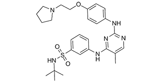
N-叔丁基-3-[[5-甲基-2-[4-[2-(1-吡咯烷基)乙氧基]苯胺基]-4-嘧啶基]氨基]苯磺酰胺是一种磺酰胺类药物。
Fedratinib,也称为SAR302503和TG101348,是一种酪氨酸激酶抑制剂,用于治疗中危-2和高危原发性及继发性骨髓纤维化。它是一种苯胺基嘧啶衍生物。Fedratinib于2019年8月16日获得FDA批准。 Fedratinib是一种口服选择性Janus激酶2 (JAK-2)和FMS样酪氨酸激酶3 (FLT3)抑制剂,用于治疗中危或高危原发性或继发性骨髓纤维化。 Fedratinib治疗期间血清酶升高发生率较高,但临床上明显的急性肝损伤病例罕见。 Fedratinib是一种口服生物利用度高的小分子ATP竞争性抑制剂,可抑制Janus激酶2 (JAK2)和FMS样酪氨酸激酶3 (FLT3; CD135; STK1; FLK2),具有潜在的抗肿瘤活性。口服后,Fedratinib可与野生型JAK2及其突变体竞争ATP结合,从而抑制JAK2活化、JAK-STAT信号通路、抑制肿瘤细胞增殖并诱导肿瘤细胞凋亡。JAK2是bcr-abl阴性骨髓增生性疾病(MPD)中最常见的突变基因。此外,Fedratinib还能靶向FLT3并抑制其活性。这抑制了不受控制的 FLT3 信号传导,从而抑制了过度表达 FLT3 的肿瘤细胞的增殖。 FLT3 是一种 III 类受体酪氨酸激酶 (RTK),在大多数 B 系肿瘤和急性髓系白血病中过度表达或发生突变,并在肿瘤细胞增殖中发挥关键作用。 另见:盐酸菲德拉替尼(活性成分)。 药物适应症 菲德拉替尼适用于治疗中危-2 或高危原发性或继发性(真性红细胞增多症后或原发性血小板增多症后)骨髓纤维化的成年患者。 因瑞替尼适用于治疗原发性骨髓纤维化、真性红细胞增多症后骨髓纤维化或原发性血小板增多症后骨髓纤维化的成年患者的疾病相关脾肿大或症状,这些患者既往未接受过 Janus 激酶 (JAK) 抑制剂治疗,或曾接受过鲁索替尼治疗。 作用机制作用机制 Fedratinib 是一种 Janus 激酶 2 (JAK2) 和 FMS 样酪氨酸激酶 3 的抑制剂。JAK2 在骨髓增生性肿瘤(如骨髓纤维化)中高度活跃。Fedratinib 通过抑制 JAK2 来抑制信号转导和转录激活因子 (STAT) 3 和 5 的磷酸化,从而阻止细胞分裂并诱导细胞凋亡。 药效学 Fedratinib 是一种激酶抑制剂,可抑制细胞分裂并诱导细胞凋亡。服用 fedratinib 的患者可能会出现贫血、血小板减少症、胃肠道毒性、肝毒性或淀粉酶和脂肪酶升高。这些不良反应应根据具体情况通过降低剂量、暂时停药或输血来控制。 据报道,TG101348 是一种选择性小分子 JAK2 抑制剂,其体外 IC50 值约为 3 nM,在 JAK2V617F 突变诱导的骨髓增生性疾病小鼠模型中显示出治疗效果。在接受治疗的动物中,血细胞比容和白细胞计数均出现统计学意义上的显著降低,髓外造血作用呈剂量依赖性降低/消除,并且至少在某些情况下,骨髓纤维化症状有所减轻。未观察到明显的毒性,且对 T 细胞数量无影响。体内反应与替代终点相关,包括通过定量基因组PCR评估的JAK2V617F疾病负荷的减少/消除、内源性红系集落形成的抑制以及通过流式细胞术测量磷酸化Stat5评估的体内JAK-STAT信号转导的抑制。[1]真性红细胞增多症(PV)是一种骨髓增生性疾病(MPD),其常见特征是JAK2(JAK2V617F)信号突变、红细胞过度生成以及易发生血栓形成、进展为骨髓纤维化或急性白血病。在本研究中,人造血祖细胞表达JAK2V617F促进了生物发光异种免疫缺陷小鼠移植模型中的红系集落形成和红系植入。选择性JAK2抑制剂TG101348(300 nM)显著抑制了JAK2V617F+祖细胞来源的集落形成以及异种移植研究中的植入(120 mg/kg)。TG101348治疗降低了GATA-1的表达,而GATA-1的表达与JAK2V617F+祖细胞分化向红系偏倚相关,TG101348还抑制了STAT5以及GATA S310的磷酸化。因此,TG101348可能是一种有效的JAK2V617F+骨髓增生性疾病(MPD)抑制剂,可用于临床试验。[2] 作用机制:Fedratinib(SAR302503)通过与ATP竞争结合激酶结构域,选择性地抑制JAK2(包括致癌的JAK2V617F突变体)。这可以阻断JAK2介导的STAT5磷酸化,从而抑制驱动真性红细胞增多症(PV)中JAK2突变造血细胞增殖和存活的下游信号通路[1,2] - 治疗重点:临床前数据支持Fedratinib(SAR302503)用于治疗JAK2V617F驱动的骨髓增生性肿瘤(MPN),特别是真性红细胞增多症(PV),因为它对JAK2具有高度选择性,并且对JAK1/JAK3的脱靶效应极小[1,2] - 药物研发背景:Fedratinib(SAR302503)(TG101348)基于其在PV小鼠模型中的临床前疗效和对JAK2的选择性,被推进为MPN的临床候选药物,以满足JAK2突变型MPN患者的未满足医疗需求[1] |
| 分子式 |
C27H36N6O3S
|
|---|---|
| 分子量 |
524.6781
|
| 精确质量 |
524.256
|
| 元素分析 |
C, 61.81; H, 6.92; N, 16.02; O, 9.15; S, 6.11
|
| CAS号 |
936091-26-8
|
| 相关CAS号 |
Fedratinib hydrochloride hydrate;1374744-69-0
|
| PubChem CID |
16722836
|
| 外观&性状 |
White to light yellow solid
|
| 密度 |
1.2±0.1 g/cm3
|
| 沸点 |
713.7±70.0 °C at 760 mmHg
|
| 闪点 |
385.5±35.7 °C
|
| 蒸汽压 |
0.0±2.3 mmHg at 25°C
|
| 折射率 |
1.611
|
| LogP |
3.27
|
| tPSA |
120.09
|
| 氢键供体(HBD)数目 |
3
|
| 氢键受体(HBA)数目 |
9
|
| 可旋转键数目(RBC) |
11
|
| 重原子数目 |
37
|
| 分子复杂度/Complexity |
787
|
| 定义原子立体中心数目 |
0
|
| SMILES |
O=S(C1C=C(NC2C(C)=CN=C(NC3C=CC(OCCN4CCCC4)=CC=3)N=2)C=CC=1)(NC(C)(C)C)=O
|
| InChi Key |
JOOXLOJCABQBSG-UHFFFAOYSA-N
|
| InChi Code |
InChI=1S/C27H36N6O3S/c1-20-19-28-26(30-21-10-12-23(13-11-21)36-17-16-33-14-5-6-15-33)31-25(20)29-22-8-7-9-24(18-22)37(34,35)32-27(2,3)4/h7-13,18-19,32H,5-6,14-17H2,1-4H3,(H2,28,29,30,31)
|
| 化学名 |
N-(tert-butyl)-3-((5-methyl-2-((4-(2-(pyrrolidin-1-yl)ethoxy)phenyl)amino)pyrimidin-4-yl)amino)benzenesulfonamide
|
| 别名 |
Brand name Inrebic; SAR302503, TG101348; TG101348; TG 101348; TG-101348; SAR-302503; Fedratinib; 936091-26-8; Tg-101348; TG101348; N-(tert-butyl)-3-((5-methyl-2-((4-(2-(pyrrolidin-1-yl)ethoxy)phenyl)amino)pyrimidin-4-yl)amino)benzenesulfonamide; Inrebic; SAR-302503; SAR 302503
|
| HS Tariff Code |
2934.99.9001
|
| 存储方式 |
Powder -20°C 3 years 4°C 2 years In solvent -80°C 6 months -20°C 1 month |
| 运输条件 |
Room temperature (This product is stable at ambient temperature for a few days during ordinary shipping and time spent in Customs)
|
| 溶解度 (体外实验) |
DMSO: 100 mg/mL (190.56mM)
Water: <1 mg/mL Ethanol: <1 mg/mL |
|---|---|
| 溶解度 (体内实验) |
配方 1 中的溶解度: ≥ 2.87 mg/mL (5.47 mM) (饱和度未知) in 5% DMSO + 40% PEG300 + 5% Tween80 + 50% Saline (这些助溶剂从左到右依次添加,逐一添加), 澄清溶液。
*生理盐水的制备:将 0.9 g 氯化钠溶解在 100 mL ddH₂O中,得到澄清溶液。 配方 2 中的溶解度: 2.87 mg/mL (5.47 mM) in 5% DMSO + 95% (20% SBE-β-CD in Saline) (这些助溶剂从左到右依次添加,逐一添加), 悬浊液; 超声助溶。 *20% SBE-β-CD 生理盐水溶液的制备(4°C,1 周):将 2 g SBE-β-CD 溶解于 10 mL 生理盐水中,得到澄清溶液。 View More
配方 3 中的溶解度: ≥ 2.08 mg/mL (3.96 mM) (饱和度未知) in 10% DMSO + 40% PEG300 + 5% Tween80 + 45% Saline (这些助溶剂从左到右依次添加,逐一添加), 澄清溶液。 配方 4 中的溶解度: ≥ 2.08 mg/mL (3.96 mM) (饱和度未知) in 10% DMSO + 90% (20% SBE-β-CD in Saline) (这些助溶剂从左到右依次添加,逐一添加), 澄清溶液。 例如,若需制备1 mL的工作液,可将100μL 20.8mg/mL澄清的DMSO储备液加入到900μL 20%SBE-β-CD生理盐水中,混匀。 *20% SBE-β-CD 生理盐水溶液的制备(4°C,1 周):将 2 g SBE-β-CD 溶解于 10 mL 生理盐水中,得到澄清溶液。 配方 5 中的溶解度: ≥ 2.08 mg/mL (3.96 mM) (饱和度未知) in 10% DMSO + 90% Corn Oil (这些助溶剂从左到右依次添加,逐一添加), 澄清溶液。 例如,若需制备1 mL的工作液,可将100 μL 20.8 mg/mL 澄清 DMSO 储备液加入900 μL 玉米油中,混合均匀。 配方 6 中的溶解度: 1% DMSO+30% polyethylene glycol+1% Tween 80:30mg/mL 1、请先配制澄清的储备液(如:用DMSO配置50 或 100 mg/mL母液(储备液)); 2、取适量母液,按从左到右的顺序依次添加助溶剂,澄清后再加入下一助溶剂。以 下列配方为例说明 (注意此配方只用于说明,并不一定代表此产品 的实际溶解配方): 10% DMSO → 40% PEG300 → 5% Tween-80 → 45% ddH2O (或 saline); 假设最终工作液的体积为 1 mL, 浓度为5 mg/mL: 取 100 μL 50 mg/mL 的澄清 DMSO 储备液加到 400 μL PEG300 中,混合均匀/澄清;向上述体系中加入50 μL Tween-80,混合均匀/澄清;然后继续加入450 μL ddH2O (或 saline)定容至 1 mL; 3、溶剂前显示的百分比是指该溶剂在最终溶液/工作液中的体积所占比例; 4、 如产品在配制过程中出现沉淀/析出,可通过加热(≤50℃)或超声的方式助溶; 5、为保证最佳实验结果,工作液请现配现用! 6、如不确定怎么将母液配置成体内动物实验的工作液,请查看说明书或联系我们; 7、 以上所有助溶剂都可在 Invivochem.cn网站购买。 |
| 制备储备液 | 1 mg | 5 mg | 10 mg | |
| 1 mM | 1.9059 mL | 9.5296 mL | 19.0592 mL | |
| 5 mM | 0.3812 mL | 1.9059 mL | 3.8118 mL | |
| 10 mM | 0.1906 mL | 0.9530 mL | 1.9059 mL |
1、根据实验需要选择合适的溶剂配制储备液 (母液):对于大多数产品,InvivoChem推荐用DMSO配置母液 (比如:5、10、20mM或者10、20、50 mg/mL浓度),个别水溶性高的产品可直接溶于水。产品在DMSO 、水或其他溶剂中的具体溶解度详见上”溶解度 (体外)”部分;
2、如果您找不到您想要的溶解度信息,或者很难将产品溶解在溶液中,请联系我们;
3、建议使用下列计算器进行相关计算(摩尔浓度计算器、稀释计算器、分子量计算器、重组计算器等);
4、母液配好之后,将其分装到常规用量,并储存在-20°C或-80°C,尽量减少反复冻融循环。
计算结果:
工作液浓度: mg/mL;
DMSO母液配制方法: mg 药物溶于 μL DMSO溶液(母液浓度 mg/mL)。如该浓度超过该批次药物DMSO溶解度,请首先与我们联系。
体内配方配制方法:取 μL DMSO母液,加入 μL PEG300,混匀澄清后加入μL Tween 80,混匀澄清后加入 μL ddH2O,混匀澄清。
(1) 请确保溶液澄清之后,再加入下一种溶剂 (助溶剂) 。可利用涡旋、超声或水浴加热等方法助溶;
(2) 一定要按顺序加入溶剂 (助溶剂) 。
| NCT Number | Recruitment | interventions | Conditions | Sponsor/Collaborators | Start Date | Phases |
| NCT05177211 | Recruiting | Drug: Fedratinib Pill | Myeloproliferative Neoplasm Chronic Neutrophilic Leukemia |
H. Lee Moffitt Cancer Center and Research Institute |
March 1, 2022 | Phase 2 |
| NCT05524857 | Recruiting | Drug: Fedratinib Oral Capsule 300 mg Drug: Decitabine 20 mg/m2 |
RMyeloproliferative Neoplasm | Joseph Jurcic | January 28, 2022 | Phase 1 |
| NCT04446650 | Active, not recruiting | Drug: Fedratinib | Primary Myelofibrosis | Celgene | October 12, 2020 | Phase 1 Phase 2 |
| NCT06073847 | Recruiting | Drug: Fedratinib | Primary Myelofibrosis Post-polycythemia Vera Myelofibrosis |
Bristol-Myers Squibb | July 13, 2023 |
InvivoChem的所有产品仅用于作科学研究,不面向患者销售
Copyright 2020 InvivoChem LLC | All Rights Reserved 粤ICP备20063088号-1


 COA
COA
 463611831
