| 规格 | 价格 | 库存 | 数量 |
|---|---|---|---|
| 5mg |
|
||
| 10mg |
|
||
| 25mg |
|
||
| 50mg |
|
||
| 100mg |
|
||
| 250mg |
|
||
| Other Sizes |
|
| 靶点 |
Gyrase ( IC50 = 1.25 μg/mL ); TOPO IV ( IC50 = 1.5-2.5 μg/mL ); Quinolone
|
|---|---|
| 体外研究 (In Vitro) |
Garenoxacin (BMS284756)(0-8 天)抑制支原体和解脲支原体测试菌株的生长,MIC90 ≤0.25 μg/mL[1]。
Garenoxacin (48 h) 的 MIC 为 0.0128-4.0 μg/mL,这可抑制野生型和突变型金黄色葡萄球菌[2]。 Garenoxacin 对拓扑异构酶 IV 的 IC50 为 1.25 至 2.5 μg/mL,对金黄色葡萄球菌促旋酶的 IC50 为 1.25 μg/mL。 Garenoxacin 具有从对环丙沙星敏感的金黄色葡萄球菌分离株中选择性富集氟喹诺酮类耐药突变体的趋势较低[3]。 对63株肺炎支原体、45株人型支原体、15株发酵支原体和68株脲原体进行了加雷沙星(BMS-284756)、去氟喹诺酮类药物及其他8种药物的体外敏感性测定。加兰诺沙星是活性最强的喹诺酮类药物,在<或=1 μ g/ml时抑制所有分离株。加列诺克星的MIC, 90%的分离株被抑制(MIC(90)s);<或=0.008 μ g/ml)比莫西沙星和克林霉素低至少4倍,比斯帕沙星低8倍,比左氧氟沙星和环丙沙星低64倍。加诺沙星MIC(90) <或=0.008 μ g/ml,比克林霉素和莫西沙星低4倍,比斯帕沙星低8倍,比左氧氟沙星和环丙沙星低64倍。所有15株发酵分枝杆菌分离株均被浓度<或=0.008微克/毫升的加林诺沙星抑制,使其成为对该菌最有效的药物。对于脲原体,加诺沙星MIC(90) (0.25 μ g/ml)与莫西沙星、多西环素相当,比左氧氟沙星、斯帕沙星低4倍,比阿奇霉素低8倍,比环丙沙星低32倍。加诺沙星和其他氟喹诺酮类药物通过最小杀菌活性测量和时间杀伤研究证明对肺炎支原体和人支原体具有杀菌活性。加兰诺沙星在治疗支原体和脲原体感染方面有很大的潜力,因此需要进一步的研究。[1] 研究人员通过遗传和生化研究确定了新型地氟喹诺酮类药物加雷沙星(BMS-284756, T-3811ME)在金黄色葡萄球菌中的靶酶相互作用。我们发现加诺沙星比环丙沙星对野生型金黄色葡萄球菌的活性高4到8倍。单个拓扑异构酶IV或回转酶突变仅导致加雷沙星MIC增加2- 4倍,而两个位点的突变组合导致MIC大幅增加(128倍)。诺拉外排泵的过表达对加诺沙星耐药的影响很小。加诺沙星为MIC的两倍时,耐药突变体(<7.4 × 10(-12)至4.0 × 10(-11))的选择比环丙沙星少5至6个对数单位。在单步突变中,拓扑异构酶IV或gyrase的喹诺酮耐药决定区(QRDR)内外的突变被选中,表明拓扑异构酶IV和gyrase具有双重靶向性。基因实验表明,其中三种新突变是产生耐药性的原因。对金黄色葡萄球菌中纯化的拓扑异构酶IV和gyrase的研究也表明,加兰诺沙星对拓扑异构酶IV和gyrase具有相似的活性(50%的抑制浓度,分别为1.25 ~ 2.5和1.25微g/ml),虽然其对拓扑异构酶IV的活性是环丙沙星的2倍,但对gyrase的活性是环丙沙星的10倍。本研究提供了第一个支持喹诺酮类药物双重靶向金黄色葡萄球菌拓扑异构酶IV和gyrase的遗传学和生化数据,并为qrdr扩展到grlB的5‘端和gyrA的3’端提供了遗传学证据。[2] 新的喹诺酮加雷沙星(BMS-284756)缺乏C-6氟,对其阻断金黄色葡萄球菌生长的能力进行了检测。MIC和突变体预防浓度(MPC)的测定显示,加诺沙星对多种环丙沙星敏感的分离株的效力是环丙沙星的20倍,其中一些对甲氧西林耐药。90%分离株的MPC(MPC(90))低于使用推荐剂量的加诺沙星所达到的公布的血清药物浓度。这些体外观察表明,在环丙沙星敏感的金黄色葡萄球菌分离株中,加诺沙星选择性富集氟喹诺酮耐药突变体的倾向较低。对于环丙沙星耐药菌株,90%的测试菌株被抑制的MIC低于血清药物浓度,而MPC(90)则没有。因此,对于这些菌株,加诺沙星浓度预计在大部分治疗时间内落在突变体选择窗口内(在MIC和MPC之间)。因此,加诺克星有望选择性地富集易感性更低的突变体。[3] |
| 体内研究 (In Vivo) |
Garenoxacin(12.5-50 mg/kg;皮下注射;一次)在肺炎链球菌感染的小鼠肺炎模型中对野生型菌株和携带单突变的突变体非常有效[4]。
Garenoxacin(10 和30 mg/kg;口服;一次)显着增加 BALB/c 雌性小鼠在由肺炎链球菌 D-979 引起的实验性继发性肺炎球菌肺炎中的存活时间[5],同时也减少了肺部活细胞的数量。 在肺炎链球菌感染的小鼠肺炎模型中,针对野生型菌株和携带单一突变的突变体,格尔德诺辛(12.5-50mg/kg;皮下注射;一次)表现出显着的疗效[4]。 当BALB/c雌性小鼠暴露于由肺炎链球菌 D-979 引起的实验性继发性肺炎球菌肺炎,给予加雷沙星(10 和 30 mg/kg;口服;一次)时,肺部活细胞计数减少,存活时间显着延长。 P-4241菌株感染小鼠经加列诺沙星或TVA (25 mg/kg体重)处理后的肺药动学参数如下:血清中最大药物浓度C(max);分别为17.3和21.2微g/ml), C(max)/MIC比分别为288和170,浓度-时间曲线下面积(AUC;48.5和250微克。h/ml), AUC/MIC比(分别为808和2000)。加诺沙星25和50 mg/kg对野生型菌株和携带单一突变的突变体非常有效(存活率为85%至100%)。TVA对这些菌株的治疗效果与加诺克星相同。TVA 200 mg/kg和加诺克星50 mg/kg对parC和gyrA双突变体和gyrA、parC和parE三突变体无效。只有当菌株发生喹诺酮类药物耐药的几个突变时,加诺克星的疗效才会降低。[4] 在流感病毒感染后的肺炎球菌肺炎小鼠模型中,加诺沙星比其他氟喹诺酮类药物更有效,并表现出高水平的肺部细菌根除,低死亡率和有效的组织病理学改善。加诺沙星可能用于治疗继发性肺炎球菌性肺炎后流感。[5] |
| 酶活实验 |
拓扑异构酶IV测定。[2]
十癸烯化试验的反应混合物(20 μl)含有50 mM Tris-HCl (pH 7.7)、5 mM MgCl2、5 mM二硫索糖醇、50 μg牛血清白蛋白/ ml 250 mM谷氨酸钾、1 mM ATP、100 ng动质体DNA和不同量的GrlA和GrlB。37℃孵育1 h后,加入EDTA至终浓度50 mM终止反应,产物在1%琼脂糖中电泳分析。凝胶电泳后用溴化乙锭染色。 DNA回转酶测定。[2] 在含有75 mM Tris-HCl (pH 7.5)、7.5 mM MgCl2、7.5 mM二硫苏糖醇、2mM ATP、75 μg牛血清白蛋白/ ml、30 mM KCl、250 mM谷氨酸钾和2 μg tRNA(以0.5 μg松弛的pBR322为底物)的缓冲液中测定DNA超卷活性,缓冲液的总容积为20 μl。反应在30°C下进行1 h,加入EDTA至终浓度为50 mM停止反应,产物在1%琼脂糖中进行电泳分析,用于拓扑异构酶IV检测。 |
| 细胞实验 |
细胞系:解脲支原体、肺炎支原体、发酵支原体和人支原体。
培养时间:人支原体 48 小时,解脲支原体 24 小时,肺炎支原体 4-8 天< br> 结果:对肺炎支原体、发酵支原体、人型支原体和解脲支原体菌株具有抑制作用。 MIC90 分别为 0.031 μg/mL、≤0.008 μg/mL、≤0.008 μg/mL 和 0.25 μg/mL。 药物敏感性测定。[2] 在含有连续两倍稀释抗生素的Trypticase大豆琼脂上至少重复两次测定mic,并在37°C孵育24和48 h后进行生长评分。用钠啶酸的mic筛选gyrA突变,用新生物素的mic筛选grlB突变,用溴化乙啶的mic筛选NorA过表达。在基因测试中,当遇到双重差异时,它们通过重复测试来证实。 突变体选择的频率。[2] 通过将适当稀释的金黄色葡萄球菌ISP794隔夜培养物在不加抗生素或加加诺克星或环丙沙星的情况下,以每种药物MIC的1倍、2倍、4倍和8倍的浓度,在脑心脏输注琼脂上镀上突变体。为了选择加诺克星,使用大的(150 × 15毫米)培养皿对1011 ~ 1012 CFU进行培养皿。每次电镀一式两份,至少重复两次。选择板37℃孵育。抗性突变体的选择频率以48 h时抗性菌落数与接种细胞数之比计算。选择的菌落在含有选定浓度的加诺沙星的脑心灌注琼脂板上传代一次,如有必要,在不含任何抗生素的脑心灌注琼脂上传代一次,然后在- 70°C的10%甘油中保存于脑心灌注肉汤中。 逐步选择抗药突变体。[2] 将金黄色葡萄球菌ISP794连续传代于含有加诺沙星浓度增加两倍的脑心灌注琼脂上,以确定可达到的最高耐药水平。从加诺克星治疗ISP794的MIC开始选择。在每一步中,将几个突变菌落传代于含有加诺克星选择浓度的脑心输注琼脂板上,然后在- 70°C保存并在高两倍抗生素浓度下传代。[2] 抗生素治疗[4] 使用野生型强毒青霉素敏感菌株(P-4241)和喹诺酮耐药突变体(parC、gyrA和外排单突变体以及parC和gyrA双突变体)攻击18小时后开始治疗。用parE和parC gyrA parE临床菌株攻毒后3小时开始治疗。加诺沙星和TVA分别以12.5、25和50 mg/kg的剂量皮下注射6次。TVA以50、100和200 mg/kg的剂量给具有双重突变的突变小鼠。感染的、未治疗的对照组小鼠接受相同体积的等渗盐水。每个治疗组15只动物。观察期为10 d。每天记录死亡率,并比较累积存活率。 |
| 动物实验 |
动物模型:感染肺炎链球菌的瑞士小鼠[4]。
剂量:12.5、25 和 50 mg/kg 给药途径:皮下注射,单次 结果:显著提高存活率。 体内杀菌活性[4] 体内杀菌活性研究方案与小鼠存活率研究方案相同。在首次给药后 6 小时(细菌感染后 18 小时开始)以及第二次、第四次和第六次给药后 12 小时,分别测定全肺匀浆中回收的总菌落形成单位 (CFU) 数,给药剂量为 12.5 和 25 mg/kg 的加雷诺沙星。每个剂量和时间点使用三只小鼠。小鼠通过腹腔注射戊巴比妥钠处死,并经心脏穿刺放血;血液用于培养。取出肺脏,在1 ml生理盐水中匀浆。将匀浆液进行10倍系列稀释,然后涂布于哥伦比亚琼脂平板上。血液在脑心浸液肉汤中培养。过夜培养后,在接种了肺脏样本的琼脂平板上计数菌落,并检查血液培养的浊度。结果以每肺脏的平均菌落形成单位(CFU) ± 标准差(log10)表示,并以每组三只小鼠的阳性或阴性血液培养数量表示。 血清和肺脏中加雷诺沙星浓度的测定及药代动力学分析[4] 对感染和未感染的小鼠均单次皮下注射25 mg/kg的加雷诺沙星或TVA。感染小鼠在感染后18小时接受治疗。分别于给药后0.25、0.5、1、2、4、6、8、12和24小时,从每组6只小鼠中采集血清和肺组织样本。所有样本均储存于-20℃并避光保存,以避免分析过程中加雷诺沙星降解。肺组织样本在液氮中用磁力研磨机研磨。血清样本(100 μl)和肺组织样本(精确称取20至50 mg肺粉)的制备方法为:分别将内标物与甲醇酸(100 μl和500 μl)混合。经沉淀或扩散、涡旋或超声混合以及离心后,取50 μl上层相注入高效液相色谱系统进行分析。采用十八烷基硅烷化柱(Novapak C18;4.6 mm × 150 mm)联用荧光分光光度计测定总药物浓度,激发波长和发射波长分别为 280 nm 和 415 nm。流动相为乙腈、柠檬酸钠缓冲溶液(pH 3.5)和水(体积比 22/15/63)的混合物,并加入 0.2% 三乙胺调节 pH 至 4。流速为 1.0 ml/min。血清和肺组织样本的定量限分别为 0.02 μg/ml 和 0.05 μg/g,线性范围分别为 0.2~10.0 μg/ml 和 0.5~50.0 μg/g。血清和肺组织样本的质量控制变异系数均低于 10%。 TVA的药代动力学(PK)参数评估方法已在其他文献中描述。 |
| 药代性质 (ADME/PK) |
24小时内游离血清浓度-时间曲线下面积除以最小抑菌浓度(fAUC0–24/MIC)是氟喹诺酮类药物临床疗效最重要的预测指标之一(Craig,1998)。在本研究建立的流感病毒感染后继发性肺炎球菌肺炎模型中,口服加雷诺沙星(10和30 mg/kg)在血清中的fAUC0–24/MIC比值分别为71.7和288,在肺部的fAUC0–24/MIC比值分别为106和381,表明其能有效清除细菌并具有优异的疗效(表1)。尽管目前尚不清楚这三种喹诺酮类药物在继发性肺炎球菌肺炎模型中是否具有相似的fAUC0-24/MIC疗效,但加雷诺沙星在临床剂量下对肺炎链球菌的fAUC/MIC90比值≥352,高于左氧氟沙星(15.5)和莫西沙星(107)(Chein等,1997;Takagi等,2008;Watanabe等,2012;Zeitlinger等,2003)。加雷诺沙星强大的抗菌活性和良好的药代动力学特征被认为反映了其对流感病毒感染后继发性肺炎球菌肺炎的优异治疗效果。尽管莫西沙星(30 mg/kg)与加雷沙星在降低肺部活细胞方面效果相似,且其 fAUC0-24/MIC 值低于加雷沙星,但其死亡率(40%)却高于加雷沙星(表 1)。需要进一步研究来阐明喹诺酮类药物 fAUC/MIC 目标值的差异。[5]
|
| 参考文献 |
|
| 其他信息 |
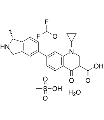
加雷诺沙星是一种喹啉单羧酸,即1,4-二氢喹啉-3-羧酸,其1位被环丙基取代,4位被氧代基取代,7位被(1R)-1-甲基-2,3-二氢-1H-异吲哚-5-基取代,8位被二氟甲氧基取代。它是一种抗菌药物和非甾体类抗炎药。它是一种喹诺酮类抗生素、喹啉单羧酸、有机氟化合物、环丙烷类化合物、芳香醚类化合物和异吲哚类化合物。
加雷诺沙星是一种喹诺酮类抗生素,目前正在研究其治疗革兰氏阳性菌和革兰氏阴性菌感染的疗效。 药物适应症 已研究用于治疗细菌感染。 除了测定部分微生物的最低杀菌浓度 (MBC) 外,我们还试图评估加雷诺沙星对肺炎支原体和人型支原体各代表性分离株的杀菌动力学,因为 MBC 已表明加雷诺沙星对这些微生物具有杀菌作用。由于支原体(尤其是肺炎支原体)生长速度较慢,其世代时间为 6 小时,因此通常 24 小时的时间-杀菌曲线研究必须延长才能证实其疗效。我们证实,加雷诺沙星在24至96小时的孵育后对肺炎支原体具有浓度依赖性的杀菌活性。加雷诺沙星在4至8倍MIC浓度下孵育24小时后,以及在2倍MIC浓度下孵育48小时后,也对人型支原体表现出杀菌活性。在某些较低浓度的加雷诺沙星存在下观察到≤2 log10 CFU的再生长,可能是由于极少量存活的微生物得以存活并随时间增殖所致,而对于肺炎支原体而言,在长时间孵育数日后,加雷诺沙星的降解和失活可能促进了这些微生物的增殖。这是首次通过改进自常用抗菌药物对其他细菌作用评估方法的时效杀菌实验,证实抗菌药物对肺炎支原体的杀菌作用。本研究表明,加雷诺沙星是一种有前景的治疗支原体和脲原体感染的药物。应进行进一步的临床评估。[1] 总之,加雷诺沙星与DNA促旋酶和拓扑异构酶IV的相互作用相似,并产生了新的突变,将喹诺酮耐药决定区(QRDR)的范围扩展到GrlB的氨基末端结构域和GyrA的羧基末端结构域。这种新型去氟喹诺酮类药物具有高效力和低耐药突变体选择频率,因此在临床应用中可能具有优势,降低新耐药突变体出现的可能性。然而,对于先前已选择出多种喹诺酮类耐药突变的菌株(例如目前常见的耐甲氧西林金黄色葡萄球菌临床分离株),加雷诺沙星存在交叉耐药性,这可能会限制该抗生素在治疗已对早期喹诺酮类药物产生耐药性的菌株方面的应用。 [2] 对于已对环丙沙星耐药的分离株,加雷诺沙星的MIC90为3.2 μg/ml,约为敏感分离株MPC90的8倍。这一观察结果表明,部分耐药菌株存在多种突变,这与其他研究的报道一致。由于耐药分离株的MIC低于可达到的血清药物浓度(图2C),因此可以推测,使用加雷诺沙星治疗环丙沙星耐药的金黄色葡萄球菌有时可能治愈感染。然而,已对环丙沙星耐药分离株的MPC90>19.6 μg/ml,即使将每日剂量提高至600 mg,也远高于加雷诺沙星所能达到的血清药物浓度(图2C)。事实上,基于MPC的加雷诺沙星及其突变株的药效学(图2C)与环丙沙星及其完全敏感菌株的药效学(图2A)相似。由于环丙沙星能迅速筛选出耐药突变株,我们预测,如果将加雷诺沙星用于治疗环丙沙星耐药菌株,金黄色葡萄球菌中将会固定出更多的突变。这些突变将使加雷诺沙星无法用于联合治疗。[3] 体内实验表明,在感染野生型菌株和单突变耐药菌株的小鼠中,加雷诺沙星的存活率与TVA相当,并且对携带parC和gyrA双突变的突变株的疗效略优于TVA:每公斤体重50毫克加雷诺沙星可延长小鼠的存活时间,而每公斤体重200毫克TVA则无效。将加雷诺沙星的活性与环丙沙星(一种特性明确且广泛使用的喹诺酮类药物)进行比较,结果表明加雷诺沙星的疗效远优于环丙沙星。考虑到环丙沙星体外活性较差,这一结果符合预期。加雷诺沙星的体内活性源于其对野生型和氟喹诺酮类耐药肺炎链球菌菌株的体外活性优于环丙沙星,以及其对双突变和三突变菌株的活性优于维甲酸(TVA)。然而,喹诺酮类药物的体内疗效还受到其他因素的影响,特别是药代动力学/药效学(PK-PD)参数。Forrest等人和Hyatt等人报道,AUC/MIC比值是与医院获得性肺炎患者的细菌清除和临床治愈相关的主要参数,其最低临床有效比值为125。因此,加雷诺沙星良好的PK-PD参数有助于其发挥疗效。与环丙沙星(CIP)相比,加雷沙星(garenoxacin)具有更长的半衰期、更大的AUC值和更优异的体外活性,尤其对肺炎链球菌(S. pneumoniae)的活性更强;在小鼠血清和肺组织样本中,加雷沙星的AUC/MIC比值最高。这些药代动力学(PK)和药效学(PD)参数对维甲酸(TVA)也非常有利,这解释了为什么这种喹诺酮类药物与加雷沙星一样有效。我们获得的加雷沙星药代动力学数据与小鼠生存数据高度吻合,表明血清蛋白结合对治疗结果的影响很小,即使在小鼠体内血清蛋白结合率高达约80%(DR Andes和WA Craig,第43届抗菌药物和化疗跨学科会议摘要,摘要A-309,第10页,2003年)。这可能是由于加雷沙星与血清蛋白的结合较弱所致。此外,肺部炎症细胞可能作为药物储存库,将加雷沙星释放到血清中。 TVA 也表现出较高的血清蛋白结合率,其疗效与其良好的药代动力学行为相关。TVA 在本肺炎球菌肺炎小鼠模型中是一个有趣的对照药物,但由于其已从市场上撤回,因此其临床相关性不如加雷诺沙星。总之,加雷诺沙星在由喹诺酮类敏感和耐药肺炎链球菌菌株引起的肺炎小鼠模型中均表现出高效性。因此,加雷诺沙星可能成为社区获得性呼吸道感染经验性治疗的有效选择。[4] 既往研究表明,尽管β-内酰胺类药物能有效清除细菌,但并未提高继发性细菌性肺炎的生存率(McCullers,2004),而大环内酯类药物治疗提高生存率是通过减轻炎症实现的(Karlström 等,2009)。Hara 等。 (2011)年的一项研究报告指出,加雷诺沙星具有抗炎活性,其作用机制是通过改变人肺上皮细胞系和人单核细胞系(尽管是在脂多糖刺激的细胞中)分泌白细胞介素8。加雷诺沙星疗效的改善可能不仅与其更高的fAUC0-24/MIC值带来的细菌清除作用有关,还可能与其抑制炎症反应有关。因此,这些数据提示加雷诺沙星可能在治疗流感后继发性肺炎球菌性肺炎方面具有潜在作用。需要进一步的研究来更好地了解加雷诺沙星对继发性肺炎球菌性肺炎患者炎症反应和临床疗效的影响。[5] |
| 分子式 |
C24H26F2N2O8S
|
|---|---|
| 分子量 |
540.53
|
| 精确质量 |
540.137
|
| 元素分析 |
C, 53.33; H, 4.85; F, 7.03; N, 5.18; O, 23.68; S, 5.93
|
| CAS号 |
223652-90-2
|
| 相关CAS号 |
194804-75-6; 223652-90-2 (mesylate hydrate); 223652-82-2 (mesylate)
|
| PubChem CID |
157690
|
| 外观&性状 |
Off-white to gray solid powder
|
| 沸点 |
581.5ºC at 760 mmHg
|
| 蒸汽压 |
2.29E-14mmHg at 25°C
|
| LogP |
5.316
|
| tPSA |
152.54
|
| 氢键供体(HBD)数目 |
4
|
| 氢键受体(HBA)数目 |
12
|
| 可旋转键数目(RBC) |
5
|
| 重原子数目 |
37
|
| 分子复杂度/Complexity |
863
|
| 定义原子立体中心数目 |
1
|
| SMILES |
O=C(C1=CN(C2CC2)C3=C(C=CC(C4=CC5=C([C@@H](C)NC5)C=C4)=C3OC(F)F)C1=O)O.CS(=O)(O)=O.O
|
| InChi Key |
IGTHEWGRXUAFKF-NVJADKKVSA-N
|
| InChi Code |
InChI=1S/C23H20F2N2O4.CH4O3S.H2O/c1-11-15-5-2-12(8-13(15)9-26-11)16-6-7-17-19(21(16)31-23(24)25)27(14-3-4-14)10-18(20(17)28)22(29)30;1-5(2,3)4;/h2,5-8,10-11,14,23,26H,3-4,9H2,1H3,(H,29,30);1H3,(H,2,3,4);1H2/t11-;;/m1../s1
|
| 化学名 |
1-cyclopropyl-8-(difluoromethoxy)-7-[(1R)-1-methyl-2,3-dihydro-1H-isoindol-5-yl]-4-oxoquinoline-3-carboxylic acid;methanesulfonic acid;hydrate
|
| 别名 |
BMS-284756-01; BMS284756-01; T-3811ME; 223652-90-2; Garenoxacin mesylate; Garenoxacin mesilate; Geninax; T3811ME; Garenoxacin mesylate; Garenoxacin mesylate hydrate
|
| HS Tariff Code |
2934.99.9001
|
| 存储方式 |
Powder -20°C 3 years 4°C 2 years In solvent -80°C 6 months -20°C 1 month 注意: 请将本产品存放在密封且受保护的环境中,避免吸湿/受潮。 |
| 运输条件 |
Room temperature (This product is stable at ambient temperature for a few days during ordinary shipping and time spent in Customs)
|
| 溶解度 (体外实验) |
DMSO: 2~100 mg/mL (4.7~185.0 mM)
|
|---|---|
| 溶解度 (体内实验) |
配方 1 中的溶解度: ≥ 2.5 mg/mL (4.63 mM) (饱和度未知) in 10% DMSO + 90% (20% SBE-β-CD in Saline) (这些助溶剂从左到右依次添加,逐一添加), 澄清溶液。
例如,若需制备1 mL的工作液,可将100 μL 25.0 mg/mL澄清DMSO储备液加入900 μL 20% SBE-β-CD生理盐水溶液中,混匀。 *20% SBE-β-CD 生理盐水溶液的制备(4°C,1 周):将 2 g SBE-β-CD 溶解于 10 mL 生理盐水中,得到澄清溶液。 配方 2 中的溶解度: ≥ 2.5 mg/mL (4.63 mM) (饱和度未知) in 10% DMSO + 90% Corn Oil (这些助溶剂从左到右依次添加,逐一添加), 澄清溶液。 例如,若需制备1 mL的工作液,可将 100 μL 25.0 mg/mL 澄清 DMSO 储备液添加到 900 μL 玉米油中并混合均匀。 请根据您的实验动物和给药方式选择适当的溶解配方/方案: 1、请先配制澄清的储备液(如:用DMSO配置50 或 100 mg/mL母液(储备液)); 2、取适量母液,按从左到右的顺序依次添加助溶剂,澄清后再加入下一助溶剂。以 下列配方为例说明 (注意此配方只用于说明,并不一定代表此产品 的实际溶解配方): 10% DMSO → 40% PEG300 → 5% Tween-80 → 45% ddH2O (或 saline); 假设最终工作液的体积为 1 mL, 浓度为5 mg/mL: 取 100 μL 50 mg/mL 的澄清 DMSO 储备液加到 400 μL PEG300 中,混合均匀/澄清;向上述体系中加入50 μL Tween-80,混合均匀/澄清;然后继续加入450 μL ddH2O (或 saline)定容至 1 mL; 3、溶剂前显示的百分比是指该溶剂在最终溶液/工作液中的体积所占比例; 4、 如产品在配制过程中出现沉淀/析出,可通过加热(≤50℃)或超声的方式助溶; 5、为保证最佳实验结果,工作液请现配现用! 6、如不确定怎么将母液配置成体内动物实验的工作液,请查看说明书或联系我们; 7、 以上所有助溶剂都可在 Invivochem.cn网站购买。 |
| 制备储备液 | 1 mg | 5 mg | 10 mg | |
| 1 mM | 1.8500 mL | 9.2502 mL | 18.5004 mL | |
| 5 mM | 0.3700 mL | 1.8500 mL | 3.7001 mL | |
| 10 mM | 0.1850 mL | 0.9250 mL | 1.8500 mL |
1、根据实验需要选择合适的溶剂配制储备液 (母液):对于大多数产品,InvivoChem推荐用DMSO配置母液 (比如:5、10、20mM或者10、20、50 mg/mL浓度),个别水溶性高的产品可直接溶于水。产品在DMSO 、水或其他溶剂中的具体溶解度详见上”溶解度 (体外)”部分;
2、如果您找不到您想要的溶解度信息,或者很难将产品溶解在溶液中,请联系我们;
3、建议使用下列计算器进行相关计算(摩尔浓度计算器、稀释计算器、分子量计算器、重组计算器等);
4、母液配好之后,将其分装到常规用量,并储存在-20°C或-80°C,尽量减少反复冻融循环。
计算结果:
工作液浓度: mg/mL;
DMSO母液配制方法: mg 药物溶于 μL DMSO溶液(母液浓度 mg/mL)。如该浓度超过该批次药物DMSO溶解度,请首先与我们联系。
体内配方配制方法:取 μL DMSO母液,加入 μL PEG300,混匀澄清后加入μL Tween 80,混匀澄清后加入 μL ddH2O,混匀澄清。
(1) 请确保溶液澄清之后,再加入下一种溶剂 (助溶剂) 。可利用涡旋、超声或水浴加热等方法助溶;
(2) 一定要按顺序加入溶剂 (助溶剂) 。
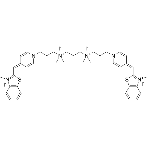 BODi-1
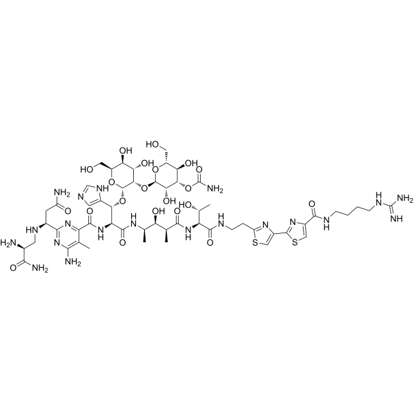 Bleomycin B2
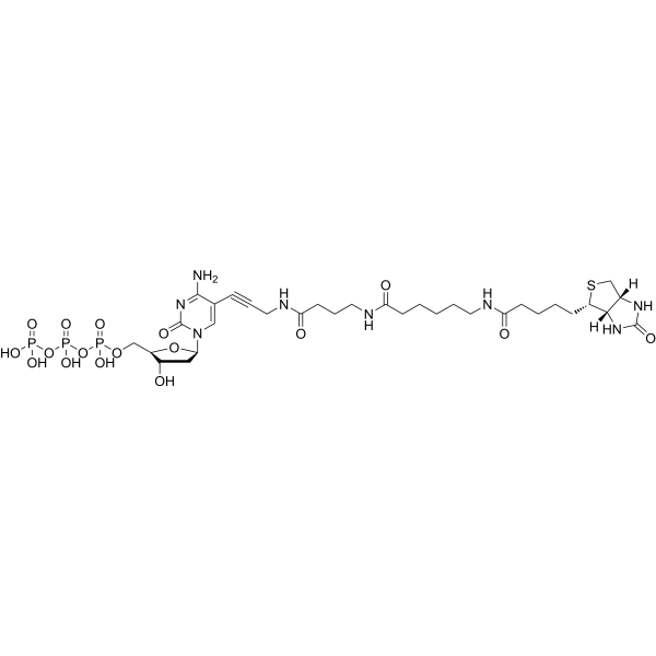 Biotin-16-dCTP
 1-Nitropyrene
InvivoChem的所有产品仅用于作科学研究,不面向患者销售
Copyright 2020 InvivoChem LLC | All Rights Reserved 粤ICP备20063088号-1


 COA
COA
 463611831
