| 规格 | 价格 | 库存 | 数量 |
|---|---|---|---|
| 100mg |
|
||
| 1g |
|
||
| 5g |
|
||
| 10g |
|
||
| 25g |
|
||
| Other Sizes |
|

| 靶点 |
Endogenous Metabolite; Microbial Metabolite
|
|---|---|
| 体外研究 (In Vitro) |
胆汁淤积症代表病理生理综合征,定义为肝脏胆汁流动受损。结果,胆汁酸积聚并促进肝细胞损伤,随后是肝硬化和肝功能衰竭。甘鹅脱氧胆酸(GCDCA)毒性相对较大,胆汁淤积后胆汁和血清中浓度较高。然而,GCDCA诱导肝毒性的机制尚不清楚。在这项研究中,我们发现GCDCA通过抑制溶酶体蛋白水解和增加溶酶体pH值来抑制自噬体形成并损害溶酶体功能,导致自噬清除缺陷,进而导致L02人肝细胞死亡。值得注意的是,通过基于串联质量标签(TMT)的定量蛋白质组学分析和数据库搜索,鉴定出313种差异表达的蛋白质,其中与对照组相比,GCDCA组有71种增加,242种减少。京都基因和基因组百科全书(KEGG)通路分析显示,GCDCA抑制了转录因子E3(TFE3)的信号通路,这与自噬通量损伤最密切相关。相比之下,GCDCA抑制溶酶体功能,TFE3过表达有效地减弱了自噬通量。具体而言,TFE3表达的降低与活性氧(ROS)稳态的破坏密切相关,这可以通过用N-乙酰半胱氨酸(NAC)抑制细胞内ROS来预防。总之,我们的研究首次证明,操纵ROS/TFE3信号传导可能是拮抗GCDCA诱导的肝毒性的一种治疗方法。[1]
方法:记录暴露于不同浓度有毒胆汁酸的胆管上皮细胞中TGF-βmRNA的表达;甘鹅脱氧胆酸(GCDCA)±PC。 结果:在这些实验中,以及在胆管上皮细胞与外周血单核细胞和肌成纤维细胞共同培养的共培养实验中,TGF-βmRNA的表达在PC存在或不存在的情况下保持不变。此外,肌成纤维组织的I型胶原α1 mRNA表达也保持不变。[2] 糖鹅脱氧胆酸/GCDC诱导的细胞凋亡对PKC活性的影响以及PKC在GCDCA诱导的肝细胞凋亡中的作用尚不清楚。本研究的具体目的是确定GCDC诱导的细胞凋亡是否会改变细胞内PKC活性,以及PKC活性的调节是否会影响GCDC引导的肝细胞凋亡。使用GCDC在分离的肝细胞中诱导凋亡。检测PKC活性,并使用特异性PKC和钙蛋白酶抑制剂研究PKC和钙粘蛋白调节对GCDC诱导的细胞凋亡的影响。暴露4小时后,50微M GCDC诱导42%的肝细胞凋亡。肝细胞暴露于GCDC 2小时后,细胞内PKC活性降至对照组的44%(p<0.001)。将肝细胞与钙蛋白酶抑制剂预孵育,使暴露于GCDC的肝细胞中的PKC活性恢复到91+/-5%的对照细胞。用钙蛋白酶抑制剂预孵育肝细胞可以减少GCDC诱导的凋亡,用激活PKC的佛波醇酯(PMA)预孵育也可以。钙蛋白酶抑制和PMA的联合使用进一步减少了GCDC诱导的细胞凋亡,但导致了低水平的肝细胞凋亡。白屈菜红碱抑制PKC也显著减少了GCDC诱导的肝细胞凋亡。GCDC诱导的细胞凋亡与总细胞PKC活性的降低有关,这似乎取决于细胞内钙蛋白酶样蛋白酶的活性。蛋白酶抑制和佛波酯预处理的组合保留了总细胞PKC活性,降低了GCDC诱导的凋亡,但在没有GCDC暴露的情况下诱导了低水平的凋亡。PKC抑制也降低了GCDC诱导的肝细胞凋亡,突显了PKC和蛋白酶在GCDC诱导凋亡过程中的复杂相互作用。[3] 据报道,胆汁酸(BA)的一种成分甘鹅脱氧胆酸(GCDC)可诱导原代人肝细胞坏死。在本研究中,我们探讨了GCDC在HCC化疗耐药性中的作用。我们发现GCDC通过分别下调和上调凋亡和抗凋亡基因的表达来促进HCC细胞的化疗耐药性。此外,GCDC诱导HCC细胞的EMT表型和干性,并激活STAT3信号通路。这些发现表明,GCDC通过STAT3途径诱导干性来促进HCC的化疗耐药性,并可能成为HCC化疗的潜在靶点[4]。 |
| 体内研究 (In Vivo) |
然后,我们评估了将这些选定的胆汁酸直接注射到脑室中是否也可以在体内抑制HPA轴。胆汁酸对HPA轴的抑制作用仅限于结合胆汁酸TCA和甘氨鹅脱氧胆酸(GCDA),而非结合胆汁酸CA、DCA和CDCA在注射后6小时对循环皮质酮水平没有显著影响(图3A)。同时,TCA和甘氨鹅脱氧胆酸(GCDA)注射液降低了下丘脑CRH mRNA表达和循环CRH蛋白水平(图3,B和C),而胆汁酸CA、DCA和CDCA没有显著影响(数据未显示)。[5]
为了评估胆汁酸的体内作用是否也依赖于ASBT,我们将ASBT vivo Morpholino或不匹配的对照序列注射到第三脑室。这显著抑制了下丘脑神经元中ASBT蛋白的翻译,如免疫荧光所示(图5A)。直接向第三脑室注射甘氨鹅脱氧胆酸(GCDA)显著抑制了下丘脑CRH mRNA表达(图5B)、蛋白质含量(图5C)和循环皮质酮水平(图5D),ASBT Vivo Morpholino注射可以减弱或逆转这一作用。[5] 然后,我们评估了胆汁酸激活GR的能力是否也与体内有关。大鼠侧脑室注射GR Vivo Morpholino(或不匹配的对照序列),通过免疫荧光评估下丘脑中GR的表达。正如预期的那样,通过免疫荧光(图7A)和免疫印迹(图7B)评估,与不匹配的对照组相比,中央GR Vivo Morpholino注射后GR免疫反应性受到显著抑制。此外,随后向第三脑室注射甘氨鹅脱氧胆酸(GCDA)显著抑制了CRH mRNA表达(图7C)、蛋白质含量(图7D)和循环皮质酮水平(图7E),这一作用在GR Vivo Morpholino注射后减弱。[5] |
| 酶活实验 |
GR萤光素酶测定[5]
此外,按照前面描述的程序,使用与含有糖皮质激素反应元件(GRE)共有序列的启动子区域偶联的萤光素酶报告构建体,在下丘脑神经元中评估GR转录活性。将神经元以10000个细胞/孔的密度铺在96孔板上,并让其粘附过夜。然后用GRE荧光素酶报告构建体(0.1-μg DNA/孔)和0.28μL TransIT-LT1转染试剂在37°C下转染细胞过夜。在此之后,用糖鹅脱氧胆酸(GCDA)或TCA(10μM)刺激细胞,并在刺激后24小时使用萤光素酶检测试剂盒检测萤光素酶活性。治疗至少进行了四次,结果以每微克蛋白质的萤光素酶活性变化程度表示。 |
| 细胞实验 |
细胞培养[1]
L02人正常肝细胞在1640培养基(HyClone)中培养,该培养基补充了10%热灭活FCS和1%(v/v)青霉素/链霉素(Sigma,St Louis,MO,USA),在37°C的5%CO2加湿气氛中。在80%融合时,细胞用不同浓度(50、75或100μM)的GCDCA处理6小时,或用100μM GCDCA处理不同时期(0、1、3或6小时),如我们之前的研究所述(Chen等人,20132015;Xu等人,2012)。将GCDCA溶解在无菌磷酸盐缓冲盐水(PBS)中以产生100mM储备溶液,然后在施用前用细胞培养基进行连续稀释。 细胞样本制备和胆汁酸检测[1] L02细胞(5×10⁶个细胞)用100μMGCDCA处理6小时,细胞样品在冰上用500μl氯仿、甲醇和水(1:2.5:1,v/v/v)的混合物均质化。然后将样品在4°C下以13000 rpm离心10分钟,并将150-μl等分上清液转移到含有IS(10μl l-4-氯苯丙氨酸水溶液,5μg/mL)。用500μl甲醇对沉积物进行再均质化,并将150-μl等分上清液加入同一小瓶中干燥,然后用乙腈/H2O(6:4,v/v)复溶至最终体积为500μl。用流动相复溶后,用Waters ACQUITY超高效液相色谱法结合Waters XEVO TQ-S质谱仪和ESI源对提取物和胆汁酸参考标准品进行分析。整个UPLC-MS/MS系统由MassLynx 4.1软件控制。所有色谱分离均使用ACQUITY BEH C18柱(1.7μm,100 mm×2.1 mm内部尺寸),注射体积为5μL。使用TargetLynx应用程序管理器4.1版分析以阴性模式获得的UPLC-MS原始数据,以获得校准方程和样品中每种胆汁酸的定量浓度。使用Pierce BCA™蛋白质检测试剂盒测量的赖氨酸样品以ng/孔为单位进行测量,并按mg蛋白质进行标度。 细胞死亡试验[1] L02细胞被放置在6孔板中(每孔5×10⁵个细胞)。用GCDCA处理后,用300μl胰蛋白酶-EDTA溶液分离细胞。将分离的细胞悬浮液在300g下离心5分钟。然后,将沉淀物与800μl台盼蓝溶液混合并分散。染色3分钟后,使用自动细胞计数器(Bio-Rad,TC10)对细胞进行计数。死细胞被染成蓝色。细胞死亡率(%)表示为死细胞/总细胞的百分比(Chang等人,2011)。 细胞培养[2] KMBC细胞在添加了110mg/L丙酮酸钠、10%胎牛血清(FBS)、100U/ml青霉素和100μg/ml链霉素的DMEM中培养。LX-2细胞在添加了0.1 mmol/L非必需氨基酸、2 mmol/L谷氨酰胺、110 mg/L丙酮酸钠、10%胎牛血清(FBS)、100 U/ml青霉素和100μg/ml链霉素的DMEM/F12中培养。所有细胞均在37°C、5%CO2和95%空气的加湿环境中孵育。在实验中,当细胞暴露于GCDCA时,暴露时间为24小时。 KMBC和LX-2细胞的共培养[2] KMBC和LX-2细胞在37°C下,在5%CO2和95%空气的加湿气氛中,在含有3.0um多孔膜的插入物的共培养板中培养。对于每个板,8×106 KMBC细胞在上室培养,8×10^6 LX-2细胞在下室培养。在处理前一天移除培养基,并在上述培养基中孵育细胞。在细胞收获前,将指示浓度的GCDCA加入上室浴溶液中24小时。 KMBC外周血单个核细胞(PBMC)和LX-2细胞的共培养[2] KMBC、PBMC(来源于健康供体)和LX-2细胞在37°C、5%CO2和95%空气的加湿气氛中,在含有3.0um多孔膜的插入物的共培养板中培养。对于每个孔,在上腔培养5×10⁶ KMBC和3×10⁶ PBMC细胞,在下腔培养8×10⁶ LX-2细胞。在GCDCA暴露前一天移除培养基,并在上述培养基中孵育细胞。 细胞增殖和细胞毒性试验[4] 通过细胞计数试剂盒-8测定化疗诱导的细胞死亡。Huh7和LM3细胞以8×10³个细胞/孔的密度接种在96孔板中,与GCDCA一起孵育,并分别用化疗药物(5-FU和顺铂)处理24小时和48小时。接下来,用磷酸缓冲盐水(PBS)洗涤细胞,并加入细胞计数试剂盒-8(CCK-8)溶液(培养基体积的1/10)1小时。使用微孔板读数器在450nm处检测细胞活力。 细胞凋亡测定[4] 将总共1×10⁵个细胞接种在6孔板中,分别用GCDCA和化疗药物处理24小时和48小时。接下来,用PBS洗涤细胞,重新悬浮在PBS中,并根据制造商的说明用膜联蛋白V和碘化丙啶(PI)染色。流式细胞术用于分析凋亡细胞的比例。 |
| 动物实验 |
雄性Sprague Dawley大鼠(150–175 g)饲养于温度控制在20°C–22°C、光照/黑暗周期为12小时/12小时的环境中。除非另有说明,动物可自由饮水和摄取标准大鼠饲料。在胆管结扎术(BDL)或假手术前3天,大鼠分别饲喂含2%考来烯胺的饲料或对照饲料AIN-93G。术后3天,于上午8点至9点之间采集组织和血清样本,以尽量减少糖皮质激素水平的昼夜节律变化。在另一项实验中,将20 pmol的胆汁酸胆酸(CA)、脱氧胆酸(DCA)、鹅脱氧胆酸(CDCA)、甘氨鹅脱氧胆酸(GCDA)或三氯乙酸(TCA)注射到大鼠的第三脑室(内侧/外侧0 mm,前/后-1.8 mm,背/腹+4.5 mm),并在6小时后收集血清和组织。同时,在单次注射甘氨鹅脱氧胆酸(GCDA)或三氯乙酸(TCA)前3天,使用连接皮下植入微型泵的脑内输注装置,将1 mg/kg·d的Vivo-Morpholino序列输注到大鼠的侧脑室(坐标:内侧/外侧-1.3 mm,前/后-0.2 mm,背/腹+3.5 mm)。注射方法与上述相同。采用免疫荧光和免疫印迹法评估 Vivo-Morpholino 输注抑制靶基因表达的程度,如前所述 [5]。
|
| 参考文献 |
|
| 其他信息 |
甘氨酸鹅脱氧胆酸是一种胆汁酸甘氨酸结合物,其胆汁酸成分为3α,7α-二羟基-5β-胆烷-24-酰基。它是一种人体代谢产物,在功能上与鹅脱氧胆酸相关,是甘氨酸鹅脱氧胆酸盐的结合酸。已有关于甘氨酸鹅脱氧胆酸在智人体内存在的报道,并有相关数据。它是一种胆汁酸盐,由鹅脱氧胆酸盐和甘氨酸在肝脏中形成,通常以钠盐的形式存在。它作为一种去污剂,能够溶解脂肪以促进吸收,自身也能被吸收。它具有利胆作用。最近的实验证据表明,肝脏自噬通量的失调在肝外胆汁淤积的发病机制中起着重要作用。自噬功能受损会促进胆汁酸诱导的肝损伤和泛素化蛋白的积累,而自噬激活则可保护肝脏免受胆汁淤积引起的肝损伤(Gao et al., 2014; Kim et al., 2018)。此外,Khambu B 等人报道,肝脏自噬缺陷会损害法尼醇X受体的功能,并导致胆汁淤积性损伤(Khambu et al., 2019)。与这些发现一致,我们的研究结果证实,GCDCA通过抑制溶酶体蛋白水解和升高溶酶体pH值来抑制自噬体的形成并损害溶酶体功能,从而导致体外自噬清除缺陷。
bHLH-亮氨酸拉链蛋白TFE3属于MiTF/TFE家族,是自噬和溶酶体生物合成的关键调控因子,并能促进细胞的整体降解(Fan et al., 2018)。TFE3正逐渐成为细胞存活和能量代谢的全局调控因子,其作用机制既包括促进溶酶体基因的表达,也包括通过新发现的靶点,例如氧化代谢和氧化应激反应(Pi et al., 2019a; Wang et al., 2019)。近年来,其他MiTF/TFE蛋白,例如黑素细胞诱导转录因子(MITF)、转录因子EB(TFEB)和转录因子EC(TFEC),作为自噬和溶酶体生物合成的主要调控因子,已成为人类疾病病理学中的关键因素(Martina等,2014)。在本研究中,GCDCA处理抑制了TFE3的表达和TFE3报告基因的活性,从而降低了自噬相关基因的表达。MITF、TFEB和TFE3的表达模式广泛,已在多种细胞类型中检测到,而TFEC的表达则局限于髓系细胞(Slade和Pulinilkunnil,2017)。基于这些结果,我们进一步研究了其他MiTF/TFE蛋白是否参与了GCDCA在L02细胞中的作用。与蛋白质组学分析结果一致,TFE3 mRNA表达在不同浓度GCDCA处理6小时后显著降低,而MITF或TFEB的水平未见显著变化(图S6)。这些结果证实了TFE3在GCDCA介导的自噬中发挥重要作用。然而,GCDCA介导的TFE3表达和活性抑制机制尚不清楚。近期研究表明,ROS的积累与乳腺癌或黑色素瘤细胞的侵袭和迁移过程中TFE3的激活有关(Deng et al., 2018; Tan et al., 2018)。ROS可能在GCDCA处理的L02细胞中TFE3的抑制过程中发挥重要作用。我们发现GCDCA以剂量依赖的方式诱导ROS的产生,而NAC预处理的细胞则消除了这种效应。此外,在用GCDCA处理前,先用NAC孵育L02细胞2小时,结果显示ROS生成受到抑制,从而消除了GCDCA对TFE3通路的影响。这一结果与之前的研究(Deng et al., 2018; Tan et al., 2018)相矛盾。我们提出了两种可能的原因。首先,我们的结果是从正常细胞系而非癌细胞系中获得的。其次,我们的研究测试的条件范围非常有限。ROS和TFE3之间的确切关系可能取决于细胞模型,其机制细节的阐明需要进一步研究。 总之,我们的数据表明,ROS/TFE3信号通路可能作为开发预防肝外胆汁淤积患者肝损伤的新疗法的治疗靶点(图7)。尽管有上述发现,但本研究仍存在一些局限性,需要强调。首先,本研究仅使用了一种细胞系来评估GCDCA诱导肝毒性的机制,未来我们将研究其他肝细胞系和/或原代肝细胞。其次,本研究仅考察了GCDCA,而未考察其他毒性胆汁酸。最重要的是,我们的结果来自体外培养细胞,因此在将体外培养实验结果外推至人类患者群体时应谨慎。我们期望通过进一步改进,包括在未来开展动物实验和严格的临床试验,来克服当前系统存在的问题。[1]尽管有上述发现,但本研究仍存在一些值得强调的局限性。首先,本研究使用的细胞系为胆管上皮细胞系和肌成纤维细胞系,而非源自人肝脏的原代细胞。这些细胞系是否比原代细胞对GCDCA的毒性作用和促纤维化细胞因子刺激的敏感性更低,仍有待确定。其次,本研究仅考察了GCDCA,而未考察其他毒性胆汁酸。第三,GCDCA的浓度范围来源于先前报道的人血中GCDCA浓度14。人胆汁中,特别是原发性硬化性胆管炎(PSC)患者的胆汁中是否存在更高浓度的GCDCA,目前尚不清楚。第四,共培养实验将胆管上皮细胞和外周血单核细胞与肌成纤维细胞物理隔离,从而阻止了细胞间接触和可能的细胞间通讯。第五,或许需要更长的GCDCA暴露时间才能诱导胆管上皮细胞损伤。然而,急性胆管结扎模型中快速的生化和组织学变化否定了这种可能性15。第六,需要注意的是,PBMC由外周血单核细胞组成,而非组织巨噬细胞。或许后一类细胞的额外特征对于此种情况下促纤维化细胞因子的表达和释放至关重要。16 最后,我们并未探讨将胆汁中PC浓度恢复至正常水平是否可能通过除保护胆道上皮细胞免受毒性胆汁酸损伤以外的其他途径,对PSC的病程产生有利影响。 总之,本研究结果不支持PC缺乏会导致毒性胆汁酸损伤胆道上皮细胞并随后激活邻近肌成纤维细胞,从而导致PSC中纤维化加剧的假设。因此,目前看来,将PSC胆汁中低水平的PC恢复至正常值并非治疗PSC的有效方法。[2] 研究表明,JAK/STAT3信号通路有助于癌细胞存活和化疗耐药。此外,它还能促进CSC样特征的形成。例如,肝癌干细胞标志物Nanog在肝肿瘤起始细胞中受STAT3通路诱导表达。类似地,SOCS和PTPN家族成员已被证实对JAK/STAT信号通路具有负调控作用。我们发现,在肝细胞癌(HCC)细胞中,GCDC通过抑制STAT3信号通路多个负调控因子(包括SOCS2、SOCS5、PTPN1和PTPN11)的表达来激活STAT3信号通路。当使用siRNA抑制HCC细胞中STAT3的表达时,GCDA诱导的耐药性受到抑制。这些结果表明,STAT3信号通路参与了GCDC诱导的HCC细胞化疗耐药性。综上所述,我们的研究结果表明,GCDC处理通过诱导CSC样特征和EMT表型,并抑制SOCS2、SOCS5、PTPN1和PTPN1的表达来激活STAT3信号通路,从而增强HCC细胞的化疗耐药性。因此,GCDC可能成为HCC预后和治疗的潜在靶点。[3] 胆汁淤积性肝损伤期间,下丘脑-垂体-肾上腺(HPA)轴功能受到抑制。此外,我们已证实,在胆汁淤积模型中,血清胆汁酸可通过血脑屏障渗漏进入大脑,导致下丘脑胆汁酸含量增加。因此,本研究旨在探讨胆汁酸信号对HPA轴的影响。本文数据表明,胆汁淤积性肝损伤期间下丘脑-垂体-肾上腺轴(HPA轴)的抑制,特别是循环皮质酮水平和下丘脑促肾上腺皮质激素释放激素(CRH)表达的降低,可通过给予胆汁酸螯合剂考来烯胺而减轻。其次,体外实验表明,用多种胆汁酸处理下丘脑神经元可抑制CRH的表达和分泌。然而,体内实验表明,仅在中枢注射牛磺胆酸或甘氨胆酸(GCDA)后才观察到HPA轴的抑制,而其他研究的胆汁酸则未观察到此现象。此外,我们证实,牛磺胆酸和甘氨胆酸(GCDA)是通过顶端钠依赖性胆汁酸转运蛋白摄取并激活糖皮质激素受体,从而对下丘脑CRH表达产生影响。结合之前的研究,我们的数据支持以下假设:在胆汁淤积性肝损伤期间,胆汁酸进入大脑,通过顶端钠依赖性胆汁酸转运蛋白转运至神经元,并激活糖皮质激素受体以抑制下丘脑-垂体-肾上腺轴(HPA轴)。这些数据也支持更广泛的假设,即胆汁酸可能作为下丘脑肽的中枢调节剂,而这些肽在肝病期间可能会发生改变。[5] |
| 分子式 |
C26H42NNAO5
|
|---|---|
| 分子量 |
471.6052
|
| 精确质量 |
471.296
|
| CAS号 |
16564-43-5
|
| 相关CAS号 |
Glycochenodeoxycholic acid;640-79-9;Glycochenodeoxycholic acid-d7 sodium;Glycochenodeoxycholic acid-d4;1201918-16-2
|
| PubChem CID |
12544
|
| 外观&性状 |
White to off-white solid powder
|
| 沸点 |
655.6ºC at 760mmHg
|
| 闪点 |
350.3ºC
|
| 蒸汽压 |
5.74E-20mmHg at 25°C
|
| LogP |
2.65
|
| tPSA |
109.69
|
| 氢键供体(HBD)数目 |
4
|
| 氢键受体(HBA)数目 |
5
|
| 可旋转键数目(RBC) |
6
|
| 重原子数目 |
32
|
| 分子复杂度/Complexity |
727
|
| 定义原子立体中心数目 |
10
|
| SMILES |
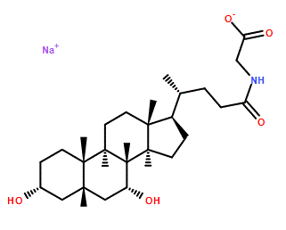
C[C@H](CCC(=O)NCC(=O)O)[C@H]1CC[C@@H]2[C@@]1(CC[C@H]3[C@H]2[C@@H](C[C@H]4[C@@]3(CC[C@H](C4)O)C)O)C
|
| InChi Key |
GHCZAUBVMUEKKP-GYPHWSFCSA-N
|
| InChi Code |
InChI=1S/C26H43NO5/c1-15(4-7-22(30)27-14-23(31)32)18-5-6-19-24-20(9-11-26(18,19)3)25(2)10-8-17(28)12-16(25)13-21(24)29/h15-21,24,28-29H,4-14H2,1-3H3,(H,27,30)(H,31,32)/t15-,16+,17-,18-,19+,20+,21-,24+,25+,26-/m1/s1
|
| 化学名 |
2-[[(4R)-4-[(3R,5S,7R,8R,9S,10S,13R,14S,17R)-3,7-dihydroxy-10,13-dimethyl-2,3,4,5,6,7,8,9,11,12,14,15,16,17-tetradecahydro-1H-cyclopenta[a]phenanthren-17-yl]pentanoyl]amino]acetic acid
|
| 别名 |
16564-43-5; Glycochenodeoxycholic acid sodium salt; Sodium glycochenodeoxycholate; Sodium glycylchenodeoxycholate; NSC 681056; CHENYLGLYCINE SODIUM; Glycochenodeoxycholic acid (sodium salt); OK5NH65A9B;
|
| HS Tariff Code |
2934.99.9001
|
| 存储方式 |
Powder -20°C 3 years 4°C 2 years In solvent -80°C 6 months -20°C 1 month 注意: 请将本产品存放在密封且受保护的环境中,避免吸湿/受潮。 |
| 运输条件 |
Room temperature (This product is stable at ambient temperature for a few days during ordinary shipping and time spent in Customs)
|
| 溶解度 (体外实验) |
DMSO : ~250 mg/mL (~530.10 mM)
H2O : ≥ 100 mg/mL (~212.04 mM) |
|---|---|
| 溶解度 (体内实验) |
配方 1 中的溶解度: ≥ 2.08 mg/mL (4.41 mM) (饱和度未知) in 10% DMSO + 40% PEG300 + 5% Tween80 + 45% Saline (这些助溶剂从左到右依次添加,逐一添加), 澄清溶液。
例如,若需制备1 mL的工作液,可将100 μL 20.8 mg/mL澄清DMSO储备液加入400 μL PEG300中,混匀;然后向上述溶液中加入50 μL Tween-80,混匀;加入450 μL生理盐水定容至1 mL。 *生理盐水的制备:将 0.9 g 氯化钠溶解在 100 mL ddH₂O中,得到澄清溶液。 配方 2 中的溶解度: ≥ 2.08 mg/mL (4.41 mM) (饱和度未知) in 10% DMSO + 90% (20% SBE-β-CD in Saline) (这些助溶剂从左到右依次添加,逐一添加), 澄清溶液。 例如,若需制备1 mL的工作液,可将 100 μL 20.8 mg/mL澄清DMSO储备液加入900 μL 20% SBE-β-CD生理盐水溶液中,混匀。 *20% SBE-β-CD 生理盐水溶液的制备(4°C,1 周):将 2 g SBE-β-CD 溶解于 10 mL 生理盐水中,得到澄清溶液。 View More
配方 3 中的溶解度: ≥ 2.08 mg/mL (4.41 mM) (饱和度未知) in 10% DMSO + 90% Corn Oil (这些助溶剂从左到右依次添加,逐一添加), 澄清溶液。 配方 4 中的溶解度: 50 mg/mL (106.02 mM) in PBS (这些助溶剂从左到右依次添加,逐一添加), 澄清溶液; 超声助溶. 1、请先配制澄清的储备液(如:用DMSO配置50 或 100 mg/mL母液(储备液)); 2、取适量母液,按从左到右的顺序依次添加助溶剂,澄清后再加入下一助溶剂。以 下列配方为例说明 (注意此配方只用于说明,并不一定代表此产品 的实际溶解配方): 10% DMSO → 40% PEG300 → 5% Tween-80 → 45% ddH2O (或 saline); 假设最终工作液的体积为 1 mL, 浓度为5 mg/mL: 取 100 μL 50 mg/mL 的澄清 DMSO 储备液加到 400 μL PEG300 中,混合均匀/澄清;向上述体系中加入50 μL Tween-80,混合均匀/澄清;然后继续加入450 μL ddH2O (或 saline)定容至 1 mL; 3、溶剂前显示的百分比是指该溶剂在最终溶液/工作液中的体积所占比例; 4、 如产品在配制过程中出现沉淀/析出,可通过加热(≤50℃)或超声的方式助溶; 5、为保证最佳实验结果,工作液请现配现用! 6、如不确定怎么将母液配置成体内动物实验的工作液,请查看说明书或联系我们; 7、 以上所有助溶剂都可在 Invivochem.cn网站购买。 |
| 制备储备液 | 1 mg | 5 mg | 10 mg | |
| 1 mM | 2.1204 mL | 10.6020 mL | 21.2040 mL | |
| 5 mM | 0.4241 mL | 2.1204 mL | 4.2408 mL | |
| 10 mM | 0.2120 mL | 1.0602 mL | 2.1204 mL |
1、根据实验需要选择合适的溶剂配制储备液 (母液):对于大多数产品,InvivoChem推荐用DMSO配置母液 (比如:5、10、20mM或者10、20、50 mg/mL浓度),个别水溶性高的产品可直接溶于水。产品在DMSO 、水或其他溶剂中的具体溶解度详见上”溶解度 (体外)”部分;
2、如果您找不到您想要的溶解度信息,或者很难将产品溶解在溶液中,请联系我们;
3、建议使用下列计算器进行相关计算(摩尔浓度计算器、稀释计算器、分子量计算器、重组计算器等);
4、母液配好之后,将其分装到常规用量,并储存在-20°C或-80°C,尽量减少反复冻融循环。
计算结果:
工作液浓度: mg/mL;
DMSO母液配制方法: mg 药物溶于 μL DMSO溶液(母液浓度 mg/mL)。如该浓度超过该批次药物DMSO溶解度,请首先与我们联系。
体内配方配制方法:取 μL DMSO母液,加入 μL PEG300,混匀澄清后加入μL Tween 80,混匀澄清后加入 μL ddH2O,混匀澄清。
(1) 请确保溶液澄清之后,再加入下一种溶剂 (助溶剂) 。可利用涡旋、超声或水浴加热等方法助溶;
(2) 一定要按顺序加入溶剂 (助溶剂) 。
 L-Histidinol-d3
 D-myo-Inositol-3-phosphate sodium
 (±)19(20)-DiHDPA
 4-Hydroxy-2-butanone
InvivoChem的所有产品仅用于作科学研究,不面向患者销售
Copyright 2020 InvivoChem LLC | All Rights Reserved 粤ICP备20063088号-1


 463611831
