| 规格 | 价格 | 库存 | 数量 |
|---|---|---|---|
| 50mg |
|
||
| 100mg |
|
||
| 250mg |
|
||
| 500mg |
|
||
| Other Sizes |
|

| 靶点 |
IC50: 0.94 nM (CBP), 6.2 nM (BRET), 5100 nΜ (BRD4(1))[1]
GNE-781 is a highly developed, strong, and specific bromodomain inhibitor of the CBP (cyclic adenosine monophosphate response element-binding protein). The transcript levels of FOXP3 (forkhead box P3) are decreased by GNE-781. Analysis of a subset of bromodomains revealed that GNE-781 has significant selectivity for both CBP (5425-fold) and P300 (4250-fold), and is very selective for CBP/P300. GNE-781 exhibits cellular potency and selectivity in the ideal ratio—5425 times higher than BRD4 (1)—[1]. |
|---|---|
| 体外研究 (In Vitro) |
GNE-781 是一种高度发达、强效且特异性的 CBP(环磷酸腺苷反应元件结合蛋白)溴结构域抑制剂。 GNE-781 降低了 FOXP3(叉头盒 P3)的转录水平。对溴结构域子集的分析表明,GNE-781 对 CBP(5425 倍)和 P300(4250 倍)具有显着的选择性,并且对 CBP/P300 具有很高的选择性。 GNE-781 表现出理想比例的细胞效力和选择性 — 比 BRD4 高 5425 倍 (1) — [1]。
在生化 TR-FRET 实验中,GNE-781 抑制 CBP 溴结构域的 IC50 为 0.94 nM。 在用于靶点结合的细胞 BRET 实验中,IC50 为 6.21 nM。 它能强效下调细胞中 Myc 蛋白表达,EC50 为 6.63 nM。 BROMOscan 分析证实了其高选择性,显示对大多数脱靶溴结构域的 Kd >10,000 nM,仅对 BRD4(全长,短异构体,Kd=5300 nM)和 BRDT(1)(Kd=4900 nM)有较弱活性。 [1] |
| 体内研究 (In Vivo) |
在携带 MOLM-16 AML 异种移植物的小鼠中,GNE-781(3-30 mg/kg;口服;每天两次,持续 21 天)在 3、10 和 30 mg/kg 剂量下抑制肿瘤生长抑制 (%TGI) 73% 、 71% 和 89% ,分别 [1]。 GNE-781 以剂量依赖性方式降低 Foxp3 转录水平。在剂量低至 3 mg/kg 时,GNE-781 (3-30 mg/kg) 在 2 小时和 8 小时抑制 MYC;在 10 和 30 mg/kg 浓度下,2 小时时达到最大抑制(87% 和 88% 抑制)[1]。
在 AML MOLM-16 皮下异种移植模型中,单次口服 GNE-781 可迅速且深度抑制肿瘤中的 MYC 表达。 给药后 2 小时,3 mg/kg 剂量将 MYC 水平降至对照组的 16%。更高剂量在相同时间点达到最大抑制,降至对照组的 12-13%。 MYC 抑制是短暂的,在所有测试剂量下,水平在 16-24 小时内恢复至接近基线。 [1] |
| 酶活实验 |
时间分辨荧光共振能量转移分析[1]
在一组生化溴结构域结合试验中评估了化合物的效力。通过时间分辨荧光共振能量转移(TR-FRET)评估生物素化小分子配体与重组His标记的溴结构域的结合。与生物素化配体竞争溴结构域结合的测试化合物会降低TR-FRET信号。所有生化测定方案均如前所述进行。 细胞检测方案[1] CBP BRET测定如前所述进行。为了确定MYC表达的抑制作用,将MV-4-11细胞(ATCC)以每孔10000个细胞的速度接种在96孔板中,培养基中补充有10%胎牛血清和2 mM l-谷氨酰胺。将稀释在DMSO中的试验化合物转移到细胞板上,使DMSO的最终浓度保持在0.1%,并在37°C下孵育4小时。使用QuantiGene 2.0试剂并按照供应商的说明进行MYC表达的裂解和分析。使用EnVision平板阅读器读取发光,并使用四参数非线性回归拟合在XLFit中生成EC50s。 使用 BROMOscan 平台测定了 GNE-781 与广谱溴结构域的结合亲和力。该实验测量测试化合物与固定化配体竞争结合可溶的、DNA 标记的溴结构域蛋白,从中得出解离常数 (Kd)。 [1] 使用时间分辨荧光共振能量转移 结合实验测定了 GNE-781 对 CBP 溴结构域的抑制效力 (IC50)。 [1] |
| 细胞实验 |
化合物19(GNE-781)对Tregs的体外评价[1]
使用天然CD4+T细胞分离试剂盒II从健康供体的外周血单个核细胞中分离出人类天然CD4+T淋巴细胞,并在完全RPMI-1640培养基(10%FCS、50μM 2-巯基乙醇、10%青霉素/链霉素、10%NEAA、10%丙酮酸钠)中使用平板结合抗CD3(5μg/mL)、可溶性抗CD28(3μg/mL)加rTGFβ(5 ng/mL)和rIL-2(10 ng/mL。化合物19以2μM的浓度使用,并以2倍稀释度滴定 使用针对表面标记CD4 FITC(克隆OKT-4)和CD25 Pacific Blue(克隆BC96)的抗体对iTreg进行染色,用Foxp3/转录因子染色缓冲液组固定/渗透,并标记细胞内Foxp3 APC(克隆259D/C7)。使用可固定的活性染料efluor 781对iTreg进行活性染色。使用FACSDiva软件在BD LSR Fortessa上采集样本。使用FlowJo软件分析数据 使用RNeasy从iTregs中分离总RNA,包括柱上DNase I消化。使用高容量cDNA逆转录酶试剂盒制备cDNA。使用ABI 7900 HT快速实时PCR系统进行定量RT-PCR以确定Foxp3基因表达水平。基因表达数据被标准化为B2M作为管家基因。 使用生物发光共振能量转移 实验评估细胞内的靶点结合情况,该实验报告 GNE-781 在细胞中对 CBP 的 IC50 为 6.21 nM。 通过免疫实验量化 Myc 蛋白表达的下调来测量 CBP 溴结构域抑制的功能性后果,得出的 EC50 为 6.63 nM。 [1] |
| 动物实验 |
小鼠[1] [1]
本研究使用12只雌性CD-1小鼠。所有动物在研究时均为6-9周龄,体重在20-35克之间。每组给药途径(n=3)分别给予10或GNE-781,剂量为1 mg/kg,静脉注射(溶于35% v/v丙二醇400和65% v/v水)或5 mg/kg,口服(悬浮于0.5% w/v甲基纤维素和0.2% w/v吐温80溶液中)。所有动物均可自由摄取食物和水。分别于静脉给药后 0.033、0.083、0.25、0.5、1、3、8 和 24 小时,以及口服给药后 0.083、0.25、0.5、1、3、8 和 24 小时,通过尾部采血法采集系列血样(15 μL)。所有血样均用 60 μL 含 1.7 mg/mL EDTA 的水溶液稀释,并保存于 -80 °C 直至分析[1]。实验动物[1]:本研究使用 12 只雄性 Sprague-Dawley 大鼠。所有动物在实验时均为 6-9 周龄,体重在 200 至 300 g 之间。每组动物(每组3只)分别静脉注射10或GNE-781(1 mg/kg,溶于35% v/v丙二醇400和65% v/v水的混合溶液中)或口服5 mg/kg(溶于0.5% w/v甲基纤维素和0.2% w/v吐温80的混合溶液中)。静脉注射组动物可自由摄取食物和水。口服组动物禁食过夜,并在给药后4小时停止进食。分别于静脉或口服给药后0.033、0.083、0.25、0.5、1、2、4、8和24小时,通过导管采集约250 μL血液。所有血液样本均收集于含有5 μL 0.5 M K2EDTA的试管中,并进行血浆分离处理。样品采集后1小时内进行离心(4°C,2500g,15分钟),血浆样品保存于-80°C直至分析[1]。 化合物10和19(GNE-781)的体内药代动力学[1] 小鼠药代动力学:使用12只雌性CD-1小鼠。所有动物在研究时均为6-9周龄,体重在20-35克之间。每组给药途径各3只动物,分别以1 mg/kg的剂量静脉注射化合物10或19(溶于35% v/v丙二醇400和65% v/v水)或以5 mg/kg的剂量口服(溶于0.5% w/v甲基纤维素和0.2% w/v吐温80的混合溶液中)。所有动物均可自由摄取食物和水。分别于静脉给药后 0.033、0.083、0.25、0.5、1、3、8 和 24 小时,以及口服给药后 0.083、0.25、0.5、1、3、8 和 24 小时,通过尾部采血法采集系列血样(15 μL)。所有血样均用 60 μL 含 1.7 mg/mL EDTA 的水溶液稀释,并保存于 -80 °C 直至分析。 大鼠药代动力学:[1] 本研究使用了 12 只雄性 Sprague-Dawley 大鼠。所有动物在研究时均为 6-9 周龄,体重在 200-300 g 之间。每组动物(每组3只)分别以1 mg/kg的剂量静脉注射(溶于35% v/v丙二醇400和65% v/v水)或以5 mg/kg的剂量口服(溶于0.5% w/v甲基纤维素和0.2% w/v吐温80的混合溶液中)给药10或19。静脉注射组动物可自由摄取食物和水。口服组动物禁食过夜,并在给药后4小时停止进食。分别于静脉或口服给药后0.033、0.083、0.25、0.5、1、2、4、8和24小时,通过导管采集约250 μL血液。所有血液样本均收集于含有5 μL 0.5 M K2EDTA的试管中,并进行血浆分离处理。样本采集后1小时内进行离心(2500g,4℃,15分钟),血浆样本保存在-80℃直至分析。 查看更多犬类PK:[1] 在携带皮下 MOLM-16 AML 肿瘤的雌性 SCID 小鼠中评估了GNE-781的体内药效学 (PD) 和药代动力学 (PK) 特性。 该化合物配制于含有 0.5% 甲基纤维素和 0.2% Tween-80 的载体中。 小鼠单次灌胃给予 3、10 或 30 mg/kg 的化合物(每组每个时间点 n=3)。 分别在给药后 2、4、8、16 和 24 小时收集肿瘤和血浆。 分析肿瘤裂解物中的 MYC 蛋白。采用 ELISA 法测定血浆和肿瘤匀浆中 GNE-781 的浓度,以评估其药效学效应。 采用 LC-MS/MS 法测定血浆和肿瘤匀浆中 GNE-781 的浓度。[1] |
| 药代性质 (ADME/PK) |
在荷瘤小鼠中口服给药后,GNE-781的总血浆和肿瘤暴露量均达到低至中等水平,呈剂量依赖性,但持续时间相对较短。
例如,30 mg/kg剂量后,2小时的总血浆浓度为2.23 µM,肿瘤浓度为0.46 µM。 校正血浆蛋白结合率(PPB,根据相关化合物假设为94%)后,2小时(30 mg/kg)的游离血浆浓度为0.13 µM,游离肿瘤浓度为0.027 µM。 暴露量迅速下降,给药后16小时游离药物浓度降至可定量水平以下。[1] |
| 毒性/毒理 (Toxicokinetics/TK) |
在浓度为 10 µM 的条件下,对 GNE-781 进行了针对 44 种非靶向受体、离子通道和酶(CEREP 靶标库)的筛选。结果显示,其对 NK1(神经激肽 1)受体的抑制作用最为显著(抑制率为 39%)。对 hERG 和罗利普兰位点(PDE4)的抑制作用较弱(23-30%)。大多数其他靶点的抑制率低于 20%。在浓度为 1 µM 的广谱激酶选择性靶标库(包含 215 种激酶)中,GNE-781 仅对 CamKIV(抑制率为 25.3%)、Cot(抑制率为 23.7%)和 MAP4K4(抑制率为 25.3%)的抑制率超过 25%。对大多数其他激酶的抑制率低于 20%。[1]
|
| 参考文献 | |
| 其他信息 |
抑制转录调控因子CBP/P300的溴结构域是肿瘤治疗领域一个特别引人关注的新策略。研究人员近期公开了一种针对CBP溴结构域的体内化学工具1(GNE-272),该工具具有中等的效力,且对BRD4具有选择性(1)。为了寻找效力更强、选择性更高的CBP抑制剂,我们采用了基于结构的药物设计方法。将化合物1中的苯胺基团改造为四氢喹啉结构,在保持原有效力的同时,将选择性提高了2倍。结构-活性关系研究结合进一步的基于结构的靶向LPF平台、BC环和KAc区域的设计,显著提高了效力和选择性,最终鉴定出非中枢神经系统穿透性化合物19(GNE-781,TR-FRET IC50 = 0.94 nM,BRET IC50 = 6.2 nM;BRD4(1) IC50 = 5100 nM),该化合物在多种动物体内均保持了良好的药代动力学性质。化合物19在急性髓系白血病(AML)肿瘤模型中显示出抗肿瘤活性,并且能够以剂量依赖的方式降低Foxp3转录水平。[1] 研究人员已鉴定出一种高效且选择性的CBP溴结构域体内探针(19,GNE-781),该探针适用于研究CBP的生物学特性,且避免了BET抑制剂带来的复杂性。我们的研究始于近期公开的化合物 1(TR-FRET IC50 = 20 nM,BRET IC50 = 410 nM,BRD4 IC50 = 13,000 nM),该化合物对 CBP 的溴结构域具有中等活性,且对 BRD4 的选择性高 650 倍。将化合物 1 中的苯胺基团限制为四氢喹啉 3 后,化合物 3 的活性得以保持,且选择性比化合物 1 提高了 2 倍。通过结构-活性关系研究以及基于结构的、针对 LPF 平台、BC 环和 KAc 区域的优化设计,我们鉴定出了化合物 10(TR-FRET IC50 = 1.1 nM,BRET IC50 = 12 nM,BRD4 IC50 = 4200 nM)。对该化合物的进一步分析表明,它能够穿透中枢神经系统,导致中枢神经系统不良反应。后续的优化工作重点在于通过引入氢键供体来提高其总前列腺特异性抗原(tPSA)。通过将化合物 10 的 Asn 结合乙酰胺转化为甲基脲,成功鉴定出非中枢神经系统穿透性化合物 19(TR-FRET IC50 = 0.94 nM,BRET IC50 = 6.2 nM,BRD4(1) IC50 = 5100 nM)。该化合物在细胞效力、选择性(对 BRD4 的选择性高 5425 倍)和体内药代动力学方面表现出适当的平衡。化合物 19 的卓越效力和选择性使其能够清晰地区分其药理作用与对 CBP 和 BET 溴结构域的抑制作用。在体内,化合物 19 可调节 MYC 的表达,这与 AML 肿瘤模型中的抗肿瘤活性相一致。体外研究表明,化合物 19 可抑制 FOXP3 表达和 Treg 功能,进一步提示 CBP 溴结构域抑制剂可能是一种新型的小分子癌症免疫治疗方法。[1]
GNE-781 是一种高效、选择性强的 CBP/EP300 溴结构域抑制剂,由早期先导化合物开发而来。 解析了 GNE-781 与 CBP 溴结构域 (PDB: 5W0E) 和 BRD4(1) 溴结构域 (PDB: 5VZS) 的共晶结构,揭示了其结合模式,并为其对 BRD4 的高选择性提供了结构基础。 通过计算二面角扫描,研究了化合物核心骨架的构象偏好以及邻位取代基对平面性和结合能的影响。[1] |
| 分子式 |
C27H33F2N7O2
|
|---|---|
| 分子量 |
525.593432188034
|
| 精确质量 |
525.27
|
| 元素分析 |
C, 61.70; H, 6.33; F, 7.23; N, 18.65; O, 6.09
|
| CAS号 |
1936422-33-1
|
| PubChem CID |
132275066
|
| 外观&性状 |
Light yellow to yellow solid powder
|
| LogP |
2.7
|
| tPSA |
80.4Ų
|
| 氢键供体(HBD)数目 |
1
|
| 氢键受体(HBA)数目 |
7
|
| 可旋转键数目(RBC) |
4
|
| 重原子数目 |
38
|
| 分子复杂度/Complexity |
833
|
| 定义原子立体中心数目 |
0
|
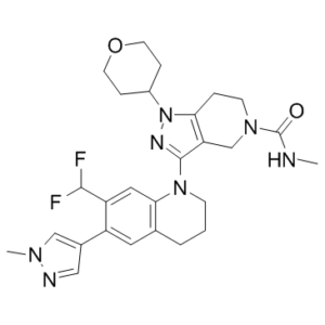
| SMILES |
CNC(=O)N1CCC2=C(C1)C(=NN2C3CCOCC3)N4CCCC5=CC(=C(C=C54)C(F)F)C6=CN(N=C6)C
|
| InChi Key |
CQCWHSDMJBAGDC-UHFFFAOYSA-N
|
| InChi Code |
InChI=1S/C27H33F2N7O2/c1-30-27(37)34-9-5-23-22(16-34)26(32-36(23)19-6-10-38-11-7-19)35-8-3-4-17-12-20(18-14-31-33(2)15-18)21(25(28)29)13-24(17)35/h12-15,19,25H,3-11,16H2,1-2H3,(H,30,37)
|
| 化学名 |
3-[7-(difluoromethyl)-6-(1-methylpyrazol-4-yl)-3,4-dihydro-2H-quinolin-1-yl]-N-methyl-1-tetrahydropyran-4-yl-6,7-dihydro-4H-pyrazolo[4,3-c]pyridine-5-carboxamide
|
| 别名 |
GNE781; GNE 781; 3-(7-(Difluoromethyl)-6-(1-methyl-1H-pyrazol-4-yl)-3,4-dihydroquinolin-1(2H)-yl)-N-methyl-1-(tetrahydro-2H-pyran-4-yl)-1,4,6,7-tetrahydro-5H-pyrazolo[4,3-c]pyridine-5-carboxamide; CHEMBL4097025; 3-[7-(difluoromethyl)-6-(1-methyl-1H-pyrazol-4-yl)-3,4-dihydroquinolin-1(2H)-yl]-N-methyl-1-(oxan-4-yl)-1,4,6,7-tetrahydro-5H-pyrazolo[4,3-c]pyridine-5-carboxamide; GNE-781.
|
| HS Tariff Code |
2934.99.03.00
|
| 存储方式 |
Powder -20°C 3 years 4°C 2 years In solvent -80°C 6 months -20°C 1 month 注意: 请将本产品存放在密封且受保护的环境中(例如氮气保护),避免吸湿/受潮。 |
| 运输条件 |
Room temperature (This product is stable at ambient temperature for a few days during ordinary shipping and time spent in Customs)
|
| 溶解度 (体外实验) |
DMSO : ~100 mg/mL (~190.26 mM)
|
|---|---|
| 溶解度 (体内实验) |
配方 1 中的溶解度: ≥ 2.87 mg/mL (5.46 mM) (饱和度未知) in 5% DMSO + 40% PEG300 + 5% Tween80 + 50% Saline (这些助溶剂从左到右依次添加,逐一添加), 澄清溶液。
*生理盐水的制备:将 0.9 g 氯化钠溶解在 100 mL ddH₂O中,得到澄清溶液。 配方 2 中的溶解度: 2.87 mg/mL (5.46 mM) in 5% DMSO + 95% (20% SBE-β-CD in Saline) (这些助溶剂从左到右依次添加,逐一添加), 悬浊液; 超声助溶。 *20% SBE-β-CD 生理盐水溶液的制备(4°C,1 周):将 2 g SBE-β-CD 溶解于 10 mL 生理盐水中,得到澄清溶液。 View More
配方 3 中的溶解度: ≥ 1.67 mg/mL (3.18 mM) (饱和度未知) in 10% DMSO + 40% PEG300 + 5% Tween80 + 45% Saline (这些助溶剂从左到右依次添加,逐一添加), 澄清溶液。 配方 4 中的溶解度: ≥ 1.67 mg/mL (3.18 mM) (饱和度未知) in 10% DMSO + 90% (20% SBE-β-CD in Saline) (这些助溶剂从左到右依次添加,逐一添加), 澄清溶液。 例如,若需制备1 mL的工作液,可将100μL 16.7mg/mL澄清的DMSO储备液加入到900μL 20%SBE-β-CD生理盐水中,混匀。 *20% SBE-β-CD 生理盐水溶液的制备(4°C,1 周):将 2 g SBE-β-CD 溶解于 10 mL 生理盐水中,得到澄清溶液。 配方 5 中的溶解度: ≥ 1.67 mg/mL (3.18 mM) (饱和度未知) in 10% DMSO + 90% Corn Oil (这些助溶剂从左到右依次添加,逐一添加), 澄清溶液。 例如,若需制备1 mL的工作液,可将100 μL 16.7 mg/mL 澄清 DMSO 储备液加入900 μL 玉米油中,混合均匀。 1、请先配制澄清的储备液(如:用DMSO配置50 或 100 mg/mL母液(储备液)); 2、取适量母液,按从左到右的顺序依次添加助溶剂,澄清后再加入下一助溶剂。以 下列配方为例说明 (注意此配方只用于说明,并不一定代表此产品 的实际溶解配方): 10% DMSO → 40% PEG300 → 5% Tween-80 → 45% ddH2O (或 saline); 假设最终工作液的体积为 1 mL, 浓度为5 mg/mL: 取 100 μL 50 mg/mL 的澄清 DMSO 储备液加到 400 μL PEG300 中,混合均匀/澄清;向上述体系中加入50 μL Tween-80,混合均匀/澄清;然后继续加入450 μL ddH2O (或 saline)定容至 1 mL; 3、溶剂前显示的百分比是指该溶剂在最终溶液/工作液中的体积所占比例; 4、 如产品在配制过程中出现沉淀/析出,可通过加热(≤50℃)或超声的方式助溶; 5、为保证最佳实验结果,工作液请现配现用! 6、如不确定怎么将母液配置成体内动物实验的工作液,请查看说明书或联系我们; 7、 以上所有助溶剂都可在 Invivochem.cn网站购买。 |
| 制备储备液 | 1 mg | 5 mg | 10 mg | |
| 1 mM | 1.9026 mL | 9.5131 mL | 19.0262 mL | |
| 5 mM | 0.3805 mL | 1.9026 mL | 3.8052 mL | |
| 10 mM | 0.1903 mL | 0.9513 mL | 1.9026 mL |
1、根据实验需要选择合适的溶剂配制储备液 (母液):对于大多数产品,InvivoChem推荐用DMSO配置母液 (比如:5、10、20mM或者10、20、50 mg/mL浓度),个别水溶性高的产品可直接溶于水。产品在DMSO 、水或其他溶剂中的具体溶解度详见上”溶解度 (体外)”部分;
2、如果您找不到您想要的溶解度信息,或者很难将产品溶解在溶液中,请联系我们;
3、建议使用下列计算器进行相关计算(摩尔浓度计算器、稀释计算器、分子量计算器、重组计算器等);
4、母液配好之后,将其分装到常规用量,并储存在-20°C或-80°C,尽量减少反复冻融循环。
计算结果:
工作液浓度: mg/mL;
DMSO母液配制方法: mg 药物溶于 μL DMSO溶液(母液浓度 mg/mL)。如该浓度超过该批次药物DMSO溶解度,请首先与我们联系。
体内配方配制方法:取 μL DMSO母液,加入 μL PEG300,混匀澄清后加入μL Tween 80,混匀澄清后加入 μL ddH2O,混匀澄清。
(1) 请确保溶液澄清之后,再加入下一种溶剂 (助溶剂) 。可利用涡旋、超声或水浴加热等方法助溶;
(2) 一定要按顺序加入溶剂 (助溶剂) 。
InvivoChem的所有产品仅用于作科学研究,不面向患者销售
Copyright 2020 InvivoChem LLC | All Rights Reserved 粤ICP备20063088号-1


 COA
COA
 463611831
