| 规格 | 价格 | 库存 | 数量 |
|---|---|---|---|
| 10 mM * 1 mL in DMSO |
|
||
| 1mg |
|
||
| 5mg |
|
||
| 10mg |
|
||
| 25mg |
|
||
| 50mg |
|
||
| 100mg |
|
||
| 250mg |
|
||
| 500mg |
|
||
| Other Sizes |
|

| 靶点 |
FGFR1 (IC50 = 1.2 nM); FGFR2 (IC50 = 2.5 nM); FGFR3 (IC50 = 3.0 nM); FGFR4 (IC50 = 5.7 nM)
FGFR1 (IC50 = 1.2 nM); FGFR2 (IC50 = 2.5 nM); FGFR3 (IC50 = 3.0 nM); FGFR4 (IC50 = 5.7 nM); VEGFR2 (IC50 = 120 nM); PDGFRβ (IC50 = 200 nM); EGFR (IC50 = 450 nM) [1] |
|---|---|
| 体外研究 (In Vitro) |
体外活性:JNJ-42756493 是一种有效的口服泛 FGFR 酪氨酸激酶抑制剂,对 FGFR 家族的所有成员(FGFR1 至 FGFR4)具有低纳摩尔范围内的半最大抑制浓度值,对血管内皮生长的活性极小因子受体 (VEGFR) 激酶与 FGFR 激酶相比(效力差异约 20 倍)。在体外,用 JNJ-42756493 处理的细胞增殖减少,与细胞凋亡增加和细胞存活率降低相关。激酶测定:Erdafitinib (JNJ-42756493) 是一种有效的口服 FGFR 家族抑制剂;抑制 FGFR1/2/3/4,IC50 分别为 1.2、2.5、3.0 和 5.7 nM。细胞测定:在 72 小时时,使用磺罗丹明 B (SRB) 测定法评估贴壁细胞(HCT116、HCA7、Caco2 和 NCI-H716 细胞)和台盼蓝染料排除悬浮液,评估不同药物浓度对细胞生长和存活的影响细胞,NCI-H716。
JNJ-42756493(Erdafitinib)抑制FGFR1扩增的H1581肺癌细胞增殖,IC50为9.5 nM [1] 它抑制FGFR2融合的SNU-16胃癌细胞生长,IC50为14 nM [1] 在FGFR3突变的RT112膀胱癌细胞中,该化合物的抗增殖IC50为7.8 nM [1] Western blot检测显示,JNJ-42756493(Erdafitinib)在H1581细胞中阻断FGFR介导的下游信号(AKT和ERK磷酸化),在100 nM时达到最大抑制效果 [1] 它在FGFR依赖性癌细胞中诱导G1期细胞周期阻滞,伴随p27表达增加和周期蛋白D1表达降低 [1] Annexin V染色和caspase-3/7活性检测显示,该化合物促进RT112细胞凋亡(50 nM时诱导2.8倍活性增加)[1] |
| 体内研究 (In Vivo) |
在体内,仅通过药物治疗,NCI-H716肿瘤的生长就会延迟5天,尽管当停止药物递送时,相对肿瘤体积与对照相比有所增加。 JNJ-42756493显示出良好的药物样特性,并且在肺、肝和肾组织中表现出高分布。 JNJ-42756493 在有效剂量下具有良好的耐受性,并产生有效的剂量依赖性抗肿瘤活性,同时对肿瘤 FGFR 和下游途径成分进行药效调节。
小鼠口服JNJ-42756493(Erdafitinib),剂量为10 mg/kg,每日一次,治疗21天后,对H1581(FGFR1扩增)异种移植瘤的生长抑制率达72% [1] 在SNU-16(FGFR2融合)异种移植模型中,每日口服15 mg/kg剂量与溶媒对照组相比,肿瘤体积减少68% [1] 接受每日12 mg/kg JNJ-42756493(Erdafitinib)治疗的RT112(FGFR3突变)异种移植瘤小鼠,肿瘤生长抑制率达75% [1] 药效学分析显示,治疗组小鼠肿瘤组织中FGFR和ERK的磷酸化水平降低,证实靶点结合有效 [1] |
| 酶活实验 |
Time-resolved fluorescence kinase assays for FGFR1-4 and KDR/FGFR1-4和KDR的时间分辨荧光激酶测定[1]
在384孔黑色Optiplates中进行FGFR1-4和KDR的时间分辨荧光能量转移分析。 通过向含有化合物的混合物中加入酶(分别为0.1、0.8、0.8、0.4和0.7 nmol/L的FGFR 1、2、3、4和KDR)来引发激酶反应,ATP的米氏常数(Km)浓度为每种激酶(分别为5、0.4、25、5和3μmol/L)和500 nmol/L FLT3底物,最终测定体积为30μL。在室温下孵育FGFR1、FGFR3和KDR 60分钟、FGFR2 30分钟和FGFR4 45分钟后,通过加入10μL检测试剂停止酶反应。在室温下孵育1小时后,在Envision阅读器上以337nm的激发和620nm(Eu信号)和665nm(FRET信号)的双发射测量荧光。 Kinase binding assays/激酶结合测定[1] 使用KINOMEscan平台评估JNJ-42756493与397种野生型激酶的结合亲和力。 Cellular kinase assays/细胞激酶测定[1] 用编码TEL(ETV6)-激酶的pcDNA3.1质粒转染IL3依赖性(终浓度为10ng/mL)小鼠BaF3-pro-B细胞(20),并选择与遗传霉素的稳定整合。 采用重组FGFR家族激酶(FGFR1-4)及其他激酶(VEGFR2、PDGFRβ、EGFR)评估抑制活性。实验在含有ATP、MgCl2和荧光肽底物的缓冲液中进行,将系列稀释的测试化合物与酶、底物和ATP在30°C下孵育60分钟。用终止液终止反应后,通过荧光偏振法检测磷酸化底物,计算IC50值 [1] 采用表面等离子体共振(SPR)检测结合亲和力:将FGFR1胞外域固定在传感器芯片上,注入系列稀释的JNJ-42756493(Erdafitinib),通过传感图计算结合动力学参数(ka、kd、KD),FGFR1的KD值为0.8 nM [1] |
| 细胞实验 |
在DMSO中,Erdafitinib被溶解。Erdafitinib用于治疗KATO III、RT-112、A-204、RT-4、DMS-114、A-427和MDA-MB-453细胞(终浓度:2%DMSO;范围为10μM至0.01 nM)。MTT试剂用于评估4天孵育期后细胞的存活率。在540nm处进行光密度测量[1]。
抑制FGFR家族受体磷酸化和下游信号传导[1] 将携带活化FGFR1、2、3或4的细胞系(分别为NCI-H1581、SNU-16、KMS-11和MDA-MB453)用不同浓度的JNJ-42756493处理4小时。移除培养基,用冰冷的磷酸盐缓冲盐水(PBS)洗涤细胞,并将其悬浮在裂解缓冲液中进行蛋白质印迹分析。在用含有FGF2(40ng/mL)的培养基替换之前,用含有100nmol/L JNJ-42756493或DMSO的培养基预处理NCI-H1581 NSCLC细胞系30分钟。用FGF2处理的细胞孵育0分钟(对照组,未用FGF2治疗)、5分钟、10分钟、30分钟、2小时、4小时或8小时。吸出培养基,用冰冷的PBS洗涤细胞,裂解并处理以进行蛋白质印迹分析。 溶酶体化合物积累[1] 在530 nm成像之前,用50 nmol/L LysoTracker red和1μmol/L JNJ-42756493处理GAMG人胶质母细胞瘤细胞30分钟。GAMG细胞用巴非霉素(75 nmol/L)处理1小时,用PBS洗涤,然后在有或没有75 nmol/L巴非霉素时加入补充有1μmol/L JNJ-42756493或JNJ-42883919的培养基。在InCell Analyzer 2000仪器上,每5分钟在Texas Red和CFP通道中获得一次连续图像。将4幅不同图像的感兴趣区域(ROI)密度与T=0进行比较,并将平均差异绘制为百分比变化(%ROI)。 癌细胞系(H1581、SNU-16、RT112)以3×10³个细胞/孔接种到96孔板中,过夜贴壁。加入系列稀释的JNJ-42756493(Erdafitinib),在37°C、5% CO2环境中孵育72小时。采用比色法检测细胞活力,从剂量-反应曲线计算IC50值 [1] Western blot分析流程:H1581细胞用0.1-100 nM JNJ-42756493(Erdafitinib)处理4小时,制备细胞裂解液,经SDS-PAGE分离后转移至膜上,用抗磷酸化FGFR、AKT、ERK抗体及总蛋白对照抗体孵育,化学发光法显影条带 [1] 细胞周期分析:RT112细胞经化合物处理24小时后,用乙醇固定,碘化丙啶染色,流式细胞术检测细胞周期分布 [1] 凋亡检测:处理48小时后,用发光试剂盒检测caspase-3/7活性;通过Annexin V-FITC/PI染色,流式细胞术检测凋亡细胞 [1] |
| 动物实验 |
小鼠:将 Erdafitinib 以 0、3、10 或 30 mg/kg 的剂量口服给予携带 SNU-16 人胃癌(FGFR2 扩增)异种移植瘤的小鼠。分别在给药后 0.5、1、3、7、16 和 24 小时,从每个时间点(每个时间点取 3 只小鼠)提取肿瘤组织和小鼠血浆[1]。
在第 0 天,将人肿瘤细胞系直接注射到雄性裸鼠的腹股沟区域(1 × 10⁷ 个细胞/200 μL/只,与 Matrigel 1:1 混合于培养基中)。待肿瘤形成后,根据肿瘤体积将小鼠随机分为两组:一组仅注射载体(10% HP-β-CD),另一组注射含 JNJ-42756493 的载体,剂量为 5 mL/kg 体重,连续给药 21 天(每组 8-10 只小鼠)。在PDX研究中,使用了Nu/Nu裸鼠。将患者来源的肿瘤样本切碎(约1-2 mm³)后加入Matrigel基质胶中,并将约50 mm³的切碎肿瘤组织皮下植入麻醉(氯胺酮/美托咪定)小鼠的侧腹部。当肿瘤体积达到200至300 mm³时,将小鼠随机分配到各治疗组,各组间平均肿瘤体积和体重保持一致,并按照实验方案进行治疗。[1] JNJ-42756493的药效学和药代动力学分析[1] 将携带SNU-16人胃癌(FGFR2扩增)异种移植瘤的小鼠口服给予0、3、10或30 mg/kg的JNJ-42756493。分别于给药后 0.5、1、3、7、16 和 24 小时收集肿瘤组织和鼠血浆(每个时间点 3 只小鼠)。将肿瘤组织置于液氮中冷冻,研磨后悬浮于裂解缓冲液中[25 mmol/L Tris-HCl (pH 7.5)、2 mmol/L EDTA (pH 8)、2 mmol/L EGTA (pH 8)、1% Triton X-100、0.1% SDS、50 mmol/L β-甘油磷酸二钠、2 mmol/L Na3VO4、4 mmol/L 焦磷酸钠、2 倍 Thermo 蛋白酶/磷酸酶抑制剂混合物]。离心(12,000 rpm,15 分钟;RCF = 15,294)后,将上清液进行 SDS-PAGE 电泳,然后转移至 PVDF 膜上。 当肺癌患者来源的异种移植瘤体积达到约 400 mm³ 时,小鼠经口给予 12.5 mg/kg JNJ-42756493。分别于给药后 1、2、4、8 和 24 小时收集肿瘤组织和小鼠血浆(每个时间点 3 只小鼠)。 H1581 异种移植瘤模型:将 5×10⁶ 个 H1581 细胞皮下植入雌性裸鼠体内。当肿瘤体积达到 150–200 mm³ 时,将小鼠随机分为载体组和治疗组。 JNJ-42756493(厄达替尼) 以 0.5% 羟丙基纤维素 + 0.1% Tween 80 配制,每日一次口服给药,剂量为 10 mg/kg,疗程 21 天。每周测量两次肿瘤体积和体重[1] SNU-16 异种移植模型:将 1×10⁷ 个 SNU-16 细胞植入雄性裸鼠体内。当肿瘤体积达到 200 mm³ 时开始治疗,每日口服给药 15 mg/kg,疗程 28 天。定期监测肿瘤生长和体重[1] RT112 异种移植模型:将 2×10⁶ 个 RT112 细胞皮下注射到雌性裸鼠体内。当肿瘤体积达到 180 mm³ 时,小鼠每日口服 12 mg/kg 的 JNJ-42756493(厄达替尼),连续 24 天。研究结束时收集肿瘤样本进行药效学分析 [1] |
| 药代性质 (ADME/PK) |
吸收、分布和排泄
每日一次服用8 mg厄达替尼后,平均(变异系数[CV%])稳态最大血浆浓度(Cmax)、曲线下面积(AUCtau)和最小血浆浓度(Cmin)分别为1399 ng/mL (51%)、29268 ng·h/mL (60%)和936 ng/mL (65%)。单次和重复每日一次给药后,厄达替尼的暴露量(最大血浆浓度[Cmax]和血浆浓度-时间曲线下面积[AUC])在0.5至12 mg的剂量范围内呈比例增加(相当于最大批准推荐剂量的0.06至1.3倍)。每日一次给药2周后达到稳态,平均累积率为4倍。达到血浆峰值浓度的中位时间(tmax)为 2.5 小时(范围:2 至 6 小时)。在健康受试者中,摄入高脂肪高热量餐(800 至 1000 卡路里,其中约 50% 的总热量来自脂肪)后,未观察到厄达替尼药代动力学方面具有临床意义的差异。 单次口服放射性标记的厄达替尼后,约 69% 的剂量从粪便中回收(其中 19% 为原形),19% 从尿液中回收(其中 13% 为原形)。 厄达替尼在患者体内的平均表观分布容积约为 26 至 29 升。 厄达替尼的平均表观总清除率 (CL/F) 约为 0.362 升/小时,而口服清除率约为 0.26 升/小时。 代谢/代谢物 厄达替尼主要通过细胞色素代谢。 CYP2C9 和 CYP3A4 同工酶在人体内形成 O-去甲基化的主要代谢物。据估计,CYP2C9 和 CYP3A4 对厄达替尼总清除率的贡献分别为 39% 和 20%。最终,血浆中发现的主要药物相关成分为未改变的厄达替尼,未观察到循环代谢物。 生物半衰期 厄达替尼的平均有效半衰期为 59 小时,但也观察到 50 至 60 小时的情况。 小鼠单次口服 10 mg/kg 剂量的JNJ-42756493(厄达替尼)后,其生物利用度为 68% [1] 小鼠静脉注射 5 mg/kg 剂量后,该化合物的血浆半衰期 (t1/2) 为 4.2 小时 [1] 大鼠口服 10 mg/kg 剂量的厄达替尼的生物利用度为 59%,血浆 t1/2 为 5.1 小时 [1] 该药物显示出广泛的组织分布,肿瘤/血浆浓度比值较高。口服给药4小时后,H1581异种移植小鼠的血药浓度为3.2 [1] 人肝微粒体代谢稳定性研究表明,该药物的半衰期为85分钟,CYP3A4被确定为主要代谢酶 [1] |
| 毒性/毒理 (Toxicokinetics/TK) |
肝毒性
在厄达替尼治疗尿路上皮癌患者的上市前临床试验中,肝功能异常较为常见,但通常程度较轻。高达 41% 的厄达替尼治疗患者出现不同程度的 ALT 升高,但仅有 1% 至 2% 的患者 ALT 升高超过正常值上限 5 倍。在这些纳入约 400 例患者的试验中,未报告严重或临床上明显的肝损伤,也未报告肝脏相关死亡病例。自厄达替尼获批并广泛应用以来,未再有因使用而导致肝损伤的报告。然而,治疗期间血清转氨酶升高发生率较高,提示可能出现罕见的临床表现明显的肝损伤。 可能性评分:E(未经证实但怀疑是罕见的临床表现明显的肝损伤原因)。 妊娠和哺乳期影响 ◉ 哺乳期用药概述 目前尚无关于厄达替尼在哺乳期临床应用的信息。由于厄达替尼与血浆蛋白的结合率高达99.8%,因此其在乳汁中的含量可能很低。然而,其在成人体内的半衰期约为59小时,因此可能在婴儿体内蓄积。制造商建议在厄达替尼治疗期间以及末次给药后1个月内停止母乳喂养。 ◉ 对母乳喂养婴儿的影响 截至修订日期,未找到相关的已发表信息。 ◉ 对泌乳和母乳的影响 截至修订日期,未找到相关的已发表信息。 蛋白结合 厄达替尼的蛋白结合率约为99.8%,主要与α-1-酸性糖蛋白结合。 在一项为期28天的大鼠重复给药毒性研究中,口服剂量高达30 mg/kg/天的JNJ-42756493(厄达替尼)导致轻度体重减轻(≤10%)以及血清肌酐和尿素氮可逆性升高(提示轻度肾脏影响)。剂量≥20 mg/kg时[1] 人血浆蛋白结合率为91%,小鼠血浆蛋白结合率为89%,大鼠血浆蛋白结合率为87%[1] hERG通道活性测定未观察到明显的心脏毒性(IC50 > 10 μM)[1] |
| 参考文献 | |
| 其他信息 |
药效学
给药后观察到,厄达替尼会因抑制FGFR而导致血清磷酸盐水平升高。在早期治疗周期中,应将厄达替尼的剂量增加至最大推荐剂量,以达到5.5–7.0 mg/dL的目标血清磷酸盐水平,并需每日持续给药。随后,在厄达替尼的临床试验中,除非别无选择,否则禁止使用可能升高血清磷酸盐水平的药物,例如磷酸钾补充剂、维生素D补充剂、抗酸剂、含磷酸盐的灌肠剂或泻药,以及已知以磷酸盐为辅料的药物。为控制磷酸盐升高,使用了磷酸盐结合剂。此外,在根据血清磷酸盐水平进行厄达替尼初始剂量增加之前,也避免同时使用可能改变血清磷酸盐水平的药物。此外,一项纳入187例癌症患者的开放标签、剂量递增和剂量扩展研究对QTc间期进行了评估,结果表明厄达替尼对QTc间期无显著影响(即>20毫秒)。 JNJ-42756493(厄达替尼)是一种功能选择性的小分子FGFR家族抑制剂,用于治疗FGFR改变的实体瘤[1]。 它与FGFR激酶的ATP结合口袋结合,抑制其催化活性以及参与细胞增殖、存活和血管生成的下游信号通路[1]。 该化合物已进入膀胱癌、肺癌和其他FGFR依赖性恶性肿瘤的临床试验[1]。 |
| 分子式 |
C25H30N6O2
|
|
|---|---|---|
| 分子量 |
446.54
|
|
| 精确质量 |
446.243
|
|
| 元素分析 |
C, 67.24; H, 6.77; N, 18.82; O, 7.17
|
|
| CAS号 |
1346242-81-6
|
|
| 相关CAS号 |
|
|
| PubChem CID |
67462786
|
|
| 外观&性状 |
Yellow solid powder
|
|
| 密度 |
1.2±0.1 g/cm3
|
|
| 沸点 |
662.3±55.0 °C at 760 mmHg
|
|
| 熔点 |
142°C
|
|
| 闪点 |
354.4±31.5 °C
|
|
| 蒸汽压 |
0.0±2.0 mmHg at 25°C
|
|
| 折射率 |
1.618
|
|
| LogP |
3.6
|
|
| tPSA |
77.33
|
|
| 氢键供体(HBD)数目 |
1
|
|
| 氢键受体(HBA)数目 |
7
|
|
| 可旋转键数目(RBC) |
9
|
|
| 重原子数目 |
33
|
|
| 分子复杂度/Complexity |
583
|
|
| 定义原子立体中心数目 |
0
|
|
| SMILES |
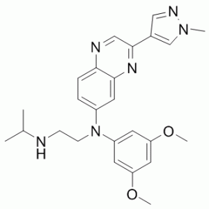
O(C([H])([H])[H])C1C([H])=C(C([H])=C(C=1[H])N(C1C([H])=C([H])C2C(C=1[H])=NC(C1C([H])=NN(C([H])([H])[H])C=1[H])=C([H])N=2)C([H])([H])C([H])([H])N([H])C([H])(C([H])([H])[H])C([H])([H])[H])OC([H])([H])[H]
|
|
| InChi Key |
OLAHOMJCDNXHFI-UHFFFAOYSA-N
|
|
| InChi Code |
InChI=1S/C25H30N6O2/c1-17(2)26-8-9-31(20-10-21(32-4)13-22(11-20)33-5)19-6-7-23-24(12-19)29-25(15-27-23)18-14-28-30(3)16-18/h6-7,10-17,26H,8-9H2,1-5H3
|
|
| 化学名 |
N'-(3,5-dimethoxyphenyl)-N'-[3-(1-methylpyrazol-4-yl)quinoxalin-6-yl]-N-propan-2-ylethane-1,2-diamine
|
|
| 别名 |
|
|
| HS Tariff Code |
2934.99.9001
|
|
| 存储方式 |
Powder -20°C 3 years 4°C 2 years In solvent -80°C 6 months -20°C 1 month |
|
| 运输条件 |
Room temperature (This product is stable at ambient temperature for a few days during ordinary shipping and time spent in Customs)
|
| 溶解度 (体外实验) |
|
|||
|---|---|---|---|---|
| 溶解度 (体内实验) |
配方 1 中的溶解度: ≥ 2.75 mg/mL (6.16 mM) (饱和度未知) in 5% DMSO + 40% PEG300 + 5% Tween80 + 50% Saline (这些助溶剂从左到右依次添加,逐一添加), 澄清溶液。
*生理盐水的制备:将 0.9 g 氯化钠溶解在 100 mL ddH₂O中,得到澄清溶液。 配方 2 中的溶解度: ≥ 2.75 mg/mL (6.16 mM) (饱和度未知) in 5% DMSO + 95% (20% SBE-β-CD in Saline) (这些助溶剂从左到右依次添加,逐一添加), 澄清溶液。 *20% SBE-β-CD 生理盐水溶液的制备(4°C,1 周):将 2 g SBE-β-CD 溶解于 10 mL 生理盐水中,得到澄清溶液。 View More
配方 3 中的溶解度: ≥ 2.33 mg/mL (5.22 mM) (饱和度未知) in 10% DMSO + 90% Corn Oil (这些助溶剂从左到右依次添加,逐一添加), 澄清溶液。 配方 4 中的溶解度: ≥ 2.08 mg/mL (4.66 mM) (饱和度未知) in 10% DMSO + 40% PEG300 + 5% Tween80 + 45% Saline (这些助溶剂从左到右依次添加,逐一添加), 澄清溶液。 例如,若需制备1 mL的工作液,可将100 μL 20.8 mg/mL澄清的DMSO储备液加入400 μL PEG300中,混匀;再向上述溶液中加入50 μL Tween-80,混匀;然后加入450 μL生理盐水定容至1 mL。 *生理盐水的制备:将 0.9 g 氯化钠溶解在 100 mL ddH₂O中,得到澄清溶液。 配方 5 中的溶解度: ≥ 2.08 mg/mL (4.66 mM) (饱和度未知) in 10% DMSO + 90% (20% SBE-β-CD in Saline) (这些助溶剂从左到右依次添加,逐一添加), 澄清溶液。 例如,若需制备1 mL的工作液,可将100μL 20.8mg/mL澄清的DMSO储备液加入到900μL 20%SBE-β-CD生理盐水中,混匀。 *20% SBE-β-CD 生理盐水溶液的制备(4°C,1 周):将 2 g SBE-β-CD 溶解于 10 mL 生理盐水中,得到澄清溶液。 配方 6 中的溶解度: 5%DMSO+40%PEG300+5%Tween80+50%ddH2O: 22.25mg/ml 1、请先配制澄清的储备液(如:用DMSO配置50 或 100 mg/mL母液(储备液)); 2、取适量母液,按从左到右的顺序依次添加助溶剂,澄清后再加入下一助溶剂。以 下列配方为例说明 (注意此配方只用于说明,并不一定代表此产品 的实际溶解配方): 10% DMSO → 40% PEG300 → 5% Tween-80 → 45% ddH2O (或 saline); 假设最终工作液的体积为 1 mL, 浓度为5 mg/mL: 取 100 μL 50 mg/mL 的澄清 DMSO 储备液加到 400 μL PEG300 中,混合均匀/澄清;向上述体系中加入50 μL Tween-80,混合均匀/澄清;然后继续加入450 μL ddH2O (或 saline)定容至 1 mL; 3、溶剂前显示的百分比是指该溶剂在最终溶液/工作液中的体积所占比例; 4、 如产品在配制过程中出现沉淀/析出,可通过加热(≤50℃)或超声的方式助溶; 5、为保证最佳实验结果,工作液请现配现用! 6、如不确定怎么将母液配置成体内动物实验的工作液,请查看说明书或联系我们; 7、 以上所有助溶剂都可在 Invivochem.cn网站购买。 |
| 制备储备液 | 1 mg | 5 mg | 10 mg | |
| 1 mM | 2.2394 mL | 11.1972 mL | 22.3944 mL | |
| 5 mM | 0.4479 mL | 2.2394 mL | 4.4789 mL | |
| 10 mM | 0.2239 mL | 1.1197 mL | 2.2394 mL |
1、根据实验需要选择合适的溶剂配制储备液 (母液):对于大多数产品,InvivoChem推荐用DMSO配置母液 (比如:5、10、20mM或者10、20、50 mg/mL浓度),个别水溶性高的产品可直接溶于水。产品在DMSO 、水或其他溶剂中的具体溶解度详见上”溶解度 (体外)”部分;
2、如果您找不到您想要的溶解度信息,或者很难将产品溶解在溶液中,请联系我们;
3、建议使用下列计算器进行相关计算(摩尔浓度计算器、稀释计算器、分子量计算器、重组计算器等);
4、母液配好之后,将其分装到常规用量,并储存在-20°C或-80°C,尽量减少反复冻融循环。
计算结果:
工作液浓度: mg/mL;
DMSO母液配制方法: mg 药物溶于 μL DMSO溶液(母液浓度 mg/mL)。如该浓度超过该批次药物DMSO溶解度,请首先与我们联系。
体内配方配制方法:取 μL DMSO母液,加入 μL PEG300,混匀澄清后加入μL Tween 80,混匀澄清后加入 μL ddH2O,混匀澄清。
(1) 请确保溶液澄清之后,再加入下一种溶剂 (助溶剂) 。可利用涡旋、超声或水浴加热等方法助溶;
(2) 一定要按顺序加入溶剂 (助溶剂) 。
| NCT Number | Recruitment | interventions | Conditions | Sponsor/Collaborators | Start Date | Phases |
| NCT02365597 | Active Recruiting |
Drug: Erdafitinib Drug: Midazolam |
Urothelial Cancer | Janssen Research & Development, LLC |
April 22, 2015 | Phase 2 |
| NCT03238196 | Active Recruiting |
Drug: Erdafitinib Drug: Palbociclib |
Metastatic Breast Cancer | Vanderbilt-Ingram Cancer Center | August 18, 2017 | Phase 1 |
| NCT04172675 | Active Recruiting |
Drug: Erdafitinib Drug: Investigator Choice (Mitomycin C) |
Urinary Bladder Neoplasms | Janssen Research & Development, LLC |
February 28, 2020 | Phase 2 |
| NCT02699606 | Active Recruiting |
Drug: Erdafitinib | Neoplasm | Janssen Research & Development, LLC |
July 8, 2016 | Phase 2 |
| NCT04083976 | Active Recruiting |
Drug: Erdafitinib | Advanced Solid Tumor | Janssen Research & Development, LLC |
November 20, 2019 | Phase 2 |
") JNJ-42756493 inhibits FGFR auto-phosphorylation in cancer cells lines with activated FGFR1-4 and FGFR-dependent signaling in NCI-H1581 cells.
Relationship betweenin vivoJNJ-42756493 plasma concentration, inhibition of pFGFR2, and efficacy in SNU-16 human gastric xenograft mouse model.Mol Cancer Ther.2017 Jun;16(6):1010-1020. |
|---|
") JNJ-42756493 antiproliferative activity against human cancer cell lines.
Relationship betweenin vivoJNJ-42756493 plasma concentration, inhibition of pERK and efficacy in LUX001 PDX with FGFR3–TACC3 fusion mouse model.Mol Cancer Ther.2017 Jun;16(6):1010-1020. |
") Lysosomal accumulation of JNJ-42756493 and sustained inhibition of FGFR following compound washout.GAMG cells showing (A) intrinsic fluorescence of JNJ-427556493 (green), fluorescence of a lysosome staining probe (LysoTracker, red), and merging of the 2 images (merged, yellow).B,Reduced lysosomal fluorescence intensity of JNJ-42756493 and LysoTracker in the presence of bafilomycin (C) absence of changes in JNJ-42883919 fluorescence intensity compared with LysoTracker in the presence of bafilomycin.Mol Cancer Ther.2017 Jun;16(6):1010-1020. |
 TYRA-200
 FGFR-IN-22
 FGFR2 M537I Recombinant Human Active Protein Kinase
 FGFR2 N549K Recombinant Human Active Protein Kinase
InvivoChem的所有产品仅用于作科学研究,不面向患者销售
Copyright 2020 InvivoChem LLC | All Rights Reserved 粤ICP备20063088号-1


 COA
COA
")
")
 463611831
