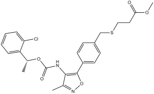
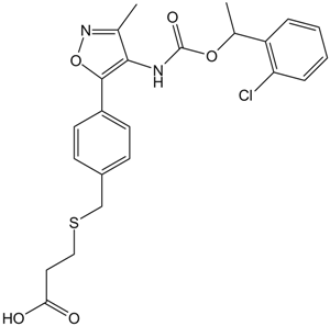
Ki16198; Ki-16198; 355025-13-7; Methyl 3-((4-(4-(((1-(2-chlorophenyl)ethoxy)carbonyl)amino)-3-methylisoxazol-5-yl)benzyl)thio)propanoate; 3-(4-(4-((1-(2-chlorophenyl)ethoxy)carbonylamino)-3-methyl-5-isoxazolyl)benzylsulfanyl)propanoic acid methyl ester; methyl 3-[({4-[4-({[1-(2-chlorophenyl)ethoxy]carbonyl}amino)-3-methyl-1,2-oxazol-5-yl]phenyl}methyl)sulfanyl]propanoate; methyl 3-[[4-[4-[1-(2-chlorophenyl)ethoxycarbonylamino]-3-methyl-1,2-oxazol-5-yl]phenyl]methylsulfanyl]propanoate; SCHEMBL709655; CHEMBL4303365; Ki 16198
| 规格 | 价格 | 库存 | 数量 |
|---|---|---|---|
| 5mg |
|
||
| 10mg |
|
||
| 25mg |
|
||
| 50mg |
|
||
| 100mg |
|
||
| 250mg |
|
||
| 500mg |
|
||
| Other Sizes |
|

| 靶点 |
LPA1 receptor ( Ki = 0.34 μM ); LPA1 receptor ( Ki = 0.34 μM )
Sphingosine-1-phosphate receptor 2 (S1P2) (Ki = 2.1 nM, human; IC50 = 3.5 nM for S1P-induced calcium mobilization inhibition) [2] - Sphingosine-1-phosphate receptor 3 (S1P3) (Ki = 3.7 nM, human; IC50 = 5.8 nM for S1P-induced RhoA activation inhibition) [2] - No significant affinity for S1P1/S1P4/S1P5 receptors (Ki > 1000 nM) [2] |
|---|---|
| 体外研究 (In Vitro) |
Ki16198 或 Ki16425 以类似的效力显着抑制 LPA1 和 LPA3 介导的反应,对 LPA2 的效力较低,对 LPA4、LPA5 和 LPA6 无活性。 Ki16198 (10 μM) 还可有效抑制 YAPC-PD 癌细胞系中 LPA 的迁移和侵袭反应,其效力与 Ki16425 相似。 Ki16198 (10 μM) 抑制 YAPC-PD 细胞中 LPA 诱导的 proMMP-9 蛋白和 mRNA 的表达。 Ki16198 (1 μM) 可抑制 lpa1Δ-1 和 lpa1Δ+-1 细胞的增殖约 70%。激酶测定:将表达 LPA1、LPA2、LPA3、LPA4 或 LPA5 的 RH7777 细胞培养在胶原包被的 12 孔培养皿中的生长培养基中,然后将培养基更换为含有 2 μCi/mL [3H]肌醇和 0.1 % (w/v) BSA(分数 V)。 24小时后,用HEPES缓冲培养基洗涤细胞3次,其组成为20 mM Hepes (pH 7.4)、134 mM NaCl、4.7 mM KCl、1.2 mM KH2PO4、1.2 mM MgSO4、2 mM CaCl2、2.5 mM NaHCO3、5 mM 葡萄糖和 0.1% (w/v) BSA,并与指定浓度的 Ki16425 或 Ki16198(含或不含 1 μM LPA)在 10 mM LiCl 存在的相同培养基中孵育 30 分钟,最终体积为0.5 毫升。通过添加 1 N HCl (0.1 mL) 并冷冻细胞来终止反应。解冻细胞的上清液(0.5 mL 酸提取物)用于分离[3H]磷酸肌醇级分。结果标准化为细胞肌醇脂质中总放射性的 105 dpm。测量三氯乙酸(5%)不溶部分的放射性作为总放射性。细胞测定:将 YAPC-PD 细胞或 Panc-1 细胞以 1 × 10⁴ 个细胞接种在 1 mL 的 12 孔板上。实验前16小时,将培养基更换为含有0.1%BSA的RPMI1640。然后,在存在或不存在 Ki16198 的情况下,在相同培养基中刺激细胞 24 小时。通过细胞减少MTT(3-(4,5-二甲基-2-噻唑基)-2,5-二苯基四唑溴化物的能力来测量增殖活性。
Ki16198 是强效、选择性S1P2/S1P3受体拮抗剂,对其他S1P受体亚型具有高特异性[2] - 在表达S1P2的HEK293细胞中,Ki16198(0.01-100 nM)剂量依赖性阻断S1P诱导的细胞内钙动员,IC50为3.5 nM,逆转S1P介导的信号传导[2] - 在表达S1P3的CHO细胞中,Ki16198(0.1-100 nM)抑制S1P诱导的RhoA激活,IC50为5.8 nM,抑制下游细胞骨架重排[2] - 在人肝癌(HepG2)细胞中,Ki16198(1-20 μM)剂量依赖性抑制细胞增殖,IC50为4.5 μM,通过激活caspase-3/7诱导凋亡(20 μM浓度下凋亡率从8%升至42%)[1] - 在人非小细胞肺癌(A549)细胞中,Ki16198(2-20 μM)减少细胞迁移50-75%,通过下调Snail和Twist表达阻断S1P介导的上皮-间质转化(EMT)[1] - 在大鼠心肌成纤维细胞中,Ki16198(1-10 μM)抑制S1P诱导的细胞增殖40-65%,减少I/III型胶原蛋白合成35-55%,减弱成纤维细胞活化[3] |
| 体内研究 (In Vivo) |
对LPA1和LPA3有效的Ki16198口服到YAPC–PD胰腺癌症细胞接种的裸鼠中,显著抑制肿瘤重量,显著减轻对肺、肝和脑的侵袭和转移,并抑制体内腹水中基质金属蛋白酶(MMP)的积聚。Ki16198在体外抑制了LPA诱导的几种癌症细胞的迁移和侵袭,这与LPA诱导MMP产生的抑制有关。总之,Ki16198是一种很有前途的口服活性LPA拮抗剂,可抑制胰腺癌症细胞的侵袭和转移。拮抗剂对体内侵袭和转移的抑制作用可以通过抑制癌症细胞的运动活性和MMP产生来部分解释。[1]
在 YAPC-PD 异种移植小鼠模型中,Ki16198 (2 mg/kg) 显着降低腹腔内转移淋巴结总重量和腹水形成 50%。 Ki16198(口服 60 毫克/千克)显着抑制乳酸诱导的大鼠肢体损伤。 在携带HepG2人肝癌异种移植瘤的裸鼠中,口服Ki16198(5-20 mg/kg/天,连续21天)剂量依赖性减少肿瘤体积35-60%,增加瘤内凋亡(TUNEL阳性细胞)2.3-3.8倍[1] - 在心肌梗死(MI)诱导的心脏纤维化大鼠中,腹腔注射Ki16198(1-5 mg/kg,每3天一次,连续4周)减少左心室纤维化30-50%,使左心室射血分数(LVEF)从42%提升至58%(5 mg/kg剂量)[3] - 在A549肺癌异种移植小鼠中,Ki16198(10 mg/kg/天,口服)抑制肺转移65%,降低肿瘤微血管密度40%[1] - 在MI大鼠中,Ki16198(5 mg/kg)下调心脏促纤维化基因(TGF-β1、α-SMA)和促炎细胞因子(TNF-α、IL-6)40-60%[3] |
| 酶活实验 |
在胶原蛋白包被的 12 孔培养皿上,表达 LPA1、LPA2、LPA3、LPA4 或 LPA5 的 RH7777 细胞在生长培养基中培养。之后,将培养基更换为含有 2 μCi/mL [ 3 H]肌醇和 0.1% (w/v) BSA(组分 V)的 TCM199。然后将细胞与指定浓度的 Ki16425 或 Ki16198(含或不含 1 μM LPA)在 10 mM LiCl 存在的相同培养基中孵育 30 分钟,最终体积为 0.5 mL。 24小时后,用HEPES缓冲培养基冲洗细胞3次,该培养基由20 mM Hepes (pH 7.4)、134 mM NaCl、4.7 mM KCl、1.2 mM KH2PO4、1.2 mM MgSO4、2.5 mM NaHCO3、5 mM 葡萄糖和 0.1% (w/v) BSA。将细胞冷冻并添加 1 N HCl (0.1 mL) 以终止反应。使用解冻细胞的上清液(0.5 mL 酸提取物)分离 [ 3 H]肌醇磷酸部分。数据标准化为整合到细胞肌醇脂质中的总放射性的 10 5 dpm。测定三氯乙酸(5%)不溶部分的总放射性。
S1P2/S1P3受体结合实验:制备表达人S1P2/S1P3的细胞膜制剂,与[³H]-S1P(0.5 nM)及不同浓度的Ki16198(0.001-1000 nM)在25°C孵育90分钟。在过量未标记S1P存在下测定非特异性结合,过滤分离结合态配体,定量放射性强度以计算Ki值[2] - S1P诱导的钙动员实验:给表达S1P2的HEK293细胞负载钙敏感染料,经Ki16198(0.01-100 nM)预处理20分钟后,用S1P(100 nM)刺激。通过流式细胞术监测钙荧光强度,确定IC50值[2] - RhoA激活实验:S1P3-CHO细胞饥饿12小时后,经Ki16198(0.1-100 nM)预处理30分钟,再用S1P(10 nM)刺激15分钟。使用RhoA特异性结合蛋白通过下拉实验测定RhoA活性[2] |
| 细胞实验 |
在 12 孔板上,YAPC-PD 或 Panc-1 细胞以每毫升 1 × 10 4 细胞的密度接种。在实验前 16 小时将培养基更换为含有 0.1% BSA 的 RPMI1640。然后在含有或不含 Ki16198 的相同培养基中刺激细胞 24 小时。细胞减少MTT(3-(4,5-二甲基-2-噻唑基)-2,5-二苯基溴化四唑)的能力用于测量增殖活性。
肿瘤细胞增殖实验:HepG2/A549细胞接种于96孔板,经Ki16198(0.1-50 μM)处理72小时。通过CCK-8法测定细胞活力,计算抗增殖活性的IC50值[1] - 肿瘤细胞凋亡实验:HepG2细胞经Ki16198(5-20 μM)处理48小时后,用膜联蛋白V-FITC和碘化丙啶染色,流式细胞术分析凋亡率。通过发光试剂盒检测caspase-3/7活性[1] - 心肌成纤维细胞活化实验:从新生大鼠心脏分离心肌成纤维细胞,接种于6孔板,经Ki16198(1-10 μM)联合S1P(1 μM)处理48小时。Western blot检测I/III型胶原蛋白表达,MTT法测定细胞增殖[3] - 肿瘤细胞迁移实验:A549细胞经Ki16198(2-20 μM)预处理30分钟后加入Transwell上室,下室加入S1P(100 nM)。24小时后计数迁移细胞[1] |
| 动物实验 |
溶于PBS/12.5% DMSO;2mg/kg;口服给药。
YAPC-PD异种移植小鼠模型:雄性BALB/c裸鼠(6周龄)购自Charles River Japan, Inc.(日本横滨),用于体内研究。所有动物实验均按照群马大学动物伦理委员会的指导原则进行。我们研究了LPA及其受体拮抗剂Ki16198对腹膜播散和转移至肝脏、肺和脑等组织的影响,具体方法如下:将YAPC-PD细胞(1×10⁷个细胞,100 μL)于第0天经小鼠右侧腹部注射入腹腔。在LPA实验中,从第0天至第7天,每天腹腔注射生物活性脂质(0.4 μmol,100 μL),并在第7天处死小鼠。在LPA拮抗剂实验中,从第0天(接种癌细胞系前)至第28天,每天给小鼠口服Ki16198(1 mg溶于500 μL PBS/12.5% DMSO溶液)。对照组小鼠则给予溶剂(LPA实验组100 μL生理盐水,Ki16198实验组500 μL 12.5% DMSO溶液)。收集腹水以测定MMP活性,并通过称量所有可见肿瘤结节来确定肿瘤体积。通过检测分离的肝脏、肺和脑组织中人甘油醛-3-磷酸脱氢酶(GAPDH)和鼠GAPDH的mRNA表达水平来评估肿瘤的侵袭或转移活性。 [1]HepG2肝细胞癌异种移植模型:将HepG2细胞(5×10⁶个细胞/只)皮下接种到雌性裸鼠(18-22 g)体内。当肿瘤体积达到100 mm³时,将Ki16198悬浮于0.5% CMC-Na溶液中,并以5、10、20 mg/kg/天的剂量灌胃给药,持续21天。评估肿瘤体积、重量和细胞凋亡情况。[1] - 心肌梗死(MI)大鼠模型:将雄性Sprague-Dawley大鼠(250-300 g)行左前降支冠状动脉结扎术以诱导MI。将Ki16198(1、3、5 mg/kg)溶于生理盐水中,每3天腹腔注射一次,持续4周。采用超声心动图和组织学方法分析心脏功能(左室射血分数,LVEF)和纤维化[3] - A549肺癌转移模型:将A549细胞(2×10⁶个细胞/只)静脉注射到裸鼠(18-22 g)体内。将Ki16198(10 mg/kg/天)悬浮于0.5% CMC-Na溶液中,连续28天口服给药。计数肺转移结节,并通过免疫组织化学方法检测微血管密度[1] |
| 药代性质 (ADME/PK) |
口服生物利用度:小鼠口服10 mg/kg后约为55% [1]
- 消除半衰期:小鼠为5.1小时 [1] |
| 毒性/毒理 (Toxicokinetics/TK) |
急性毒性:小鼠口服LD50 > 200 mg/kg [1]
- 亚慢性毒性(异种移植小鼠21天口服给药):剂量高达20 mg/kg/天时,未见明显的肝毒性或肾毒性;体重或血液学参数无变化 [1] - 慢性毒性(MI大鼠4周腹腔注射给药):剂量高达5 mg/kg时,血清肌酐、BUN、ALT/AST水平无明显异常 [3] - 治疗动物未观察到明显的不良反应(例如,胃肠道不适、器官损伤)[1][3] |
| 参考文献 | |
| 其他信息 |
胰腺癌具有高度转移性,预后极差。然而,目前尚无针对胰腺癌的有效治疗方法。溶血磷脂酸(LPA)已被证实存在于癌细胞的流出液中,并在体外参与多种癌细胞(包括胰腺癌细胞)的迁移和增殖。本研究旨在探讨口服LPA拮抗剂是否能有效抑制体内胰腺癌的肿瘤发生和转移。将对LPA(1)和LPA(3)均有效的Ki16198口服给予接种YAPC-PD胰腺癌细胞的裸鼠后,可显著抑制肿瘤重量,并显著减弱癌细胞向肺、肝和脑的侵袭和转移,同时抑制腹水中基质金属蛋白酶(MMP)的积累。 Ki16198在体外抑制了多种胰腺癌细胞中LPA诱导的迁移和侵袭,这与LPA诱导的MMP生成受到抑制有关。总之,Ki16198是一种有前景的口服活性LPA拮抗剂,可用于抑制胰腺癌细胞的侵袭和转移。该拮抗剂在体内对侵袭和转移的抑制作用可能部分归因于其对癌细胞运动活性和MMP生成的抑制。[1]溶血磷脂酸(LPA)是一种细胞外信号脂质,可调节正常细胞和癌细胞的增殖、存活和运动。这些作用是通过G蛋白偶联的LPA受体LPA(1)至LPA(5)实现的。我们构建了一个缺失C端PDZ结合域SerValVal序列的LPA(1)突变体,以研究该结构域在细胞内信号传导和其他细胞功能中的作用。表达突变型LPA(1)的B103神经母细胞瘤细胞在无血清条件下表现出快速增殖并易于形成克隆。外源表达抑制G蛋白(包括Gβγ、Gαi、Gαq或Gα12/13)的质粒,或用百日咳毒素、磷脂酰肌醇3-激酶(PI3K)抑制剂或Rho抑制剂处理,均可抑制突变细胞的增殖。我们通过Western blot分析检测磷酸化Akt的增加或直接测量Rho活性,证实了PI3K-Akt和Rho通路在突变细胞中被内在激活。有趣的是,在非肿瘤小鼠成纤维细胞中表达突变型LPA(1)可诱导克隆形成软琼脂克隆形成实验中的克隆形成,表明致癌通路被激活。综上所述,这些观察结果表明,突变体LPA(1)持续激活G蛋白信号通路,进而激活PI3K-Akt和Rho通路,最终导致细胞增殖增强。[2]
目的:我们研究了溶血磷脂酸(LPA)调节血管内皮(VE)钙黏蛋白动态和细胞间接触的作用机制。方法和结果:低浓度LPA刺激VE钙黏蛋白内化并导致细胞间解离,而高浓度LPA则掩盖了其对VE钙黏蛋白的破坏作用,并保护了人血管内皮细胞的屏障功能。利用特异性小干扰RNA对主要LPA受体亚型LPA(1)和p2y5(也称为LPA(6))进行敲低实验表明,LPA(1)和LPA(6)分别介导LPA诱导的屏障完整性破坏和保护作用。体外血管生成实验证实了LPA(6)介导的管状结构形成,反映了屏障完整性的稳定。百日咳毒素、显性失活的Rac1以及c-Jun氨基末端激酶(JNK)和p38丝裂原活化蛋白激酶(p38MAPK)抑制剂可抑制LPA(1)介导的破坏作用,但显性失活的RhoA则无此作用。相反,LPA(6)介导的保护作用与Src和Rap1的激活相关,并可通过抑制它们的活性而减弱。进一步的表征表明,Rap1位于Src的下游,并依赖于Rap1鸟嘌呤核苷酸交换因子C3G。最后,LPA拮抗剂显著抑制了体内乳酸诱导的肢体损伤,这可能归因于内皮细胞功能障碍。结论:LPA分别通过LPA(1)-G(i)蛋白-Rac1-JNK/p38MAPK和LPA(6)-G(12/13)蛋白-Src-C3G-Rap1通路诱导VE-钙黏蛋白完整性的破坏和保护。[3] Ki16198是一种选择性S1P2/S1P3受体拮抗剂,被开发为研究S1P介导的信号通路在癌症和心血管疾病中的作用的研究工具。[1][2][3] - 其核心机制是阻断S1P与S1P2/S1P3的结合,从而抑制参与细胞增殖、迁移和纤维化的下游信号通路(钙动员、RhoA、PI3K/Akt)。[2][3] - 研究应用包括抑制肿瘤生长和转移(肝细胞癌、肺癌)以及减轻心肌梗死后的心脏纤维化。 [1][3] - 它能诱导癌细胞凋亡并抑制EMT,提示其具有作为靶向S1P2/S1P3过表达肿瘤的抗肿瘤药物的潜力[1] - 在心脏纤维化中,它能抑制心脏成纤维细胞活化和胶原蛋白合成,从而改善心脏功能且无明显毒性[3] - 对S1P2/S1P3的高选择性最大限度地减少了脱靶效应,使其成为解析S1P亚型特异性生物学功能的理想工具[2] |
| 分子式 |
C24H25CLN2O5S
|
|
|---|---|---|
| 分子量 |
488.98
|
|
| 精确质量 |
488.117
|
|
| 元素分析 |
C, 58.95; H, 5.15; Cl, 7.25; N, 5.73; O, 16.36; S, 6.56
|
|
| CAS号 |
355025-13-7
|
|
| 相关CAS号 |
|
|
| PubChem CID |
9913405
|
|
| 外观&性状 |
White to off-white solid powder
|
|
| 密度 |
1.3±0.1 g/cm3
|
|
| 沸点 |
594.2±50.0 °C at 760 mmHg
|
|
| 闪点 |
313.2±30.1 °C
|
|
| 蒸汽压 |
0.0±1.7 mmHg at 25°C
|
|
| 折射率 |
1.604
|
|
| LogP |
5.19
|
|
| tPSA |
119.45
|
|
| 氢键供体(HBD)数目 |
1
|
|
| 氢键受体(HBA)数目 |
7
|
|
| 可旋转键数目(RBC) |
11
|
|
| 重原子数目 |
33
|
|
| 分子复杂度/Complexity |
634
|
|
| 定义原子立体中心数目 |
0
|
|
| SMILES |
O=C(OC)CCSCC1=CC=C(C2=C(NC(OC(C3=CC=CC=C3Cl)C)=O)C(C)=NO2)C=C1
|
|
| InChi Key |
HHVJBROTJWPHHX-UHFFFAOYSA-N
|
|
| InChi Code |
InChI=1S/C24H25ClN2O5S/c1-15-22(26-24(29)31-16(2)19-6-4-5-7-20(19)25)23(32-27-15)18-10-8-17(9-11-18)14-33-13-12-21(28)30-3/h4-11,16H,12-14H2,1-3H3,(H,26,29)
|
|
| 化学名 |
methyl 3-[[4-[4-[1-(2-chlorophenyl)ethoxycarbonylamino]-3-methyl-1,2-oxazol-5-yl]phenyl]methylsulfanyl]propanoate
|
|
| 别名 |
|
|
| HS Tariff Code |
2934.99.9001
|
|
| 存储方式 |
Powder -20°C 3 years 4°C 2 years In solvent -80°C 6 months -20°C 1 month |
|
| 运输条件 |
Room temperature (This product is stable at ambient temperature for a few days during ordinary shipping and time spent in Customs)
|
| 溶解度 (体外实验) |
|
|||
|---|---|---|---|---|
| 溶解度 (体内实验) |
配方 1 中的溶解度: 2.5 mg/mL (5.11 mM) in 10% DMSO + 40% PEG300 + 5% Tween80 + 45% Saline (这些助溶剂从左到右依次添加,逐一添加), 悬浮液;超声助溶。
例如,若需制备1 mL的工作液,可将100 μL 25.0 mg/mL澄清DMSO储备液加入到400 μL PEG300中,混匀;然后向上述溶液中加入50 μL Tween-80,混匀;加入450 μL生理盐水定容至1 mL。 *生理盐水的制备:将 0.9 g 氯化钠溶解在 100 mL ddH₂O中,得到澄清溶液。 配方 2 中的溶解度: 2.5 mg/mL (5.11 mM) in 10% DMSO + 90% (20% SBE-β-CD in Saline) (这些助溶剂从左到右依次添加,逐一添加), 悬浊液; 超声助溶。 例如,若需制备1 mL的工作液,可将 100 μL 25.0 mg/mL澄清DMSO储备液加入900 μL 20% SBE-β-CD生理盐水溶液中,混匀。 *20% SBE-β-CD 生理盐水溶液的制备(4°C,1 周):将 2 g SBE-β-CD 溶解于 10 mL 生理盐水中,得到澄清溶液。 View More
配方 3 中的溶解度: ≥ 2.5 mg/mL (5.11 mM) (饱和度未知) in 10% DMSO + 90% Corn Oil (这些助溶剂从左到右依次添加,逐一添加), 澄清溶液。 配方 4 中的溶解度: 1% DMSO +30% polyethylene glycol+1% Tween 80 : 30 mg/mL 1、请先配制澄清的储备液(如:用DMSO配置50 或 100 mg/mL母液(储备液)); 2、取适量母液,按从左到右的顺序依次添加助溶剂,澄清后再加入下一助溶剂。以 下列配方为例说明 (注意此配方只用于说明,并不一定代表此产品 的实际溶解配方): 10% DMSO → 40% PEG300 → 5% Tween-80 → 45% ddH2O (或 saline); 假设最终工作液的体积为 1 mL, 浓度为5 mg/mL: 取 100 μL 50 mg/mL 的澄清 DMSO 储备液加到 400 μL PEG300 中,混合均匀/澄清;向上述体系中加入50 μL Tween-80,混合均匀/澄清;然后继续加入450 μL ddH2O (或 saline)定容至 1 mL; 3、溶剂前显示的百分比是指该溶剂在最终溶液/工作液中的体积所占比例; 4、 如产品在配制过程中出现沉淀/析出,可通过加热(≤50℃)或超声的方式助溶; 5、为保证最佳实验结果,工作液请现配现用! 6、如不确定怎么将母液配置成体内动物实验的工作液,请查看说明书或联系我们; 7、 以上所有助溶剂都可在 Invivochem.cn网站购买。 |
| 制备储备液 | 1 mg | 5 mg | 10 mg | |
| 1 mM | 2.0451 mL | 10.2254 mL | 20.4507 mL | |
| 5 mM | 0.4090 mL | 2.0451 mL | 4.0901 mL | |
| 10 mM | 0.2045 mL | 1.0225 mL | 2.0451 mL |
1、根据实验需要选择合适的溶剂配制储备液 (母液):对于大多数产品,InvivoChem推荐用DMSO配置母液 (比如:5、10、20mM或者10、20、50 mg/mL浓度),个别水溶性高的产品可直接溶于水。产品在DMSO 、水或其他溶剂中的具体溶解度详见上”溶解度 (体外)”部分;
2、如果您找不到您想要的溶解度信息,或者很难将产品溶解在溶液中,请联系我们;
3、建议使用下列计算器进行相关计算(摩尔浓度计算器、稀释计算器、分子量计算器、重组计算器等);
4、母液配好之后,将其分装到常规用量,并储存在-20°C或-80°C,尽量减少反复冻融循环。
计算结果:
工作液浓度: mg/mL;
DMSO母液配制方法: mg 药物溶于 μL DMSO溶液(母液浓度 mg/mL)。如该浓度超过该批次药物DMSO溶解度,请首先与我们联系。
体内配方配制方法:取 μL DMSO母液,加入 μL PEG300,混匀澄清后加入μL Tween 80,混匀澄清后加入 μL ddH2O,混匀澄清。
(1) 请确保溶液澄清之后,再加入下一种溶剂 (助溶剂) 。可利用涡旋、超声或水浴加热等方法助溶;
(2) 一定要按顺序加入溶剂 (助溶剂) 。
|
|
|
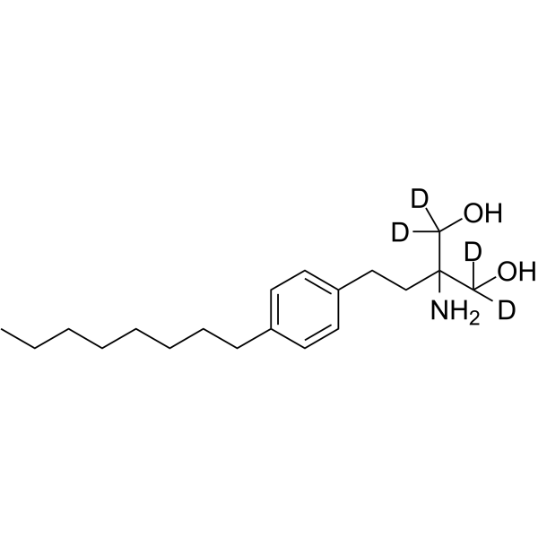 Fingolimod-d4 (FTY720 free based-d4)
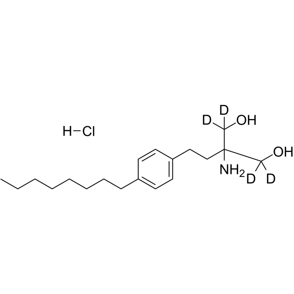 Fingolimod-d4 hydrochloride (FTY720-d4)
 Ki16425 (Debio 0719)
InvivoChem的所有产品仅用于作科学研究,不面向患者销售
Copyright 2020 InvivoChem LLC | All Rights Reserved 粤ICP备20063088号-1


 COA
COA


 463611831
