| 规格 | 价格 | 库存 | 数量 |
|---|---|---|---|
| 5mg |
|
||
| 10mg |
|
||
| 25mg |
|
||
| 50mg |
|
||
| 100mg |
|
||
| 250mg |
|
||
| 500mg |
|
||
| Other Sizes |
|

| 靶点 |
ATM ( IC50 = 12.9 nM ); DNA-PK ( IC50 = 2500 nM ); mTOR ( IC50 = 9300 nM ); PI3K ( IC50 = 16600 nM )
KU-55933 targets ataxia telangiectasia mutated (ATM) kinase (IC50 = 13 nM) [3] |
|---|---|
| 体外研究 (In Vitro) |
体外活性:KU-55933 抑制 DNA-PK 和 PI3K,IC50 分别为 2.5 μM 和 16.6 μM。此外,KU-55933 还可以抑制 mTOR 的活性,IC50 为 9.3 μM。 KU-55933 在细胞水平上活跃,可消除已充分表征的 ATM 依赖性磷酸化事件。 KU-55933 抑制这种 ATM 依赖性磷酸化事件具有剂量依赖性作用,IC50 为 300 nM。 KU-58050 在剂量为 30 μM 之前不会阻止 p53 丝氨酸 15 的 ATM 依赖性磷酸化。添加 KU-55933 对紫外线诱导的丝氨酸 139 上的 H2AX、丝氨酸 343 上的 NBS1、丝氨酸 345 上的 CHK1 和丝氨酸 966 上的 SMC1 磷酸化没有明显影响。与紫外线反应形成鲜明对比的是,KU-55933 消除了电离这些 ATM 底物的辐射诱导磷酸化。 KU-55933 使 HeLa 细胞对一系列电离辐射剂量敏感。 KU-55933 抑制癌细胞中生长因子诱导的 Akt 磷酸化。 KU-55933 抑制癌细胞的增殖。此外,KU-55933 抑制 ATM 可以提高生存率,这可能是通过阻止 TAp63α 下游激活来实现的。激酶测定:用于体外测定的 ATM 是通过用兔多克隆抗血清进行免疫沉淀从 HeLa 核提取物中获得的,该血清在含有 25 mM HEPES (pH 7.4)、2 mM MgCl2、250 的缓冲液中升至 ATM 的 COOH 末端 400 个氨基酸mM KCl、500 μM EDTA、100 μM Na3VO4、10% v/v 甘油和 0.1% v/v Igepal。通过与 Protein A-Sepharose 珠子一起孵育 1 小时,然后通过离心回收珠子,从核提取物中分离出 ATM-抗体复合物。在 96 孔板的孔中,将含有 ATM 的琼脂糖珠与 1 μg 底物谷胱甘肽 S-转移酶 -p53N66(p53 的 NH2 末端 66 个氨基酸与谷胱甘肽 S-转移酶融合)在 ATM 测定缓冲液中孵育 [25 mM HEPES (pH 7.4)、75 mM NaCl、3 mM MgCl2、2 mM MnCl2、50 μM Na3VO4、500 μM DTT 和 5% v/v 甘油],37 °C,存在或不存在抑制剂。轻轻摇动 10 分钟后,添加 ATP 至终浓度 50 μM,并在 37 °C 下继续反应 1 小时。将板以 250 × g 离心 10 分钟(4 °C),除去含有 ATM 的珠子,取出上清液,转移至白色不透明 96 孔板中,室温孵育 1.5 小时,使谷胱甘肽S-转移酶-p53N66 结合。然后用 PBS 清洗该板,吸干,并使用磷酸丝氨酸 15 p53 抗体通过标准 ELISA 技术进行分析。磷酸化谷胱甘肽 S-转移酶-p53N66 底物的检测是与山羊抗小鼠辣根过氧化物酶缀合的二抗结合进行的。使用增强化学发光溶液产生信号并进行化学发光检测。 细胞测定:U2OS 细胞暴露于电离辐射(3、5 或 15 Gy)或紫外线(5 或 50 J/m2),ATM 响应由下式确定: p53 丝氨酸 15 磷酸化和野生型 p53 稳定性的蛋白质印迹分析。从每个时间点获得全细胞提取物,通过 SDS-PAGE 分离蛋白质,并使用 p53 磷酸丝氨酸 15 特异性抗体测量磷酸化丝氨酸 15 的 ATM 特异性增加。使用 p53 特异性抗体 (DO-1) 还可以观察到 p53 随着时间的推移总体稳定。同样,为了研究 H2AX、CHK1、NBS1 和 SMC1 上的 ATM 依赖性磷酸化,使用以下抗体:CHK1 磷酸丝氨酸 345 和 NBS1 磷酸丝氨酸 343 抗体。还使用组蛋白 H2A (H-124) 和 CHK1 抗体,以及 SMC1 和 SMC1 磷酸丝氨酸 966 抗体。为了测定 KU-55933 的细胞 IC50,使用 2 小时的 p53 丝氨酸 15 磷酸化峰值响应时间来监测 ATM 的抑制。在电离辐射之前,将 KU-55933 滴定到细胞上并预孵育 1 小时。使用扫描密度测定法,计算相对于载体对照的抑制百分比,并计算体外测定的IC50值。
在人黑色素瘤细胞系(A375、SK-MEL-28、MeWo)中,KU-55933(5–20 μM)单独使用时抗增殖活性较弱(20 μM时细胞活力仅降低≤20%)。但与TRAIL(10–50 ng/mL)联合使用时,可显著增强TRAIL介导的凋亡:Annexin V-FITC/PI染色和流式细胞仪检测显示,A375细胞的凋亡率从(TRAIL单独使用组的)约15%升高至(TRAIL + 10 μM KU-55933组的)约65%[3] - Western blot检测显示,KU-55933 可抑制黑色素瘤细胞中ATM激酶活性,表现为ATM磷酸化(p-ATM)及其下游底物Chk2磷酸化(p-Chk2)水平降低。免疫荧光染色显示γ-H2AX灶点形成增加,证实双链DNA断裂(DSBs)积累[3] - KU-55933 与TRAIL的协同凋亡效应与死亡受体5(DR5)的mRNA和蛋白水平上调(RT-PCR和Western blot检测)以及半胱天冬酶级联反应激活相关:半胱天冬酶-8、半胱天冬酶-3和PARP的切割增强[3] - 在正常人黑素细胞(NHM)中,KU-55933(最高20 μM)与TRAIL(50 ng/mL)联合使用时无显著凋亡效应,表明其对癌细胞具有选择性毒性[3] |
| 体内研究 (In Vivo) |
ATM激酶消融可改善小鼠[4]
的细胞周期紊乱和足细胞损伤[4] 为了进一步证实ATM在adr相关足细胞细胞周期再进入中的作用,研究人员使用一种特异性的ATM激酶抑制剂KU-55933抑制ATM的磷酸化和活性。值得注意的是,KU-55933缓解了ADR刺激引起的MAD2B升高(P<0.05)(图6A-B)。同时,ADR诱导的已知的MAD2B底物和细胞周期关键调节因子Skp2和p27的改变被KU-55933部分逆转(P<0.05)(图6C-D)。此外,流式细胞分析显示,ATM抑制剂增加了G2/ m期的足细胞(P<0.05)(图6E-F),避免了灾难性的分裂和细胞死亡。此外,KU-55933成功地阻止了adr引发的足细胞功能障碍,从nephrin (P<0.05)和CD2AP (P<0.05)表达的恢复可以看出(图6G-H)。同样,KU-55933有效抑制ADR注射引起的小鼠MAD2B过表达(P<0.05)(图7A-B)。经KU-55933预处理后,FSGS形态学异常减少(图7C-E),蛋白尿降低(P<0.05),血清白蛋白升高(P<0.05)(图7F-G)。因此,我们的观察表明,阻断ATM激活可以有效地预防或减轻足细胞损伤。 |
| 酶活实验 |
为了获得用于体外测定的 ATM,使用涉及用 25 mM HEPES (pH 7.4)、2 mM 缓冲混合物的方法,从 HeLa 核提取物中免疫沉淀 ATM 的 COOH 末端 400 个氨基酸的兔多克隆抗血清。 MgCl2、250 mM KCl、500 μM EDTA、100 μM Na3VO4、10% v/v 甘油和 0.1% v/v Igepal。与 Protein A-Sepharose 珠子一起孵育一小时并随后离心回收珠子后,ATM-抗体复合物从核提取物中分离出来。 96 孔板的孔用于在 ATM 测定缓冲液 [25 mM HEPES ( pH 7.4)、75 mM NaCl、3 mM MgCl2、2 mM MnCl2、50 μM Na3VO4、500 μM DTT 和 5% v/v 甘油],37 °C,有或没有抑制剂。轻轻摇动 10 分钟后添加 ATP 至终浓度 50 μM,然后在 37 °C 下继续反应 1 小时。将板以 250 × g 离心 10 分钟 (4 °C),以去除含有 ATM 的珠子,从而使谷胱甘肽 S-转移酶 -p53N66 结合发生。然后取出上清液并放入白色不透明96孔板中。该孵育过程在室温下需要 1.5 小时。该板的后续步骤是 PBS 清洗、干印迹和使用磷酸丝氨酸 15 p53 抗体的标准 ELISA 分析。当使用与山羊抗小鼠辣根过氧化物酶缀合的二抗时,可以检测到磷酸化谷胱甘肽 S-转移酶-p53N66 的底物。创建信号和化学发光检测的过程涉及使用增强的化学发光溶液。进行化学发光检测并使用增强的化学发光溶液产生信号。
重组人ATM激酶与特异性肽底物(源自p53)和ATP在激酶检测缓冲液中孵育。加入浓度范围为0.1 nM–1 μM的KU-55933,混合物在30°C孵育60分钟。采用荧光偏振法检测肽底物的磷酸化水平,相对于溶媒对照组计算ATM激酶活性抑制率,通过非线性回归分析确定IC50值[3] |
| 细胞实验 |
ATM 反应通过对暴露于电离辐射(3、5 或 15 Gy)或 UV(5 或 50 J/m< sup>2)。提取每个时间点的全细胞提取物,使用 SDS-PAGE 分离蛋白质,并使用 p53 磷酸化丝氨酸 15 特异性抗体来测量磷酸化丝氨酸 15 的 ATM 特异性增加。当使用 p53 特异性抗体 (DO- 1),随着时间的推移,p53 总体稳定。同样,以下抗体用于研究 H2AX、CHK1、NBS1 和 SMC1 上的 ATM 依赖性磷酸化:NBS1 磷酸丝氨酸 343 和 CHK1 磷酸丝氨酸 345 抗体。还使用 SMC1 和 SMC1 磷酸丝氨酸 966 抗体,以及抗组蛋白 H2A (H-124) 和 CHK1 的抗体。 p53 丝氨酸 15 磷酸化的两小时峰值响应时间用于追踪 ATM 抑制,以确定 KU-55933 的细胞 IC50。在应用电离辐射之前,将 KU-55933 滴定到细胞上并预孵育一小时。 IC 50 值的测定与体外测定类似,并且使用扫描密度测定法计算相对于媒介物对照的抑制百分比。
细胞活力和凋亡检测:黑色素瘤细胞(每孔5×10³个)接种于96孔板,培养过夜后,用KU-55933(0.5–40 μM)单独处理或与TRAIL(10–50 ng/mL)联合处理48小时。CCK-8法检测细胞活力(450 nm处吸光度)。凋亡检测时,细胞用Annexin V-FITC/PI染色,流式细胞仪量化凋亡率[3] - Western blot分析:KU-55933(5–20 μM)和/或TRAIL(30 ng/mL)处理24小时的细胞裂解提取总蛋白。等量蛋白经SDS-PAGE电泳、转膜至PVDF膜,用抗ATM、p-ATM、Chk2、p-Chk2、γ-H2AX、DR5、半胱天冬酶-8、切割型半胱天冬酶-8、半胱天冬酶-3、切割型半胱天冬酶-3、PARP、切割型PARP或GAPDH(内参)抗体孵育。化学发光显影蛋白条带,ImageJ软件量化条带强度[3] - γ-H2AX灶点免疫荧光染色:A375细胞接种于盖玻片,用KU-55933(10 μM)处理12小时,多聚甲醛固定,Triton X-100透化,用抗γ-H2AX抗体(FITC标记)和DAPI染色。共聚焦显微镜捕获荧光图像,计数每个细胞的γ-H2AX灶点数(每组n ≥ 50个细胞)[3] - RT-PCR分析:TRIzol试剂提取处理后细胞的总RNA,逆转录为cDNA。用DR5和GAPDH(参考基因)的特异性引物进行实时定量PCR,2-ΔΔCt法计算DR5的相对mRNA表达水平[3] |
| 动物实验 |
阿霉素诱导的局灶节段性肾小球硬化症(FSGS)小鼠模型[4]
成年雄性小鼠(8周龄,Balb/C背景),体重21-24 g,饲养于特定病原体清除(SPF)级环境中,光照/黑暗周期为12小时,并可自由摄取食物和水。为建立FSGS动物模型,小鼠经尾静脉注射单剂量阿霉素(15 mg/kg),4周后处死。处死前收集尿液和血清样本,并使用全自动生化分析仪测定尿蛋白/肌酐比值、血清白蛋白、肌酐和血尿素氮。经主动脉导管用冰冷的Krebs-Henseleit生理盐水冲洗后,在冰上解剖肾脏。将皮质组织速冻后保存于-80℃直至使用。为抑制ATM激酶,将特异性ATM抑制剂KU-55933(500 µg/kg)溶于0.15% DMSO中,于注射ADR前24小时腹腔注射,之后每3天重复一次,直至处死。[4] BALB/c nu/nu裸鼠,携带LU1205细胞 10 μM |
| 参考文献 |
|
| 其他信息 |
ATM激酶抑制剂是指任何能够抑制共济失调毛细血管扩张突变基因(ATM)激酶的药物。化疗加速卵巢衰老的机制尚未完全阐明。我们使用广泛应用的抗癌化疗药物阿霉素,在多种体内异种移植模型和体外模型中,研究了化疗诱导的衰老对人卵巢的影响。阿霉素以剂量依赖的方式导致原始卵泡、卵母细胞和颗粒细胞中出现大量双链DNA断裂,表现为γH2AX灶的积累。这种损伤与卵母细胞凋亡相关,并导致ATM激活。修复反应似乎使一小部分卵母细胞(34.7%)和颗粒细胞(12.1%)得以存活,而大多数则发生凋亡。矛盾的是,KU-55933 对 ATM 的抑制反而提高了生存率,这可能是通过阻止下游 TAp63α 的激活实现的。此外,阿霉素会导致人卵巢血管和间质损伤,这可能会损害绝经前和绝经后的卵巢功能。化疗引起的卵巢早衰似乎是由涉及卵巢生殖细胞和非生殖细胞成分的复杂过程造成的。这些影响可能对绝经前和绝经后癌症幸存者的衰老具有临床意义。[2] 本研究旨在阐明共济失调毛细血管扩张突变基因 (ATM) 激酶对人黑色素瘤细胞中外源性肿瘤坏死因子相关凋亡诱导配体 (TRAIL) 受体 2/DR5 介导的死亡通路调控的影响。我们发现,与正常细胞相比,某些人类黑色素瘤细胞系中总ATM蛋白水平较高。在许多黑色素瘤细胞系中,活性形式的ATM磷酸化Ser(1981)的基线水平也可检测到,并且可通过γ射线照射进一步上调。在γ射线照射前,用ATM激酶抑制剂KU-55933预处理几种黑色素瘤细胞系,可抑制p53和核因子-κB (NF-κB)的激活,但显著增加辐射诱导的DR5表面表达,下调cFLIP(caspase-8抑制剂)水平,并显著增强外源性TRAIL诱导的细胞凋亡。此外,在KU-55933存在的情况下进行γ射线照射,可使TRAIL耐药的HHMSX黑色素瘤细胞对TRAIL介导的细胞凋亡敏感。此外,特异性短发夹RNA抑制ATM表达也导致cFLIP水平下调、DR5表面表达上调以及TRAIL介导的黑色素瘤细胞凋亡。除p53和NF-κB这两个DR5表达的关键调控因子外,转录因子STAT3也已知对DR5表达具有负调控作用。KU-55933抑制STAT3的Ser(727)和Tyr(705)磷酸化可降低STAT3的转录活性,并伴随DR5表达升高。显性负性STAT3β也能有效上调体外培养和体内黑色素瘤细胞中DR5的表面表达并下调cFLIP水平。综上所述,我们的数据表明存在一条ATM依赖的STAT3介导的抗凋亡通路,抑制该通路可使人黑色素瘤细胞对TRAIL介导的凋亡更加敏感。[3]
原理:局灶节段性肾小球硬化症(FSGS)的特征是“有丝分裂后”足细胞功能障碍。足细胞重新进入细胞周期最终会导致细胞死亡。有丝分裂阻滞缺陷2样蛋白2(MAD2B)是后期促进复合物(APC)/细胞周期体的抑制剂,它精确控制着中期到后期的转变以及有序的细胞周期进程。然而,MAD2B在FSGS足细胞损伤中的作用尚不清楚。方法:为了探究MAD2B在足细胞重新进入细胞周期中的功能,我们使用了在阿霉素(ADR)诱导的FSGS小鼠模型中,选择性地在足细胞中敲除MAD2B的条件性突变小鼠。此外,本研究还利用共济失调毛细血管扩张突变基因(ATM)特异性抑制剂KU-55933,在体内外实验中探讨了ATM在调控MAD2B中的作用。结果显示,FSGS患者和ADR治疗的小鼠足细胞中MAD2B的表达显著升高,并伴有足细胞周期再入。足细胞特异性敲除MAD2B可有效减轻蛋白尿和足细胞损伤,并阻止异常的细胞周期再入。生物信息学分析表明,ATM激酶是MAD2B的关键上游调控因子。此外,抑制ATM激酶可消除MAD2B驱动的细胞周期再入,并减轻FSGS小鼠模型中的足细胞损伤。通过定点诱变和免疫沉淀的体外研究,我们发现 ATM 磷酸化 MAD2B,从而以磷酸化依赖的方式阻碍 MAD2B 的泛素化。结论:ATM激酶-MAD2B轴对足细胞的细胞周期再入起着重要作用,这是FSGS的一种新的致病机制,并可能为FSGS的治疗方法开发提供思路。[4] KU-55933是一种对ATM激酶具有高选择性的合成小分子抑制剂,与其他磷脂酰肌醇3-激酶相关激酶(PIKK)如ATR(IC50 > 10 μM)和DNA-PKcs(IC50 > 10 μM)的交叉反应性极低。[3] - 其作用机制涉及与ATM的ATP结合口袋结合,抑制ATM介导的参与DNA损伤修复的下游底物的磷酸化。这会导致DSB的积累,从而通过上调DR5表达使癌细胞对TRAIL诱导的细胞凋亡更加敏感[3] -KU-55933代表了一种克服黑色素瘤TRAIL耐药性的潜在策略,因为它能特异性地增强TRAIL在癌细胞中的凋亡作用,而不影响正常黑色素细胞[3] |
| 分子式 |
C21H17NO3S2
|
|
|---|---|---|
| 分子量 |
395.49
|
|
| 精确质量 |
395.064
|
|
| 元素分析 |
C, 63.77; H, 4.33; N, 3.54; O, 12.14; S, 16.22
|
|
| CAS号 |
587871-26-9
|
|
| 相关CAS号 |
|
|
| PubChem CID |
5278396
|
|
| 外观&性状 |
White to off-white solid powder
|
|
| 密度 |
1.4±0.1 g/cm3
|
|
| 沸点 |
628.0±55.0 °C at 760 mmHg
|
|
| 熔点 |
229.98° C
|
|
| 闪点 |
333.6±31.5 °C
|
|
| 蒸汽压 |
0.0±1.8 mmHg at 25°C
|
|
| 折射率 |
1.714
|
|
| LogP |
6.13
|
|
| tPSA |
93.28
|
|
| 氢键供体(HBD)数目 |
0
|
|
| 氢键受体(HBA)数目 |
6
|
|
| 可旋转键数目(RBC) |
2
|
|
| 重原子数目 |
27
|
|
| 分子复杂度/Complexity |
643
|
|
| 定义原子立体中心数目 |
0
|
|
| SMILES |
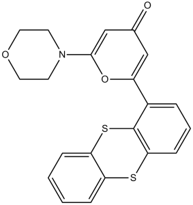
S1C2=C([H])C([H])=C([H])C([H])=C2SC2=C([H])C([H])=C([H])C(=C12)C1=C([H])C(C([H])=C(N2C([H])([H])C([H])([H])OC([H])([H])C2([H])[H])O1)=O
|
|
| InChi Key |
XRKYMMUGXMWDAO-UHFFFAOYSA-N
|
|
| InChi Code |
InChI=1S/C21H17NO3S2/c23-14-12-16(25-20(13-14)22-8-10-24-11-9-22)15-4-3-7-19-21(15)27-18-6-2-1-5-17(18)26-19/h1-7,12-13H,8-11H2
|
|
| 化学名 |
2-morpholin-4-yl-6-thianthren-1-ylpyran-4-one
|
|
| 别名 |
|
|
| HS Tariff Code |
2934.99.9001
|
|
| 存储方式 |
Powder -20°C 3 years 4°C 2 years In solvent -80°C 6 months -20°C 1 month |
|
| 运输条件 |
Room temperature (This product is stable at ambient temperature for a few days during ordinary shipping and time spent in Customs)
|
| 溶解度 (体外实验) |
|
|||
|---|---|---|---|---|
| 溶解度 (体内实验) |
配方 1 中的溶解度: ≥ 2.5 mg/mL (6.32 mM) (饱和度未知) in 10% DMSO + 40% PEG300 + 5% Tween80 + 45% Saline (这些助溶剂从左到右依次添加,逐一添加), 澄清溶液。
例如,若需制备1 mL的工作液,可将100 μL 25.0 mg/mL澄清DMSO储备液加入到400 μL PEG300中,混匀;然后向上述溶液中加入50 μL Tween-80,混匀;加入450 μL生理盐水定容至1 mL。 *生理盐水的制备:将 0.9 g 氯化钠溶解在 100 mL ddH₂O中,得到澄清溶液。 配方 2 中的溶解度: ≥ 2.5 mg/mL (6.32 mM) (饱和度未知) in 10% DMSO + 90% (20% SBE-β-CD in Saline) (这些助溶剂从左到右依次添加,逐一添加), 澄清溶液。 例如,若需制备1 mL的工作液,可将 100 μL 25.0 mg/mL澄清DMSO储备液加入900 μL 20% SBE-β-CD生理盐水溶液中,混匀。 *20% SBE-β-CD 生理盐水溶液的制备(4°C,1 周):将 2 g SBE-β-CD 溶解于 10 mL 生理盐水中,得到澄清溶液。 View More
配方 3 中的溶解度: ≥ 2.5 mg/mL (6.32 mM) (饱和度未知) in 10% DMSO + 90% Corn Oil (这些助溶剂从左到右依次添加,逐一添加), 澄清溶液。 配方 4 中的溶解度: 5% DMSO and 47.5% PEG300: 10mg/mL 1、请先配制澄清的储备液(如:用DMSO配置50 或 100 mg/mL母液(储备液)); 2、取适量母液,按从左到右的顺序依次添加助溶剂,澄清后再加入下一助溶剂。以 下列配方为例说明 (注意此配方只用于说明,并不一定代表此产品 的实际溶解配方): 10% DMSO → 40% PEG300 → 5% Tween-80 → 45% ddH2O (或 saline); 假设最终工作液的体积为 1 mL, 浓度为5 mg/mL: 取 100 μL 50 mg/mL 的澄清 DMSO 储备液加到 400 μL PEG300 中,混合均匀/澄清;向上述体系中加入50 μL Tween-80,混合均匀/澄清;然后继续加入450 μL ddH2O (或 saline)定容至 1 mL; 3、溶剂前显示的百分比是指该溶剂在最终溶液/工作液中的体积所占比例; 4、 如产品在配制过程中出现沉淀/析出,可通过加热(≤50℃)或超声的方式助溶; 5、为保证最佳实验结果,工作液请现配现用! 6、如不确定怎么将母液配置成体内动物实验的工作液,请查看说明书或联系我们; 7、 以上所有助溶剂都可在 Invivochem.cn网站购买。 |
| 制备储备液 | 1 mg | 5 mg | 10 mg | |
| 1 mM | 2.5285 mL | 12.6425 mL | 25.2851 mL | |
| 5 mM | 0.5057 mL | 2.5285 mL | 5.0570 mL | |
| 10 mM | 0.2529 mL | 1.2643 mL | 2.5285 mL |
1、根据实验需要选择合适的溶剂配制储备液 (母液):对于大多数产品,InvivoChem推荐用DMSO配置母液 (比如:5、10、20mM或者10、20、50 mg/mL浓度),个别水溶性高的产品可直接溶于水。产品在DMSO 、水或其他溶剂中的具体溶解度详见上”溶解度 (体外)”部分;
2、如果您找不到您想要的溶解度信息,或者很难将产品溶解在溶液中,请联系我们;
3、建议使用下列计算器进行相关计算(摩尔浓度计算器、稀释计算器、分子量计算器、重组计算器等);
4、母液配好之后,将其分装到常规用量,并储存在-20°C或-80°C,尽量减少反复冻融循环。
计算结果:
工作液浓度: mg/mL;
DMSO母液配制方法: mg 药物溶于 μL DMSO溶液(母液浓度 mg/mL)。如该浓度超过该批次药物DMSO溶解度,请首先与我们联系。
体内配方配制方法:取 μL DMSO母液,加入 μL PEG300,混匀澄清后加入μL Tween 80,混匀澄清后加入 μL ddH2O,混匀澄清。
(1) 请确保溶液澄清之后,再加入下一种溶剂 (助溶剂) 。可利用涡旋、超声或水浴加热等方法助溶;
(2) 一定要按顺序加入溶剂 (助溶剂) 。
|
|
|
|
InvivoChem的所有产品仅用于作科学研究,不面向患者销售
Copyright 2020 InvivoChem LLC | All Rights Reserved 粤ICP备20063088号-1


 COA
COA


 463611831
