
| 规格 | 价格 | 库存 | 数量 |
|---|---|---|---|
| 5mg |
|
||
| 10mg |
|
||
| 25mg |
|
||
| 50mg |
|
||
| 100mg |
|
||
| 250mg |
|
||
| 500mg |
|
||
| 1g |
|
||
| 2g |
|
||
| 5g |
|
||
| Other Sizes |
|

| 靶点 |
Bacterial protein synthesis; 30S subunit of the bacterial ribosome; tetracycline antibiotic; hypoxia-inducible factor (HIF)-1α
|
|---|---|
| 体外研究 (In Vitro) |
OVCAR-3、SKOV-3 和 A2780 珍珠棉系的增殖和克隆形成活性受到盐酸米诺环素(0-100 μM,24-72 小时)的抑制 [3]。米诺环素盐酸盐(0-100 μM,24-48 小时)。在示波器中,盐酸米诺环素(0-100 μM,72 小时))会引起细胞周期 [3]。除了抑制 caspase 依赖性和 caspase 非依赖性细胞死亡外,直接神经保护还可能与线粒体异常和细胞色素 c 保护有关 [2]。盐酸米诺环素诱导缺氧诱导因子(HIF)细胞增殖的检测[3]
|
| 体内研究 (In Vivo) |
在雌性裸鼠中,每天一次静脉注射盐酸卵分泌素(0-30 mg/kg),持续四个星期,可抑制 OVCAR-3 肿瘤的生长[3]。在脑损伤动物模型中,盐酸米诺环素 (IP) 是一种强效药物,在腹腔内高剂量给药时表现出神经保护作用 [1]。盐酸米诺环素(0-40 mg/kg,IP,一次)可显着抑制 METH 诱导的小鼠过度运动和行为敏化 [2]。在暂时性大脑中动脉闭塞 (TMCAO) 模型中,盐酸米诺环素(静脉注射 3 和 10 mg/kg 一次)可有效减少梗塞面积 [1]。盐酸米诺环素(3-10 mg/kg,静脉注射一次)对血液的影响可能会因电位诱发的室性心律失常而减轻。标准 200 mg 剂量对人体的这种作用可能与线粒体 KATP 通道、PI3K/Akt 信号传导和 L 型水平 (3 mg/kg) 有关[1]。
|
| 细胞实验 |
细胞增殖检测[3]
细胞类型:人卵巢癌细胞系(OVCAR-1α抑制,以及up-p53蛋白和AKT水平的调节/mTOR/p70S6K/4E-BP1失活染料 [6]。3. SKOV-3 和 A2780)和原代细胞(HEK-293、HMEC、HUVEC、ATCC) 测试浓度:0、1、10、50 和 100 μM 孵育时间:24、48或72小时 实验结果:抑制OVCAR-3、SKOV-3的增殖和A2780细胞呈浓度依赖性,IC50值分别为62.0、56.1和59.5 μM。对 HEK-293 或 HUVEC 的活力没有影响。 蛋白质印迹分析[3] 细胞类型: OVCAR-3、SKOV-3 和 A2780 细胞 测试浓度: 0、 10、50 和 100 μM 孵育时间:72 小时 实验结果: Cyclins A、B 和 E 低表达水平。 caspase- 增加 3 个水平,在 100 μM 时增加超过 3.0 倍。米诺环素激活的 caspase-3 进而导致 PARP-1 的裂解。 Caspase-3 增加 PARP-1、p89 的降解产物。 细胞周期分析[3] 细胞类型: OVCAR-3、SKOV-3 和 A2 |
| 动物实验 |
动物/疾病模型:雌性裸鼠(6周龄,每组9只,每只小鼠腹部左侧皮下注射OVCAR-3细胞)[3]
剂量:10或30 mg/kg 给药途径:从细胞接种后第8天开始,每日一次,连续4周,通过饮用水灌胃给药。 实验结果:抑制了OVCAR-3肿瘤的生长,并降低了雌性裸鼠的微血管密度。 动物/疾病模型:雄性Balb/cAnNCrICrIj小鼠(8周龄,23-30 g,皮下注射甲基苯丙胺(METH,3 mg/kg),注射体积为10 ml/kg)[2] 剂量:0、10、20或40 mg/kg 给药途径:腹腔注射(ip),单次,在METH给药前30分钟 实验结果:40 mg/kg剂量显著减弱了METH诱导的小鼠活动过度和行为敏感化。对METH诱导的小鼠高热无影响。显著减弱了纹状体中DA和DOPAC的减少。显著减弱了小鼠纹状体中DAT免疫反应性的降低。显著减弱了甲基苯丙胺(METH)给药后纹状体中MAC1免疫反应性的增加。 动物/疾病模型:雄性Sprague-Dawley大鼠(270-330 g,TMCAO模型)[1] 剂量:3 mg/kg和10 mg/kg 给药途径:静脉注射,单次,TMCAO后4、5或6小时 实验结果:3 mg/kg剂量使梗死面积减少了42%,而10 mg/kg剂量使梗死面积减少了56%; 10 mg/kg 剂量组在 5 小时内显著减少了梗死面积 40%,3 mg/kg 剂量组显著减少了梗死面积 34%。6 小时时间窗内梗死面积减少的趋势不显著。 动物/疾病模型:雄性 Sprague-Dawley 大鼠(270-330 g)[1] 剂量:3、10 或 20 mg/kg 给药途径:静脉注射,单次 实验结果:米诺环素血清峰浓度在 3、10 和 20 mg/kg 剂量组的平均浓度分别为 3.6、13.0 和 28.8 mg/L。 3 mg/kg剂量下米诺环素的血清浓度(3.6 mg/L)与人体服用标准200 mg剂量后的血清浓度相似。未对血流动力学和生理指标产生显著影响。 |
| 毒性/毒理 (Toxicokinetics/TK) |
妊娠期和哺乳期影响
◉ 哺乳期用药概述 许多评论指出,由于四环素类药物可能导致婴儿牙釉质染色或沉积于骨骼,因此哺乳期禁用。然而,仔细查阅现有文献表明,哺乳期短期使用米诺环素不太可能造成危害,因为乳汁中的药物浓度较低,且婴儿对药物的吸收会受到母乳中钙的抑制。哺乳期妇女短期使用米诺环素是可以接受的。作为一项理论上的预防措施,应避免哺乳期长期或重复用药。应密切观察婴儿是否出现皮疹以及可能对胃肠道菌群产生的影响,例如腹泻或念珠菌病(鹅口疮、尿布疹)。已有报道称,服用米诺环素会导致母乳变黑。母亲局部使用米诺环素治疗痤疮不会对母乳喂养的婴儿造成风险。 ◉ 对母乳喂养婴儿的影响 截至修订日期,未找到相关的已发表信息。 ◉ 对泌乳和母乳的影响 一名妇女每日两次服用100毫克米诺环素近4年,在服用奋乃静、阿米替林和苯海拉明后出现溢乳,且乳汁呈黑色。 另一名妇女在断奶后的18个月内哺乳,偶尔分泌少量母乳,之后每日口服150毫克米诺环素。3至4周后,挤出的乳汁变为黑色。乳汁中的铁含量比正常乳汁高出100多倍。乳房X光检查结果正常。 在这两个病例中,乳汁中均发现了含有黑色含铁色素的巨噬细胞。人们认为这种色素是米诺环素或其代谢物的铁螯合物。 |
| 参考文献 |
|
| 其他信息 |
根据州或联邦政府的标签要求,盐酸米诺环素(内服)可能引起发育毒性。
它是一种四环素类似物,具有7-二甲基氨基,缺少5个甲基和羟基,对耐四环素的葡萄球菌感染有效。 另见:盐酸米诺环素(注释已移至此处)。 治疗多重耐药(MDR)鲍曼不动杆菌感染的选择极其有限。静脉注射米诺环素对许多MDR鲍曼不动杆菌菌株有效,临床和实验室标准协会(CLSI)制定了断点,用于指导对鲍曼不动杆菌米诺环素药敏试验结果的解读。此外,静脉注射米诺环素已获得美国食品药品监督管理局(FDA)批准,用于治疗鲍曼不动杆菌感染。越来越多的文献报道了静脉注射米诺环素成功用于治疗严重的耐多药鲍曼不动杆菌感染,特别是院内肺炎。这些结果,加上静脉注射米诺环素通常良好的耐受性,支持将其作为治疗耐多药鲍曼不动杆菌感染的可行治疗方案。[5] 缺氧诱导因子 (HIF)-1α 是缺氧条件下关键的细胞存活蛋白,与肿瘤进展和血管生成相关。我们最近发现米诺环素对卵巢肿瘤生长的抑制作用与血管内皮生长因子 (VEGF) 的减弱相关,本文报告一项相关的实验室研究,旨在验证这些作用是否是 HIF-1α 抑制的结果。本研究利用人卵巢癌细胞系(A2780、OVCAR-3 和 SKOV-3)在体外研究了米诺环素对 HIF-1 及其上游通路组分的影响,以阐明米诺环素的作用机制。同时,用米诺环素治疗携带 OVCAR-3 异种移植瘤的小鼠,以评估米诺环素在体内 HIF-1 通路中的疗效。结果表明,米诺环素以浓度依赖的方式负向调控 HIF-1α 蛋白水平,并通过一种不依赖于脯氨酰羟基化的机制诱导其降解。此外,HIF-1α 的抑制与内源性 p53 的上调相关,而 p53 是一种肿瘤抑制因子,已被证实参与 HIF-1α 的降解。进一步研究表明,米诺环素的作用不仅限于蛋白酶体降解,它还能通过抑制AKT/mTOR/p70S6K/4E-BP1信号通路来下调HIF-1α的翻译。在已建立卵巢肿瘤的小鼠中,米诺环素治疗导致HIF-1α表达受到抑制,同时p53蛋白水平上调,AKT/mTOR/p70S6K/4E-BP1通路失活。这些数据揭示了米诺环素作为一种靶向促癌因子HIF-1α的药物,在卵巢癌治疗中具有潜在的治疗价值,其作用机制涉及多种途径。[6] 米诺环素已被证实具有保护心肌免受缺血再灌注损伤的作用。本研究探讨了米诺环素对大鼠缺血诱发的心室性心律失常的影响。麻醉后的雄性大鼠在缺血前1小时接受一次米诺环素(45mg/kg,腹腔注射)治疗,同时分别给予或不给予2-(4-吗啉基)-8-苯基-1(4H)-苯并吡喃-4-酮盐酸盐(LY294002,0.3mg/kg,静脉注射,一种PI3K抑制剂)和5-羟基癸酸[5-HD,10mg/kg,静脉注射,一种线粒体ATP敏感性钾通道(K(ATP))的特异性抑制剂],并在缺血前10分钟注射一次,随后进行30分钟的缺血处理。评估室性心律失常。采用膜片钳技术测量L型Ca²⁺电流。在30分钟的缺血期间,米诺环素显著降低了室颤(VF)的发生率(P<0.05)。与心肌缺血组相比,米诺环素显著降低了室性心动过速合并室颤(VT+VF)的持续时间、VT+VF发作次数以及心律失常的严重程度(所有P<0.05)。LY294002或5-HD的给药消除了米诺环素对室颤发生率、VT+VF持续时间、VT+VF发作次数以及心律失常严重程度的保护作用(所有P<0.05)。此外,米诺环素以剂量依赖的方式抑制正常心肌细胞膜的L型Ca²⁺电流。本研究提示,米诺环素可能通过PI3K/Akt信号通路、线粒体K(ATP)通道和L型Ca²⁺通道参与,从而减轻大鼠缺血诱发的室性心律失常。[7] |
| 分子式 |
C23H28CLN3O7
|
|---|---|
| 分子量 |
493.9373
|
| 精确质量 |
493.161
|
| 元素分析 |
C, 55.93; H, 5.71; Cl, 7.18; N, 8.51; O, 22.67
|
| CAS号 |
13614-98-7
|
| 相关CAS号 |
Minocycline;10118-90-8; Minocycline hydrochloride;13614-98-7;Minocycline-d6;1036070-10-6; 128420-71-3 (HCl hydrate)
|
| PubChem CID |
54685925
|
| 外观&性状 |
Light yellow to yellow solid powder
|
| 沸点 |
659.4ºC at 760mmHg
|
| 熔点 |
205-210° (dec)
|
| 闪点 |
352.6ºC
|
| 蒸汽压 |
6.33E-28mmHg at 25°C
|
| LogP |
1.688
|
| tPSA |
164.63
|
| 氢键供体(HBD)数目 |
6
|
| 氢键受体(HBA)数目 |
9
|
| 可旋转键数目(RBC) |
3
|
| 重原子数目 |
34
|
| 分子复杂度/Complexity |
971
|
| 定义原子立体中心数目 |
4
|
| SMILES |
CN(C)[C@H]1[C@@H]2C[C@@H]3CC4=C(C=CC(=C4C(=C3C(=O)[C@@]2(C(=C(C1=O)C(=O)N)O)O)O)O)N(C)C.Cl
|
| InChi Key |
KDLQIOPKJDNQIM-YKWOUSISSA-N
|
| InChi Code |
InChI=1S/C23H27N3O7.ClH/c1-25(2)12-5-6-13(27)15-10(12)7-9-8-11-17(26(3)4)19(29)16(22(24)32)21(31)23(11,33)20(30)14(9)18(15)28;/h5-6,9,11,17,27-28,32-33H,7-8,24H2,1-4H3;1H/b22-16+;/t9-,11-,17+,23-;/m1./s1
|
| 化学名 |
(4S,4aR,5aS,12aR,E)-2-(amino(hydroxy)methylene)-4,7-bis(dimethylamino)-10,11,12a-trihydroxy-4a,5a,6,12a-tetrahydrotetracene-1,3,12(2H,4H,5H)-trione hydrochloride
|
| 别名 |
NSC 141993; Minocycline HCl; NSC 141993; Mynocine hydrochloride; NSC141993; NSC-141993; Periocline; Klinomycin; Minocin; Solodyn; Mynocine; Tri-mino; Vectrin; Ximino; Minomax; Minomycin chloride; Mynocine hydrochloride
|
| HS Tariff Code |
2934.99.9001
|
| 存储方式 |
Powder -20°C 3 years 4°C 2 years In solvent -80°C 6 months -20°C 1 month 注意: 请将本产品存放在密封且受保护的环境中,避免吸湿/受潮。 |
| 运输条件 |
Room temperature (This product is stable at ambient temperature for a few days during ordinary shipping and time spent in Customs)
|
| 溶解度 (体外实验) |
DMSO : ~19.23 mg/mL (~38.93 mM)
H2O : ~9.09 mg/mL (~18.40 mM) |
|---|---|
| 溶解度 (体内实验) |
配方 1 中的溶解度: 7.69 mg/mL (15.57 mM) in PBS (这些助溶剂从左到右依次添加,逐一添加), 澄清溶液; 超声助溶。
请根据您的实验动物和给药方式选择适当的溶解配方/方案: 1、请先配制澄清的储备液(如:用DMSO配置50 或 100 mg/mL母液(储备液)); 2、取适量母液,按从左到右的顺序依次添加助溶剂,澄清后再加入下一助溶剂。以 下列配方为例说明 (注意此配方只用于说明,并不一定代表此产品 的实际溶解配方): 10% DMSO → 40% PEG300 → 5% Tween-80 → 45% ddH2O (或 saline); 假设最终工作液的体积为 1 mL, 浓度为5 mg/mL: 取 100 μL 50 mg/mL 的澄清 DMSO 储备液加到 400 μL PEG300 中,混合均匀/澄清;向上述体系中加入50 μL Tween-80,混合均匀/澄清;然后继续加入450 μL ddH2O (或 saline)定容至 1 mL; 3、溶剂前显示的百分比是指该溶剂在最终溶液/工作液中的体积所占比例; 4、 如产品在配制过程中出现沉淀/析出,可通过加热(≤50℃)或超声的方式助溶; 5、为保证最佳实验结果,工作液请现配现用! 6、如不确定怎么将母液配置成体内动物实验的工作液,请查看说明书或联系我们; 7、 以上所有助溶剂都可在 Invivochem.cn网站购买。 |
| 制备储备液 | 1 mg | 5 mg | 10 mg | |
| 1 mM | 2.0245 mL | 10.1227 mL | 20.2454 mL | |
| 5 mM | 0.4049 mL | 2.0245 mL | 4.0491 mL | |
| 10 mM | 0.2025 mL | 1.0123 mL | 2.0245 mL |
1、根据实验需要选择合适的溶剂配制储备液 (母液):对于大多数产品,InvivoChem推荐用DMSO配置母液 (比如:5、10、20mM或者10、20、50 mg/mL浓度),个别水溶性高的产品可直接溶于水。产品在DMSO 、水或其他溶剂中的具体溶解度详见上”溶解度 (体外)”部分;
2、如果您找不到您想要的溶解度信息,或者很难将产品溶解在溶液中,请联系我们;
3、建议使用下列计算器进行相关计算(摩尔浓度计算器、稀释计算器、分子量计算器、重组计算器等);
4、母液配好之后,将其分装到常规用量,并储存在-20°C或-80°C,尽量减少反复冻融循环。
计算结果:
工作液浓度: mg/mL;
DMSO母液配制方法: mg 药物溶于 μL DMSO溶液(母液浓度 mg/mL)。如该浓度超过该批次药物DMSO溶解度,请首先与我们联系。
体内配方配制方法:取 μL DMSO母液,加入 μL PEG300,混匀澄清后加入μL Tween 80,混匀澄清后加入 μL ddH2O,混匀澄清。
(1) 请确保溶液澄清之后,再加入下一种溶剂 (助溶剂) 。可利用涡旋、超声或水浴加热等方法助溶;
(2) 一定要按顺序加入溶剂 (助溶剂) 。
 α-Gurjunene
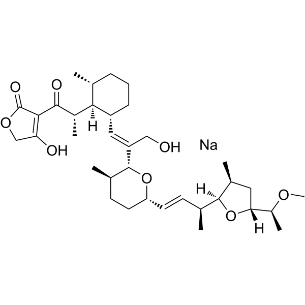 Tetronasin sodium
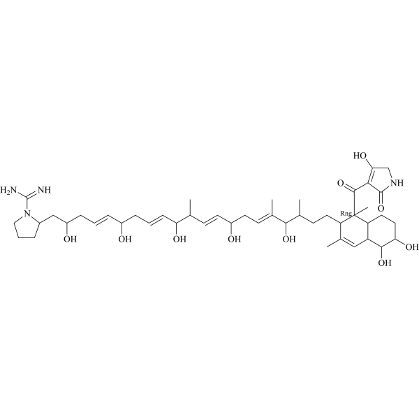 TPU-0037A
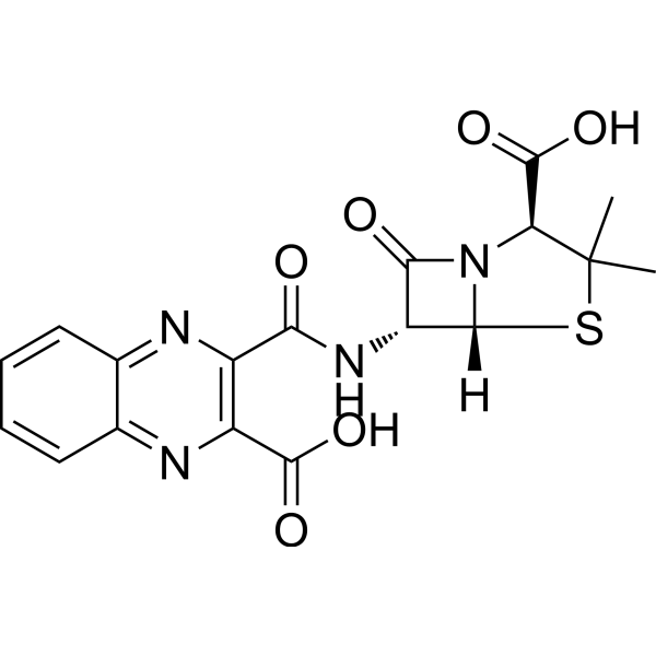 Quinacillin
InvivoChem的所有产品仅用于作科学研究,不面向患者销售
Copyright 2020 InvivoChem LLC | All Rights Reserved 粤ICP备20063088号-1


 COA
COA
 463611831
