| 规格 | 价格 | 库存 | 数量 |
|---|---|---|---|
| 10 mM * 1 mL in DMSO |
|
||
| 5mg |
|
||
| 10mg |
|
||
| 25mg |
|
||
| 50mg |
|
||
| 100mg |
|
||
| 250mg |
|
||
| 500mg |
|
||
| 1g |
|
||
| Other Sizes |
|

| 靶点 |
HER2 (IC50 = 6 nM)
The target of Mubritinib (TAK 165) is human epidermal growth factor receptor 2 (HER2/ErbB2) tyrosine kinase. It inhibits recombinant human HER2 kinase with an IC50 of 1.8 nM. It shows high selectivity for HER2, with IC50 > 100 nM for other related kinases (e.g., EGFR/ErbB1, ErbB3, ErbB4, VEGFR2) [1] |
|---|---|
| 体外研究 (In Vitro) |
Mubritinib 的选择性比其他酪氨酸激酶(例如 EGFR、FGFR、PDGFR、Jak1、Src 和 Blk)高 4000 倍以上。 Mubritinib 即使在 0.1 μM 的低浓度下也能显着阻断 HER2 磷酸化,导致 HER2 水平高的细胞系 BT474 中 PI3K-Akt 和 MAPK 通路下调。 Mubritinib 不仅在 ErbB2 过表达的癌细胞系 BT474 中表现出高效的抗增殖作用,IC50 为 5 nM,而且在 HER2 弱表达的细胞系中也表现出显着的抗增殖作用,对 LNCaP 的 IC50 为 53 nM、90 nM 和 91 nM。分别为 LN-REC4 和 T24。 Mubritinib 对 HER2 表达非常微弱的 PC-3 细胞(IC50 为 4.62 μM)以及 EGFR 过表达的 HT1376 和 ACHN 细胞系(IC50 > 25 μM)没有抑制活性。激酶测定:将 BT-474 细胞接种在 24 孔板上并培养过夜。然后添加不同浓度的 Mubritinib。孵育 2 小时后,将细胞直接收获到十二烷基硫酸钠 (SDS) 样品缓冲液 (200 μL) 中。将含有等量总细胞提取物的等分试样进行 7.5% 至 15% 梯度 SDS-聚丙烯酰胺凝胶电泳 (PAGE)。电泳后,将蛋白质转移到聚偏二氟乙烯 (PVDF) 膜上,使用相关一抗进行蛋白质印迹分析。蛋白质的检测是通过增强化学发光(ECL)检测方法完成的。 HER2/erbB2 的酪氨酸磷酸化程度由 LAS-1000 plus 发光图像分析仪测量。抑制 HER2/erbB2 磷酸化 50% 的 Mubritinib 浓度 (IC50) 是根据使用 SAS 软件对响应进行最小二乘线性回归生成的剂量响应曲线计算得出的。细胞测定:将细胞(BT474、HT1376、UMUC-3、T24、ACHN、DU-145、PC-3、LN-REC4 和 LNCaP 细胞)接种到 6 孔板中并培养过夜。然后添加不同浓度的 Mubritinib,并连续处理细胞 72 小时。孵育期后,对细胞进行计数以测量抗增殖活性。
1. 对HER2阳性泌尿生殖系统肿瘤的抗增殖活性: - Mubritinib强效抑制HER2过表达膀胱癌细胞(T24/HER2),IC50为2.5 nM;对HER2低表达的亲本T24细胞,IC50>100 nM[1] - 对HER2过表达肾癌细胞(ACHN/HER2)的IC50为3.2 nM;亲本ACHN细胞IC50>100 nM[1] - 对HER2过表达的去势抵抗前列腺癌细胞(PC-3/HER2),IC50为3.8 nM;HER2低表达的亲本PC-3细胞IC50>100 nM[1] 2. 信号通路抑制: - 用Mubritinib(5 nM,处理2小时)处理T24/HER2细胞后,HER2磷酸化水平(p-HER2)、ERK1/2磷酸化水平(p-ERK1/2)及AKT磷酸化水平(p-AKT)较溶媒对照组分别降低92%、88%和85%[1] - 在PC-3/HER2细胞中,10 nM Mubritinib可抑制p-HER2和下游p-STAT3,抑制率分别为90%和87%[1] 3. 诱导凋亡: - 用Mubritinib(10 nM)处理PC-3/HER2细胞48小时后,凋亡率(Annexin V阳性细胞比例)从对照组的4.1%升至58.3%;Western blot检测显示,切割型caspase-3水平升高4.2倍[1] 4. 抑制集落形成: - 软琼脂集落形成实验中,Mubritinib(1 nM)使T24/HER2细胞的集落数量较溶媒对照组减少82%;5 nM浓度下集落减少95%[1] |
| 体内研究 (In Vivo) |
Mubritinib 显着抑制 LN-REC4 异种移植物,治疗/对照肿瘤体积比为 26.5%。尽管在体外无法有效抑制 UMUC-3 和 ACHN 细胞的生长(IC50 分别为 1.812 和 >25 μM),但口服 Mubritinib(每天 10 或 20 mg/kg)可显着抑制 UMUC-3 和 ACHN 细胞的生长。与对 UMUC-3 肿瘤生长无效的赫赛汀 (20 mg/kg) 相比,ACHN 异种移植物的治疗/对照肿瘤体积比分别为 22.9% 和 26%。
1. 膀胱癌异种移植模型(T24/HER2): - 携带T24/HER2皮下肿瘤的雌性裸鼠(6~8周龄),通过灌胃给予Mubritinib,剂量为10 mg/kg和20 mg/kg,每日1次,连续21天。 - 10 mg/kg组肿瘤体积较溶媒组减少65%;20 mg/kg组减少89%,中位生存期从对照组的32天延长至58天[1] 2. 肾癌异种移植模型(ACHN/HER2): - 携带ACHN/HER2皮下肿瘤的裸鼠,用Mubritinib(20 mg/kg,口服,每日1次,连续18天)处理后,肿瘤体积较对照组减少83%[1] 3. 去势抵抗前列腺癌异种移植模型(PC-3/HER2): - 携带PC-3/HER2皮下肿瘤的SCID雄性小鼠,用Mubritinib(20 mg/kg,口服,每日1次,连续21天)处理后,肿瘤体积减少86%;肿瘤组织分析证实,p-HER2水平较对照组降低91%[1] |
| 酶活实验 |
在 24 孔板上,将 BT-474 细胞铺板并使其生长过夜。然后,添加不同剂量的mubritinib。孵育两小时后,将细胞直接收获到 200 μL 十二烷基硫酸钠 (SDS) 样品缓冲液中。在 7.5% 至 15% 梯度 SDS-聚丙烯酰胺凝胶电泳 (PAGE) 上,运行等体积总细胞提取物的等分试样。电泳过程结束后,使用特定于所需蛋白质印迹分析的一抗将蛋白质印迹到 PVDF 膜上。一种称为增强化学发光 (ECL) 检测的技术用于检测蛋白质。 LAS-1000 plus 发光图像分析仪可测量 HER2/erbB2 的酪氨酸磷酸化程度。 Mubritinib 对 HER2/erbB2 磷酸化的 50% 抑制浓度 (IC50) 通过使用 SAS 软件对响应的最小二乘线性回归分析生成的剂量响应曲线来确定。
1. HER2激酶活性实验: - 制备反应体系:含重组人HER2激酶结构域、Mubritinib(浓度0.01 nM~100 nM)、10 μM [γ-32P]ATP及合成肽底物(对应HER2自身磷酸化位点),溶于50 mM Tris-HCl缓冲液(pH 7.5)。 - 37°C孵育60分钟后,加入50%三氯乙酸终止反应。 - 磷酸化肽通过P81磷酸纤维素滤膜捕获,用液体闪烁计数器测定放射性强度。 - 将激酶活性百分比(相对于溶媒对照)拟合四参数逻辑模型,计算IC50[1] 2. 激酶选择性实验: - 采用上述相同的激酶实验方案,检测Mubritinib(100 nM)对40种人源激酶(包括EGFR、ErbB3、VEGFR2、KIT)的抑制率。 - 计算每种激酶的抑制率,仅HER2的抑制率>90%[1] |
| 细胞实验 |
将细胞接种到 6 孔板中并培养过夜。然后添加不同浓度的 Mubritinib,并连续处理细胞 72 小时。孵育期后,对细胞进行计数以测量抗增殖活性。
1. 细胞增殖实验(MTT法): - 将HER2过表达细胞(T24/HER2、ACHN/HER2、PC-3/HER2)及其HER2低表达亲本细胞以4×10³个细胞/孔的密度接种于96孔板,过夜孵育。 - 加入浓度为0.1 nM~1000 nM的Mubritinib,培养72小时。 - 每孔加入10 μL MTT试剂(5 mg/mL),孵育4小时后去除培养基,加入150 μL DMSO溶解甲臜结晶。 - 用酶标仪在570 nm处测定吸光度,将抑制细胞增殖50%的药物浓度定义为IC50[1] 2. Western blot实验: - 用Mubritinib(1 nM~50 nM)处理T24/HER2或PC-3/HER2细胞2小时至24小时,收集细胞并用冷PBS洗涤,加入含蛋白酶和磷酸酶抑制剂的RIPA裂解液裂解细胞。 - BCA法测定蛋白浓度,取30 μg蛋白进行10% SDS-PAGE电泳,转移至PVDF膜。 - 膜用5%脱脂牛奶封闭1小时后,4°C过夜孵育一抗(抗p-HER2、HER2、p-ERK1/2、p-AKT、切割型caspase-3或GAPDH内参)。 - TBST洗涤后,加入辣根过氧化物酶标记的二抗室温孵育1小时,用增强化学发光(ECL)试剂检测信号[1] 3. 凋亡实验(Annexin V/PI染色法): - 用Mubritinib(10 nM)处理PC-3/HER2细胞24小时或48小时,收集细胞并用冷PBS洗涤,重悬于结合缓冲液中,加入Annexin V-FITC和碘化丙啶(PI)避光孵育15分钟,通过流式细胞仪分析凋亡率[1] 4. 软琼脂集落形成实验: - 在6孔板中制备0.6%琼脂(RPMI 1640培养基配制)底层。 - 将T24/HER2细胞(1×10³个细胞/孔)与Mubritinib(0.1 nM~10 nM)及0.3%琼脂(顶层)混合,铺于底层之上。 - 37°C、5% CO₂培养箱中孵育14天,计数>50个细胞的集落,计算较溶媒对照组的抑制率[1] |
| 动物实验 |
小鼠:将 50% Matrigel 溶液植入 UMUC-3 和 LN-REC4 细胞上。当 LN-REC4 和 UMUC-3 细胞的肿瘤体积达到 200–300 mm³,ACHN 细胞的肿瘤体积达到 100–200 mm³ 时,小鼠接受为期 14 天的口服治疗,每日两次,分别给予载体(对照组)或每日 10 或 20 mg/kg 的 mubritinib[1]。
1. 膀胱癌异种移植模型 (T24/HER2): - 动物:雌性裸鼠(6–8 周龄),每组 n=6。 - 肿瘤诱导:将 5×10⁶ 个 T24/HER2 细胞(悬浮于 0.2 mL PBS 中,与 Matrigel 按 1:1 比例混合)皮下注射到每只小鼠的右侧腹部。 - 药物制剂:Mubritinib溶于0.5%甲基纤维素+0.2% Tween 80溶液中,浓度分别为10 mg/mL和20 mg/mL。 - 给药途径:每日一次灌胃给药,连续21天,剂量分别为10 mg/kg和20 mg/kg;对照组给予溶剂。 - 监测:每2天测量一次肿瘤体积(长×宽²/2),每周记录体重,并追踪生存时间直至出现并发症[1]。 2. 肾癌异种移植模型(ACHN/HER2): - 动物:雌性裸鼠(6-8周龄),每组n=6。 - 肿瘤诱导:将4×10⁶个ACHN/HER2细胞(0.2 mL PBS/Matrigel 1:1)皮下注射到右侧腹部。 - 给药方案:Mubritinib(20 mg/kg,口服,每日一次,持续18天);对照组给予赋形剂。 - 终点:治疗结束时切除肿瘤,称重并测量体积[1] 3. 前列腺癌异种移植模型 (PC-3/HER2): - 动物:雄性 SCID 小鼠(6-8 周龄),每组 n=6。 - 肿瘤诱导:将 6×10⁶ 个 PC-3/HER2 细胞(0.2 mL PBS/Matrigel 1:1)皮下注射到小鼠右侧腹部。 - 给药方案:Mubritinib(20 mg/kg,口服,每日一次,持续21天);对照组给予赋形剂。 - 肿瘤组织分析:处死小鼠后,提取肿瘤蛋白,并通过 Western blot 检测 p-HER2 水平[1] |
| 药代性质 (ADME/PK) |
1. 小鼠口服药代动力学:
- 雄性 C57BL/6 小鼠(每个时间点 n=3)经口灌胃给予 20 mg/kg 的 Mubritinib。 - 分别于给药后 0.25、0.5、1、2、4、8、12 和 24 小时采集血样;离心分离血浆(3500 rpm,4°C,10 分钟)。 - 采用经验证的 LC-MS/MS 方法分析血浆药物浓度。关键参数: - 血浆峰浓度 (Cmax) = 826 ng/mL - 达峰时间 (Tmax) = 1.2 小时 - 血浆浓度-时间曲线下面积 (AUC0-24h) = 5480 ng·h/mL - 消除半衰期 (t1/2) = 7.1 小时 - 口服生物利用度 = 46% [1] 2. 组织分布: - 口服给药 (20 mg/kg) 2 小时后,处死小鼠并收集组织(肝脏、肾脏、肿瘤、脑、脾脏)。 - 药物浓度最高的组织是肝脏 (3280 ng/g),其次是肿瘤 (2940 ng/g) 和肾脏 (2650 ng/g);脑组织浓度较低(42 ng/g),表明其血脑屏障穿透性差[1] 3. 血浆蛋白结合率: - 使用超滤法测定蛋白结合率:将Mubritinib分别以10 ng/mL和1000 ng/mL的浓度加入小鼠、大鼠、犬和人血浆中。 - 在37°C下孵育1小时,然后使用超滤装置(截留分子量30 kDa)以3000 rpm离心30分钟。 - 通过LC-MS/MS测定滤液中的游离药物和血浆中的总药物;在所有物种和浓度下,蛋白结合率均>99%[1] |
| 毒性/毒理 (Toxicokinetics/TK) |
1. 小鼠急性毒性:
- 雄性和雌性 C57BL/6 小鼠(每性别每剂量组 n=3)通过灌胃给予 Mubritinib,剂量分别为 50 mg/kg、100 mg/kg 和 200 mg/kg。 - 50 mg/kg 和 100 mg/kg 剂量组未观察到死亡;200 mg/kg 剂量组,6 只小鼠中有 1 只在 72 小时内死亡,存活小鼠出现短暂性体重下降(第 4 天最大下降 13%),并在第 8 天恢复 [1] 2. 亚急性毒性(小鼠 28 天研究): - 剂量:10 mg/kg 和 20 mg/kg(口服,每日一次)。 - 10 mg/kg 组未观察到体重、食物摄入量或血清生化指标(ALT、AST、肌酐、血尿素氮)的显著变化。 - 20 mg/kg 组 ALT 略有升高(较对照组升高 1.5 倍),但未观察到肝脏或肾脏的组织病理学损伤[1] 3. 血液学毒性: - 在为期 28 天的亚急性研究中,两个剂量组的白细胞计数、血小板计数或血红蛋白水平均未观察到显著变化[1] |
| 参考文献 | |
| 其他信息 |
莫布替尼已用于研究肺癌、肾癌、乳腺癌、卵巢癌和胰腺癌治疗的临床试验。
1. 治疗背景:莫布替尼 (TAK 165) 是一种选择性 HER2 酪氨酸激酶抑制剂,用于治疗 HER2 过表达的泌尿生殖系统肿瘤,包括 HER2 阳性膀胱癌、肾细胞癌和雄激素非依赖性前列腺癌——这些肿瘤通常预后不良且治疗选择有限 [1] 2. 作用机制:莫布替尼 通过与 HER2 的 ATP 结合口袋竞争性结合,抑制 HER2 的自身磷酸化以及随后下游信号通路(RAS-ERK、PI3K-AKT、JAK-STAT)的激活,从而发挥其抗肿瘤作用。这导致肿瘤细胞增殖受到抑制、细胞凋亡被诱导以及集落形成受到抑制[1] 3. 临床前优势:与非选择性EGFR/HER2抑制剂(例如拉帕替尼)相比,Mubritinib对HER2具有更高的选择性,减少了脱靶效应(例如,与EGFR抑制相关的皮疹、腹泻),并在临床前模型中提高了耐受性[1] |
| 分子式 |
C25H23F3N4O2
|
|
|---|---|---|
| 分子量 |
468.47
|
|
| 精确质量 |
468.177
|
|
| 元素分析 |
C, 64.10; H, 4.95; F, 12.17; N, 11.96; O, 6.83
|
|
| CAS号 |
366017-09-6
|
|
| 相关CAS号 |
|
|
| PubChem CID |
6444692
|
|
| 外观&性状 |
white solid powder
|
|
| 密度 |
1.3±0.1 g/cm3
|
|
| 沸点 |
620.9±65.0 °C at 760 mmHg
|
|
| 熔点 |
158.0 to 162.0 °C
|
|
| 闪点 |
329.3±34.3 °C
|
|
| 蒸汽压 |
0.0±1.8 mmHg at 25°C
|
|
| 折射率 |
1.578
|
|
| LogP |
5.91
|
|
| tPSA |
65.97
|
|
| 氢键供体(HBD)数目 |
0
|
|
| 氢键受体(HBA)数目 |
8
|
|
| 可旋转键数目(RBC) |
10
|
|
| 重原子数目 |
34
|
|
| 分子复杂度/Complexity |
620
|
|
| 定义原子立体中心数目 |
0
|
|
| SMILES |
FC(C1C([H])=C([H])C(=C([H])C=1[H])/C(/[H])=C(\[H])/C1=NC(=C([H])O1)C([H])([H])OC1C([H])=C([H])C(=C([H])C=1[H])C([H])([H])C([H])([H])C([H])([H])C([H])([H])N1C([H])=C([H])N=N1)(F)F
|
|
| InChi Key |
ZTFBIUXIQYRUNT-MDWZMJQESA-N
|
|
| InChi Code |
InChI=1S/C25H23F3N4O2/c26-25(27,28)21-9-4-20(5-10-21)8-13-24-30-22(18-34-24)17-33-23-11-6-19(7-12-23)3-1-2-15-32-16-14-29-31-32/h4-14,16,18H,1-3,15,17H2/b13-8+
|
|
| 化学名 |
4-[[4-[4-(triazol-1-yl)butyl]phenoxy]methyl]-2-[(E)-2-[4-(trifluoromethyl)phenyl]ethenyl]-1,3-oxazole
|
|
| 别名 |
TAK-165; Mubritinib; TAK 165; TAK165
|
|
| HS Tariff Code |
2934.99.9001
|
|
| 存储方式 |
Powder -20°C 3 years 4°C 2 years In solvent -80°C 6 months -20°C 1 month |
|
| 运输条件 |
Room temperature (This product is stable at ambient temperature for a few days during ordinary shipping and time spent in Customs)
|
| 溶解度 (体外实验) |
|
|||
|---|---|---|---|---|
| 溶解度 (体内实验) |
配方 1 中的溶解度: 2.5 mg/mL (5.34 mM) (饱和度未知) in 10% DMSO + 40% PEG300 + 5% Tween80 + 45% Saline (这些助溶剂从左到右依次添加,逐一添加), 悬浮液;超声助溶。
例如,若需制备1 mL的工作液,可将100 μL 25.0 mg/mL澄清DMSO储备液加入到400 μL PEG300中,混匀;然后向上述溶液中加入50 μL Tween-80,混匀;加入450 μL生理盐水定容至1 mL。 *生理盐水的制备:将 0.9 g 氯化钠溶解在 100 mL ddH₂O中,得到澄清溶液。 配方 2 中的溶解度: ≥ 2.5 mg/mL (5.34 mM) (饱和度未知) in 10% DMSO + 90% Corn Oil (这些助溶剂从左到右依次添加,逐一添加), 澄清溶液。 例如,若需制备1 mL的工作液,可将 100 μL 25.0 mg/mL 澄清 DMSO 储备液加入到 900 μL 玉米油中并混合均匀。 View More
配方 3 中的溶解度: 1% DMSO+30% polyethylene glycol+1% Tween 80: 5mg/mL 1、请先配制澄清的储备液(如:用DMSO配置50 或 100 mg/mL母液(储备液)); 2、取适量母液,按从左到右的顺序依次添加助溶剂,澄清后再加入下一助溶剂。以 下列配方为例说明 (注意此配方只用于说明,并不一定代表此产品 的实际溶解配方): 10% DMSO → 40% PEG300 → 5% Tween-80 → 45% ddH2O (或 saline); 假设最终工作液的体积为 1 mL, 浓度为5 mg/mL: 取 100 μL 50 mg/mL 的澄清 DMSO 储备液加到 400 μL PEG300 中,混合均匀/澄清;向上述体系中加入50 μL Tween-80,混合均匀/澄清;然后继续加入450 μL ddH2O (或 saline)定容至 1 mL; 3、溶剂前显示的百分比是指该溶剂在最终溶液/工作液中的体积所占比例; 4、 如产品在配制过程中出现沉淀/析出,可通过加热(≤50℃)或超声的方式助溶; 5、为保证最佳实验结果,工作液请现配现用! 6、如不确定怎么将母液配置成体内动物实验的工作液,请查看说明书或联系我们; 7、 以上所有助溶剂都可在 Invivochem.cn网站购买。 |
| 制备储备液 | 1 mg | 5 mg | 10 mg | |
| 1 mM | 2.1346 mL | 10.6730 mL | 21.3461 mL | |
| 5 mM | 0.4269 mL | 2.1346 mL | 4.2692 mL | |
| 10 mM | 0.2135 mL | 1.0673 mL | 2.1346 mL |
1、根据实验需要选择合适的溶剂配制储备液 (母液):对于大多数产品,InvivoChem推荐用DMSO配置母液 (比如:5、10、20mM或者10、20、50 mg/mL浓度),个别水溶性高的产品可直接溶于水。产品在DMSO 、水或其他溶剂中的具体溶解度详见上”溶解度 (体外)”部分;
2、如果您找不到您想要的溶解度信息,或者很难将产品溶解在溶液中,请联系我们;
3、建议使用下列计算器进行相关计算(摩尔浓度计算器、稀释计算器、分子量计算器、重组计算器等);
4、母液配好之后,将其分装到常规用量,并储存在-20°C或-80°C,尽量减少反复冻融循环。
计算结果:
工作液浓度: mg/mL;
DMSO母液配制方法: mg 药物溶于 μL DMSO溶液(母液浓度 mg/mL)。如该浓度超过该批次药物DMSO溶解度,请首先与我们联系。
体内配方配制方法:取 μL DMSO母液,加入 μL PEG300,混匀澄清后加入μL Tween 80,混匀澄清后加入 μL ddH2O,混匀澄清。
(1) 请确保溶液澄清之后,再加入下一种溶剂 (助溶剂) 。可利用涡旋、超声或水浴加热等方法助溶;
(2) 一定要按顺序加入溶剂 (助溶剂) 。
| NCT Number | Recruitment | interventions | Conditions | Sponsor/Collaborators | Start Date | Phases |
| NCT00034281 | Completed | Drug: TAK-165 | Breast Neoplasm Lung Neoplasm |
Takeda | June 2002 | Phase 1 |
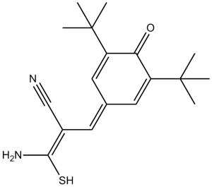 Tyrphostin AG 879
InvivoChem的所有产品仅用于作科学研究,不面向患者销售
Copyright 2020 InvivoChem LLC | All Rights Reserved 粤ICP备20063088号-1


 463611831
