| 规格 | 价格 | 库存 | 数量 |
|---|---|---|---|
| 5mg |
|
||
| 10mg |
|
||
| 25mg |
|
||
| 50mg |
|
||
| 100mg |
|
||
| 250mg |
|
||
| 500mg |
|
||
| Other Sizes |
|

| 靶点 |
FGFR1 (IC50 = 52.4 nM)
|
|---|---|
| 体外研究 (In Vitro) |
PD166866抑制FGFR1扩增的乳腺癌症细胞中的细胞增殖。
PD166866诱导FGFR1扩增的乳腺癌症细胞自噬。
靶向自噬提高了PD166866的抗癌效果。
PD166866通过抑制Akt/mTOR通路诱导自噬。[3]
体外活性:PD166866 抑制人全长 FGFR-1 酪氨酸激酶,IC50 值为 52.4 nM,是 FGFR-1 的 ATP 竞争性抑制剂。 PD 166866 是表达内源性 FGFR-1 的 NIH 3T3 细胞和过表达人 FGFR-1 酪氨酸激酶的 L6 细胞中 FGFR 自磷酸化的有效抑制剂。 PD 166866 还抑制 L6 细胞中 bFGF 诱导的 44-和 42-kDa (ERK 1/2) 丝裂原激活蛋白激酶亚型的酪氨酸磷酸化。每日将 PD 166866 以 1 至 100 nM 的浓度暴露于 L6 细胞,会连续 8 天对 bFGF 刺激的细胞生长产生浓度相关的抑制,IC50 值为 24 nM。激酶测定:PD166866 是一种新结构的酪氨酸激酶抑制剂 6-芳基-吡啶并[2,3-d]嘧啶的成员,是一种新型纳摩尔强效选择性 FGFR(成纤维细胞生长因子-1)小分子抑制剂受体)酪氨酸激酶抑制剂,IC50 为 52.4 nM。 PD 166866 是表达内源性 FGFR-1 的 NIH 3T3 细胞和过表达人 FGFR-1 酪氨酸激酶的 L6 细胞中 FGFR 自磷酸化的有效抑制剂。 PD 166866 还抑制 L6 细胞中 bFGF 诱导的 44-和 42-kDa (ERK 1/2) 丝裂原激活蛋白激酶亚型的酪氨酸磷酸化。 PD 166866 具有作为抗增殖/抗血管生成剂的潜在用途,可用于治疗肿瘤生长和动脉粥样硬化斑块的新血管形成等治疗目标。细胞测定:将 PD 166866 溶解在 DMSO 中。每天将 PD 166866 或载体(0.5% DMSO,终浓度)与 25 ng/mL bFGF 一起添加到一式三份的细胞培养物中,以刺激 FGF 驱动的生长。在一些实验中,每天将 PD 166866 与 30 ng/mL PDGF-BB 一起添加到一式三份的细胞培养物中,以刺激 PDGF 驱动的生长。药物暴露后第 1、3、6 或 8 天通过 Coulter 计数测量细胞数。 通过直接合成,我们发现了一种小分子,它是人成纤维细胞生长因子-1受体(FGFR)酪氨酸激酶的纳摩尔抑制剂。PD166866是酪氨酸激酶抑制剂新结构类别6-芳基吡啶并[2,3-d]嘧啶的成员,通过筛选具有测量蛋白酪氨酸激酶活性的测定的化合物库进行鉴定。PD 166866抑制人全长FGFR-1酪氨酸激酶,IC50值为52.4+/-0.1nM,并进一步被表征为FGFR-1的ATP竞争性抑制剂。相比之下,PD 166866在高达50微M的浓度下对c-Src、血小板衍生生长因子受体β、表皮生长因子受体或胰岛素受体酪氨酸激酶或丝裂原活化蛋白激酶、蛋白激酶c和CDK4没有影响。PD166866是表达内源性FGFR-1的NIH 3T3细胞和过表达人FGFR-1酪氨酸激酶的L6细胞中碱性成纤维细胞生长因子(bFGF)介导的受体自磷酸化的强效抑制剂,证实了酪氨酸激酶介导的机制。PD 166866还抑制了L6细胞中bFGF诱导的44和42 kDa(ERK 1/2)丝裂原活化蛋白激酶亚型的酪氨酸磷酸化,可能是通过抑制bFGF刺激的FGFR-1酪氨酸激酶活化。PD 166866分别不抑制血管平滑肌、A431或NIHIR细胞中的血小板衍生生长因子、表皮生长因子或胰岛素刺激的受体自磷酸化,进一步支持其对FGFR-1的特异性。此外,PD 166866以1至100 nM的浓度每天暴露于L6细胞,导致bFGF刺激的细胞生长连续8天受到浓度相关的抑制,IC50值为24 nM。相比之下,PD 166866对血小板源性生长因子BB刺激的L6细胞生长或血清刺激的血管平滑肌细胞增殖几乎没有影响。最后,发现PD 166866是人类胎盘培养动脉片段微血管生长(血管生成)的强效抑制剂。这些结果强调了PD 166866的发现,PD 166866是一种新的纳摩尔强效和选择性的FGFR-1酪氨酸激酶小分子抑制剂,可能用作肿瘤生长和动脉粥样硬化斑块新生血管等治疗靶点的抗增殖/抗血管生成剂。[2] 癌症约占每年诊断的癌症妇科病例的30%,是女性癌症相关死亡率的主要原因。FGFR1的扩增在乳腺癌中经常被观察到,并且与不良预后有关。尽管FGFR长期以来被认为是抗癌药物靶点,并且一组FGFR拮抗剂目前正在进行临床试验,但FGFR拮抗药治疗下的确切细胞反应仍不清楚。在这里,我们发现PD166866,一种FGFR1选择性抑制剂,在FGFR1扩增的乳腺癌症细胞系中抑制增殖并触发失活。值得注意的是,我们证明PD166866在FGFR1扩增的乳腺癌症细胞系中诱导自噬,而Atg5敲低对自噬的阻断进一步增强了PD166 866的抗增殖活性。此外,机制研究表明,PD166866通过抑制Akt/mTOR信号通路诱导自噬。总之,本研究为FGFR拮抗剂抗肿瘤活性的分子机制提供了新的见解,并可能进一步帮助基于FGFR的药物发现。 |
| 体内研究 (In Vivo) |
为了扩展我们的体外研究结果,通过皮下注射稳定表达shAtg5的MDA-MB-134亚系建立了小鼠肿瘤异种移植物模型,并用PD166866治疗小鼠。尽管前五天没有观察到差异,但与由模拟载体转染细胞组成的肿瘤相比,由Atg5沉默细胞形成的肿瘤显示出明显降低的生长速率(图3E-F)。此外,Ki-67免疫染色显示,当Atg5表达受到抑制时,肿瘤异种移植物中增殖细胞的数量大大减少,如图3G所示。这些结果表明,阻断自噬改善了PD166866在FGFR1扩增的乳腺癌症中的抗增殖作用[3]。
|
| 酶活实验 |
聚ADP核糖聚合酶(PARP)的免疫定位[1]
PARP酶在DNA断裂时被激活。PARP的免疫定位如先前发表的那样进行。简而言之,HeLa细胞用PD166866处理24小时,去除生长培养基,用PBS洗涤细胞,在25°C下固定1小时,加入新鲜制备的副甲醛溶液(4%的PBS溶液)。再次用PBS洗涤样品,在黑暗中阻断内源性氧化酶2分钟。随后用PBS进一步洗涤,在25°C下阻断非特异性位点1小时。PARP的证据是利用针对N端蛋白水解片段的多克隆抗体进行免疫定位。在4°C下孵育16小时后,通过二次抗兔抗体显示免疫反应。用PBS彻底洗涤后,将样品在ABC溶液中孵育30分钟。最终,加入DAB(3,3'-二氨基联苯胺),在黑暗中孵育样品10分钟。再次洗涤样品,密封平板,准备进行显微镜观察。 PD166866是一种新型 FGFR(成纤维细胞生长因子-1 受体)酪氨酸激酶小分子抑制剂,IC50 为 52.4 nM。它属于一种新结构的酪氨酸激酶抑制剂,6-芳基-吡啶并[2,3-d]嘧啶。在过表达人 FGFR-1 酪氨酸激酶的 L6 细胞和表达内源性 FGFR-1 的 NIH 3T3 细胞中,PD 166866 是 FGFR 自磷酸化的强抑制剂。此外,PD 166866 还可防止 L6 细胞 bFGF 诱导的 44-和 42-kDa (ERK 1/2) 丝裂原激活蛋白激酶亚型的酪氨酸磷酸化。对于肿瘤生长和动脉粥样硬化斑块新生血管等治疗靶点,PD 166866 具有作为抗增殖/抗血管生成剂的潜在应用。 |
| 细胞实验 |
HeLa 细胞暴露于PD166866一整天后,除去生长培养基,使用 PBS 洗涤细胞,并使用最近制备的多聚甲醛溶液(PBS 中的 4%)将它们在 25°C 下固定一小时。再次用 PBS 清洗样品后,在避光条件下将内源氧化酶封闭两分钟。之后,进行更多的 PBS 洗涤,并将未指定的位点在 25°C 下封闭一小时。通过使用多克隆抗体靶向 N 端蛋白水解片段进行免疫定位,证明了 PARP。 4°C 孵育 16 小时后,通过抗兔二抗进行免疫反应。将样品在 PBS 中彻底清洗,然后在溶液 ABC 中孵育半小时。添加 DAB(3,3'-二氨基联苯胺)后,将样品在黑暗中孵育 10 分钟。再次洗涤后,将板密封并准备用于显微镜检查。
HeLa细胞的生长和维持[1] 细胞在DMEM(Dulbecco's Modified Eagle's Medium-高糖)中维持,补充新生牛血清[终浓度(f.c.)10%]、青霉素-链霉素(10000 U/ml)和谷氨酰胺(2 mM);培养基的pH值为7.2,在37°C和5%CO2气氛中孵育。细胞在融合时常规传代。 细胞活力评估和脂质过氧化试验[1] 通过比色Mosmann测定法评估细胞存活率,该测定法是一种测量线粒体损伤水平的定量方法。MTT[3-(4,5-二甲基噻唑-2-基)-2,5-二芬太尼溴化四唑]是一种黄色的水溶性盐,可转化为由活细胞线粒体中存在的活性脱氢酶形成的不溶性紫色盐。在570nm处测量的吸光度值提供了活细胞的数量。细胞存活数据通过标准实验室方案用台盼蓝进行活体染色进行验证 使用商业试剂盒评估膜水平的氧化应激。简而言之,该测定基于对细胞内丙二醛(MDA)形成的定量分析,MDA来源于多不饱和脂肪酸的分解。MDA分子与显色化合物(N-甲基-2-苯基吲哚)反应,从而形成稳定的发色团。586nm处的吸光度直接转化为细胞内MDA浓度。 DNA片段的TUNEL检测和分析[1] 内源性DNA酶的激活是细胞死亡导致单链缺口形成并最终导致DNA断裂的后果之一。DNA断裂可以通过原位标记来证明。细胞核被渗透,加入荧光dUTP,末端脱氧核苷酸转移酶与糖磷酸骨架中断的核苷酸结合。荧光强度提供了DNA损伤的定性概念。 |
| 动物实验 |
雌性裸鼠
20 mg/kg 腹腔注射 肿瘤异种移植模型[3] 将健康雌性裸鼠(6-8周龄,18-20 g)皮下注射空载体或shAtg5过表达的MDA-MB-134细胞(5 × 10⁶个细胞/只)。当肿瘤体积达到100 mm³时,腹腔注射20 mg/kg的PD166866。每5天测量一次肿瘤体积,25天后处死动物。解剖肿瘤,并立即置于液氮中冷冻或用福尔马林固定。 |
| 毒性/毒理 (Toxicokinetics/TK) |
PD166866 对培养的 HeLa 细胞的细胞毒性 [1]
我们研究了 HeLa 细胞暴露于相对较宽浓度范围(0.1 - 50 μM)的 PD166866 的剂量-反应效应。细胞用药物处理 24 小时后,通过 MTT 法评估其活力。在 2.5 μM 浓度下即可观察到活细胞数量的显著减少(图 1,左图)。细胞活力的损失似乎在 25 μM 时趋于稳定(约 25% 的存活率),在 50 μM 药物浓度下未进一步下降。该结果可能表明存在对该药物具有内在耐药性的细胞亚群。台盼蓝染色活细胞计数证实了这一结果(仅显示 2.5 μM 药物浓度下获得的数据;图 1,右图)。 PD166866 对细胞生长的负面影响已在先前对 3T6 细胞(一种稳定的鼠成纤维细胞系)的研究中观察到。本文结果证实了已发表的研究结果,并且就细胞存活率而言,在本文进行的相应实验中(数据未显示),HeLa 细胞与 3T6 细胞相比未观察到差异。有趣的是,正如先前研究中所观察到的,HeLa 细胞对白藜芦醇(一种具有细胞毒性和抗病毒特性的天然产物)的敏感性显著高于鼠细胞。对此的一种解释是,这两种药物的细胞/分子靶点可能不同。 |
| 参考文献 | |
| 其他信息 |
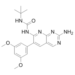
PD-166866 属于吡啶并嘧啶类化合物,其结构为吡啶并[2,3-d]嘧啶,在 2 位被氨基取代,6 位被 3,5-二甲氧基苯基取代,7 位被(叔丁基氨基甲酰基)腈基取代。它是一种选择性 ATP 竞争性人成纤维细胞生长因子-1 受体 (FGFR1) 酪氨酸激酶抑制剂,IC50 值为 52.4 nM。它具有诱导细胞凋亡、抗肿瘤、抑制 EC 2.7.10.1(受体蛋白酪氨酸激酶)和抑制血管生成的作用。它是一种二甲氧基苯、吡啶并嘧啶、脲类化合物、联芳基化合物和伯芳胺类化合物。
背景:大量实验数据表明,多种生长因子的过度表达会导致细胞增殖紊乱。成纤维细胞生长因子 (FGF) 在生长调控中的作用毋庸置疑:特别是 FGF1 及其酪氨酸激酶受体 (FGFR1) 通过极其复杂的机制和通路网络发挥作用。本研究评估了 PD166866(一种抑制 FGFR1 酪氨酸激酶活性的合成分子)的抗增殖活性。方法:细胞常规培养于添加新生儿血清和青霉素-链霉素混合物的 Dulbecco 改良 Eagle 培养基中。采用 Mosmann 法和台盼蓝染色法评估细胞活力。采用末端脱氧核苷酸转移酶dUTP缺口末端标记法(TUNEL法)进行原位荧光染色,评估DNA损伤。通过定量分析细胞内丙二酰二醛(MDA)的生成量来评估膜水平的氧化应激,MDA来源于多不饱和脂肪酸的分解。利用针对该酶N端片段的抗体,通过免疫组织化学方法检测了DNA片段化后聚ADP核糖聚合酶(PARP)的表达。结果:在Hela细胞上研究了该药物的生物活性。采用Mosmann法和台盼蓝活细胞染色法评估了其细胞毒性。细胞内丙二酰二醛浓度的显著升高表明,该分子的靶点很可能是细胞膜。 PD166866 处理后该化合物的增加提示膜脂质过氧化。TUNEL 检测结果定性但清晰地表明存在 DNA 损伤。此外,我们还证实了聚 ADP 核糖聚合酶 I (PARP I) 的细胞内积累。该酶是 DNA 链断裂的传感器,这支持了药物处理诱导细胞死亡的观点。结论:本研究数据表明 PD166866 具有明显的抗增殖作用。细胞增殖的负调控可能是通过激活凋亡途径实现的。针对这一特定点的实验结果,例如:DNA 损伤评估、细胞膜脂质过氧化以及 PARP(一种直接参与 DNA 修复的酶)表达的增加,表明暴露于 PD166866 的细胞会发生凋亡。然而,也不能排除同时存在其他细胞死亡方式的可能性。本文探讨了该药物在治疗方面的潜在用途。[1] 通过直接合成,我们发现了一种小分子,它是人成纤维细胞生长因子-1受体(FGFR)酪氨酸激酶的纳摩尔级抑制剂。PD 166866 属于一类新型酪氨酸激酶抑制剂——6-芳基-吡啶并[2,3-d]嘧啶类化合物,它是通过筛选化合物库并利用测定蛋白酪氨酸激酶活性的方法鉴定出来的。PD 166866 对人全长 FGFR-1 酪氨酸激酶的抑制 IC50 值为 52.4 ± 0.1 nM,并被进一步证实为 FGFR-1 的 ATP 竞争性抑制剂。相反,PD 166866 在浓度高达 50 μM 时,对 c-Src、血小板衍生生长因子受体-β、表皮生长因子受体或胰岛素受体酪氨酸激酶,以及丝裂原活化蛋白激酶、蛋白激酶 C 和 CDK4 均无影响。PD 166866 是表达内源性 FGFR-1 的 NIH 3T3 细胞和过表达人 FGFR-1 酪氨酸激酶的 L6 细胞中碱性成纤维细胞生长因子 (bFGF) 介导的受体自磷酸化的强效抑制剂,证实了酪氨酸激酶介导的机制。 PD 166866 还抑制了 L6 细胞中 bFGF 诱导的 44 kDa 和 42 kDa (ERK 1/2) 丝裂原活化蛋白激酶亚型的酪氨酸磷酸化,推测其作用机制是通过抑制 bFGF 刺激的 FGFR-1 酪氨酸激酶活化。PD 166866 不抑制血管平滑肌细胞、A431 细胞或 NIHIR 细胞中血小板衍生生长因子、表皮生长因子或胰岛素刺激受体的自身磷酸化,进一步证实了其对 FGFR-1 的特异性。此外,每日将 L6 细胞暴露于浓度为 1 至 100 nM 的 PD 166866 中,可连续 8 天观察到浓度依赖性的 bFGF 刺激的细胞生长抑制,IC50 值为 24 nM。相反,PD 166866 对血小板衍生生长因子-BB 刺激的 L6 细胞生长或血清刺激的血管平滑肌细胞增殖几乎没有影响。此外,PD 166866 被发现是人胎盘动脉碎片培养物中微血管生成(血管形成)的强效抑制剂。这些结果凸显了 PD 166866 的发现,它是一种新型纳摩尔级强效且选择性的小分子 FGFR-1 酪氨酸激酶抑制剂,具有作为抗增殖/抗血管生成药物的潜力,可用于治疗肿瘤生长和动脉粥样硬化斑块新生血管形成等疾病。[2] 乳腺癌约占每年所有妇科癌症病例的 30%,是女性癌症相关死亡的主要原因之一。FGFR1 扩增在乳腺癌中很常见,并且与预后不良相关。尽管FGFRs长期以来被认为是抗癌药物靶点,且目前已有多种FGFR拮抗剂正在进行临床试验,但FGFR拮抗剂治疗下细胞的确切反应仍不清楚。本研究表明,FGFR1选择性抑制剂PD166866能够抑制FGFR1扩增的乳腺癌细胞系的增殖并诱导其发生失巢凋亡。值得注意的是,我们发现PD166866能够诱导FGFR1扩增的乳腺癌细胞系发生自噬,而通过敲低Atg5阻断自噬则进一步增强了PD166866的抗增殖活性。此外,机制研究表明,PD166866通过抑制Akt/mTOR信号通路诱导自噬。本研究为FGFR拮抗剂抗肿瘤活性的分子机制提供了新的见解,并可能进一步帮助基于FGFR的药物发现。[3] |
| 分子式 |
C20H24N6O3
|
|
|---|---|---|
| 分子量 |
396.44
|
|
| 精确质量 |
396.191
|
|
| 元素分析 |
C, 60.59; H, 6.10; N, 21.20; O, 12.11
|
|
| CAS号 |
192705-79-6
|
|
| 相关CAS号 |
|
|
| PubChem CID |
5328127
|
|
| 外观&性状 |
white solid powder
|
|
| 密度 |
1.277g/cm3
|
|
| 折射率 |
1.643
|
|
| LogP |
3.545
|
|
| tPSA |
128.5
|
|
| 氢键供体(HBD)数目 |
3
|
|
| 氢键受体(HBA)数目 |
7
|
|
| 可旋转键数目(RBC) |
5
|
|
| 重原子数目 |
29
|
|
| 分子复杂度/Complexity |
545
|
|
| 定义原子立体中心数目 |
0
|
|
| SMILES |
C1=C2C(N=C(NC(=O)NC(C)(C)C)C(C3C=C(OC)C=C(OC)C=3)=C2)=NC(N)=N1
|
|
| InChi Key |
NHJSWORVNIOXIT-UHFFFAOYSA-N
|
|
| InChi Code |
InChI=1S/C20H24N6O3/c1-20(2,3)26-19(27)25-17-15(8-12-10-22-18(21)24-16(12)23-17)11-6-13(28-4)9-14(7-11)29-5/h6-10H,1-5H3,(H4,21,22,23,24,25,26,27)
|
|
| 化学名 |
1-[2-amino-6-(3,5-dimethoxyphenyl)pyrido[2,3-d]pyrimidin-7-yl]-3-tert-butylurea
|
|
| 别名 |
|
|
| HS Tariff Code |
2934.99.9001
|
|
| 存储方式 |
Powder -20°C 3 years 4°C 2 years In solvent -80°C 6 months -20°C 1 month |
|
| 运输条件 |
Room temperature (This product is stable at ambient temperature for a few days during ordinary shipping and time spent in Customs)
|
| 溶解度 (体外实验) |
|
|||
|---|---|---|---|---|
| 溶解度 (体内实验) |
配方 1 中的溶解度: ≥ 1 mg/mL (2.52 mM) (饱和度未知) in 10% DMSO + 90% (20% SBE-β-CD in Saline) (这些助溶剂从左到右依次添加,逐一添加), 澄清溶液。
例如,若需制备1 mL的工作液,可将100 μL 10.0 mg/mL澄清DMSO储备液加入900 μL 20% SBE-β-CD生理盐水溶液中,混匀。 *20% SBE-β-CD 生理盐水溶液的制备(4°C,1 周):将 2 g SBE-β-CD 溶解于 10 mL 生理盐水中,得到澄清溶液。 配方 2 中的溶解度: ≥ 1 mg/mL (2.52 mM) (饱和度未知) in 10% DMSO + 90% Corn Oil (这些助溶剂从左到右依次添加,逐一添加), 澄清溶液。 例如,若需制备1 mL的工作液,可将 100 μL 10.0 mg/mL 澄清 DMSO 储备液加入到 900 μL 玉米油中并混合均匀。 View More
配方 3 中的溶解度: 5% DMSO+40% PEG 300+5% Tween 80+50% ddH2O: 0.5mg/mL 配方 4 中的溶解度: 3.33 mg/mL (8.40 mM) in 0.5% CMC-Na/saline water (这些助溶剂从左到右依次添加,逐一添加), 悬浮液; 需要超声助溶并加热至 40°C。 *生理盐水的制备:将 0.9 g 氯化钠溶解在 100 mL ddH₂O中,得到澄清溶液。 1、请先配制澄清的储备液(如:用DMSO配置50 或 100 mg/mL母液(储备液)); 2、取适量母液,按从左到右的顺序依次添加助溶剂,澄清后再加入下一助溶剂。以 下列配方为例说明 (注意此配方只用于说明,并不一定代表此产品 的实际溶解配方): 10% DMSO → 40% PEG300 → 5% Tween-80 → 45% ddH2O (或 saline); 假设最终工作液的体积为 1 mL, 浓度为5 mg/mL: 取 100 μL 50 mg/mL 的澄清 DMSO 储备液加到 400 μL PEG300 中,混合均匀/澄清;向上述体系中加入50 μL Tween-80,混合均匀/澄清;然后继续加入450 μL ddH2O (或 saline)定容至 1 mL; 3、溶剂前显示的百分比是指该溶剂在最终溶液/工作液中的体积所占比例; 4、 如产品在配制过程中出现沉淀/析出,可通过加热(≤50℃)或超声的方式助溶; 5、为保证最佳实验结果,工作液请现配现用! 6、如不确定怎么将母液配置成体内动物实验的工作液,请查看说明书或联系我们; 7、 以上所有助溶剂都可在 Invivochem.cn网站购买。 |
| 制备储备液 | 1 mg | 5 mg | 10 mg | |
| 1 mM | 2.5224 mL | 12.6122 mL | 25.2245 mL | |
| 5 mM | 0.5045 mL | 2.5224 mL | 5.0449 mL | |
| 10 mM | 0.2522 mL | 1.2612 mL | 2.5224 mL |
1、根据实验需要选择合适的溶剂配制储备液 (母液):对于大多数产品,InvivoChem推荐用DMSO配置母液 (比如:5、10、20mM或者10、20、50 mg/mL浓度),个别水溶性高的产品可直接溶于水。产品在DMSO 、水或其他溶剂中的具体溶解度详见上”溶解度 (体外)”部分;
2、如果您找不到您想要的溶解度信息,或者很难将产品溶解在溶液中,请联系我们;
3、建议使用下列计算器进行相关计算(摩尔浓度计算器、稀释计算器、分子量计算器、重组计算器等);
4、母液配好之后,将其分装到常规用量,并储存在-20°C或-80°C,尽量减少反复冻融循环。
计算结果:
工作液浓度: mg/mL;
DMSO母液配制方法: mg 药物溶于 μL DMSO溶液(母液浓度 mg/mL)。如该浓度超过该批次药物DMSO溶解度,请首先与我们联系。
体内配方配制方法:取 μL DMSO母液,加入 μL PEG300,混匀澄清后加入μL Tween 80,混匀澄清后加入 μL ddH2O,混匀澄清。
(1) 请确保溶液澄清之后,再加入下一种溶剂 (助溶剂) 。可利用涡旋、超声或水浴加热等方法助溶;
(2) 一定要按顺序加入溶剂 (助溶剂) 。
Assessment of cell survival after treatment with PD166866.
Intracellular concentration of malonyl-dihaldehyde (MDA) after treatment with PD166866.J Exp Clin Cancer Res.2009 Dec 11;28:151. |
An extensive DNA damage is caused by treatment with PD166866.J Exp Clin Cancer Res.2009 Dec 11;28:151. |
Accumulation Poly-ADP-Ribose-Polymerase (PARP) in cells treated with PD166866 evidenced by imuno-histochemistry.J Exp Clin Cancer Res.2009 Dec 11;28:151. |
InvivoChem的所有产品仅用于作科学研究,不面向患者销售
Copyright 2020 InvivoChem LLC | All Rights Reserved 粤ICP备20063088号-1


 COA
COA


 463611831
