| 规格 | 价格 | 库存 | 数量 |
|---|---|---|---|
| 1mg |
|
||
| 5mg |
|
||
| 10mg |
|
||
| 50mg |
|
||
| Other Sizes |
|
| 靶点 |
CSF-1R (IC50 = 0.016 μM); FLT3 (IC50 = 0.39 μM); KIT (IC50 = 0.86 μM); AURKC (IC50 = 1 μM); KDR (IC50 = 1.1 μM)
|
|---|---|
| 体外研究 (In Vitro) |
PLX5622(1–20 μM;3 天)可有效减少小脑切片中的小胶质细胞,同时对少突胶质细胞或星形胶质细胞没有影响。 4 μM 浓度三天后,NG2+ 或 PDGFRα+ 细胞计数减少 30-40%,20 μM 浓度时,该数字上升至 90-95%。尽管小胶质细胞显着减少 (~95%),但暴露于 1 μM 或 2 μM PLX5622 的切片显示 NG2+ 或 PDGFRα+ OPC 没有减少[3]。
无细胞激酶抑制剂谱分析显示,PLX5622 对 CSF1R 具有高度特异性,对两个同源性最高的受体 KIT 和 FLT3 显示出 > 20 倍的选择性(补充表 2, 3)。基于晶体学分析(图 2b;补充表 4),PLX5622 和 PLX3397 之间的两个关键结构差异促成了选择性的提高。首先,PLX5622 中间吡啶环上的 2-氟取代旨在进入 Gly-795 旁边的 CSF1R 独有空间(Gly-795 是通往内部变构口袋的门控残基);而 KIT 和 FLT3 在等效位置都有一个体积更大的半胱氨酸。虽然氢原子和氟原子之间的差异看似很小(氟原子具有稍长的键长和稍大的半径,与氢原子相比,其范德华边缘延伸了约 0.5 Å),但其影响是显著的。PLX5622 两端通过氢键和疏水堆积锚定(图 2b),其氟原子处于固定位置。由于范德华斥力与两个原子之间距离的六次方成正比增加,一个更大的门控残基(如 KIT 和 FLT3 中那样)很可能会因空间位阻而产生能量损失。为了支持这一机制,一个含有与 PLX5622 相同 2-氟取代的 PLX3397 的密切类似物在抑制 CSF1R 方面具有与 PLX3397 相同的效力,但对 KIT 的选择性比 PLX3397 高 10 倍。其次,PLX5622 的末端吡啶基团经过优化,以稳定被置换的近膜域腾出的 CSF1R 变构口袋。当 PLX5622 与 KIT 或 FLT3 结合时,中间吡啶环的位置和方向会导致与门控残基半胱氨酸发生空间冲突,从而损害末端吡啶部分的最佳契合度。[1] CSF1R 激酶抑制剂 PLX5622 和 PLX3397 耗竭小脑切片中的小胶质细胞 [3] 将 PLX5622 或 PLX3397 以 1 至 20 μM 的递增浓度应用于小脑切片。治疗三天后,浓度大于 2 μM 的 PLX5622 和 PLX3397 均消除了超过 95% 的小胶质细胞(图 1A,B)。在 1 μM 浓度下,PLX5622 比 PLX3397 多耗竭 15%(Iba1+ 细胞损失:94.6±1.1% vs 79.9±2.4%)(图 1A,B)。在 2 μM 浓度下小胶质细胞损失超过 95% 时,通过 PLP-eGFP 表达或 GFAP 染色评估,未观察到少突胶质细胞或星形胶质细胞的活力或形态发生任何变化(图 1C)。 高浓度的 PLX5622 和 PLX3397 在离体和小脑切片中对 OPCs 具有细胞毒性 [3] 由于 CSF1R 抑制剂可能结合多种酪氨酸激酶,我们检测了 PLX5622 和 PLX3397 对依赖酪氨酸激酶 PDGFRα 存活的 OPCs 的影响。将小脑外植体暴露于递增的 PLX 浓度 3 天后,通过 NG2 或 PDGFRα 免疫染色评估 OPC 细胞数量。在 4 μM 浓度下,PLX5622 导致 NG2+ 或 PDGFRα+ 细胞减少 30-40%;在 20 μM 浓度下,这一减少增加到 90-95%。尽管小胶质细胞被强力耗竭(约 95%),但在暴露于 1 μM 或 2 μM PLX5622 的切片中未观察到 NG2+ 或 PDGFRα+ OPCs 的减少(图 2A,C)。相比之下,用 PLX3397 处理在所有测试浓度下均显著减少 OPC 数量。我们在 0.5 μM 浓度下观察到 NG2+ 或 PDGFRα+ 细胞损失 30-35%,尽管小胶质细胞耗竭不完全(约 70%)。PLX3397 导致的 OPC 损失呈浓度依赖性,在 1 μM 浓度下减少 75-85%,在 2 μM 和 20 μM 浓度下损失 > 90%(图 2B,D)。 在纯化的原代小鼠 OPC 培养物中也观察到对 OPCs 的类似效应。细胞类型特异性标记物染色显示,培养物由 >92% 的 OPCs(PDGFRα)、<1% 的星形胶质细胞(GFAP)和 3.7±2.1% 的小胶质细胞(Iba1)组成(补充图 S1A)。大多数少突胶质细胞是 OPCs,但偶尔(<0.5%)观察到成熟的 OLs(O4+,具有高度复杂的突起网络)(数据未显示)。低浓度(0.5 μM)的 PLX5622 在 24 小时后对活力没有影响;然而,20 μM PLX5622 导致 OPC 死亡增加。相比之下,PLX3397 处理在 0.5 μM 和 20 μM 浓度下均引起显著的细胞毒性(补充图 S1B,C)。这些结果表明,PLX CSF1R 抑制剂能够以浓度依赖的方式直接损害 OPC 的活力。 为了研究长期小胶质细胞耗竭对 OPC 存活的影响,将 2 μM PLX5622 应用于小脑切片 8 天。尽管小胶质细胞被完全清除,但与 DMSO 处理的对照组相比,我们观察到 NG2+ 细胞数量没有差异。此外,长期暴露于 PLX5622 对少突胶质细胞(通过 PLP-eGFP 评估)和星形胶质细胞(通过 GFAP 和 S100b 评估)的形态或数量没有影响(图 3A,B 和数据未显示)。髓鞘蛋白表达(通过 PLP 免疫荧光测量)没有变化(图 3A)。 综上所述,离体和小脑切片数据均表明,低浓度(≤ 2 μM)的 PLX5622 能在不直接影响 OPCs、少突胶质细胞或星形胶质细胞的情况下完全耗竭小胶质细胞。即使在低浓度下,PLX3397 也会导致存活的 OPCs 显著减少。在较高浓度下,两种 PLX 化合物在原代细胞培养物和小脑切片中都会减少 OPC 细胞数量。 在小脑切片中,PLX5622(1–20 μM;3 天)半富马酸盐可有效消耗小胶质细胞,同时对少突胶质细胞或星形胶质细胞没有影响。当暴露于 PLX5622(4 μM;3 天)半富马酸盐时,NG2+ 或 PDGFRα+ 细胞减少 30-40%;在 20 μM 时,这一比例上升至 90-95%。尽管小胶质细胞明显减少 (~95%),但经 1 μM 或 2 μM PLX5622 处理的切片显示 NG2+ 或 PDGFRα+ OPC 没有变化[3]。 |
| 体内研究 (In Vivo) |
PLX5622(1200 ppm;饲料;持续 3 周或 3 天;成年 C57/Bl6 野生型小鼠)在治疗 3 天和 3 周后分别导致约 80% 和 99% 的小胶质细胞丢失。 PLX5622 中皮质、纹状体、小脑和海马中的小胶质细胞减少(成年 C57/Bl6 野生型小鼠,3 个月大;喂养 3 周)[4]。
在总共 14 天中,PLX5622 ( 50 mg/kg;腹腔注射;新生大鼠每日一次,成年大鼠每日两次)可使小胶质细胞在 3 天内减少 80-90%,在 7 天内减少 90% 以上。 PLX5622 治疗 14 天后,成人和新生儿的小胶质细胞减少 > 96%,同时维持基线星形胶质细胞数量。 (消除新生儿的小胶质细胞只需每天注射一次悬浮在 5% 二甲亚砜中的 0.65% PLX5622 和 0.01 M PBS 中的 20% Kolliphor RH40;成人消除则需要每天注射两次。)[5]. PLX5622 (在 AIN-76A 标准饲料中配制,浓度为 1200 毫克/千克;持续 28 天)减少 14 个月大的 5xfAD 小鼠中整个 CNS 的小胶质细胞[6]。 许多阿尔茨海默病(Alzheimer's disease, AD)发展的风险基因在髓系细胞中独家或高度表达。小胶质细胞(Microglia)的生存依赖于集落刺激因子1受体(colony-stimulating factor 1 receptor, CSF1R)信号传导。我们设计并合成了一种高选择性、可穿透血脑屏障的CSF1R抑制剂(PLX5622),能够在病理发展之前和期间实现长期且特异性的小胶质细胞清除。我们发现,在AD的5xFAD小鼠模型中,小胶质细胞清除后,实质空间内无法形成斑块,除了含有存活小胶质细胞的区域。相反,Aβ沉积在皮质血管中,让人联想到脑淀粉样血管病(cerebral amyloid angiopathy)。5xFAD海马体中改变的基因表达也因小胶质细胞的缺失而逆转。对残余斑块形成小胶质细胞的转录组分析显示,它们表现出疾病相关小胶质细胞(disease-associated microglia)的特征。总之,我们描述了PLX5622的结构、配方和功效,它能够实现持续的小胶质细胞清除,并确定了小胶质细胞在启动斑块发病机制中的作用。[1] 引言 神经病理性疼痛是一种使人衰弱的疾病。神经免疫相互作用在神经病理性疼痛中的重要性已通过不同免疫细胞参与病理性疼痛的外周和中枢敏化得到证实。巨噬细胞和小胶质细胞分别是受损神经和脊髓中激活的最丰富的免疫细胞。多条证据表明,巨噬细胞/小胶质细胞的存活、激活、增殖和分化需要巨噬细胞集落刺激因子(macrophage-colony stimulating factor)的参与。在本研究中,我们研究了阻断巨噬细胞集落刺激因子/集落刺激因子1受体(macrophage-colony stimulating factor/colony stimulating factor 1 receptor)信号传导是否能有效缓解神经病理性疼痛。材料与方法 在小鼠中进行部分坐骨神经结扎(Partial sciatic nerve ligation)以诱导神经病理性疼痛行为。在预防性方案(手术前两天开始直至部分坐骨神经结扎后第14天)和逆转性方案(部分坐骨神经结扎后第28天至第33天)中,每日口服给予选择性集落刺激因子1受体抑制剂 PLX5622。使用冯弗雷细丝(von Frey hairs)和丙酮涂抹(acetone application)监测动物的神经病理性疼痛行为。在损伤后第3天和第33天,使用流式细胞术分析受损神经中巨噬细胞的表型。使用Iba-1抗体通过免疫组织化学进一步检测PLX5622对腰段脊髓中小胶质细胞活化的影响。结果 在预防性和逆转性方案中,均观察到PLX5622治疗的动物其机械性痛觉超敏(mechanical allodynia)和冷诱发性痛觉超敏(cold allodynia)显著减轻。PLX5622治疗减少了受损神经中巨噬细胞的总数,似乎集落刺激因子1受体抑制更特异地影响了CD86+(M1样)巨噬细胞。因此,各种促炎细胞因子(TNF-α, IL-1β)的表达降低。在预防性和逆转性方案中,PLX5622治疗显著抑制了部分坐骨神经结扎后腰段脊髓背角(dorsal horn)的小胶质细胞活化。结论 外周神经中的巨噬细胞和脊髓中的小胶质细胞是损伤相关神经病理性疼痛的产生和维持所必需的。阻断疼痛传导通路中这些髓系细胞上的巨噬细胞集落刺激因子/集落刺激因子1受体信号传导是缓解神经病理性疼痛的有效策略。[2] 引言 神经病理性疼痛是一种使人衰弱的疾病。神经免疫相互作用在神经病理性疼痛中的重要性已通过不同免疫细胞参与病理性疼痛的外周和中枢敏化得到证实。巨噬细胞和小胶质细胞分别是受损神经和脊髓中激活的最丰富的免疫细胞。多条证据表明,巨噬细胞/小胶质细胞的存活、激活、增殖和分化需要巨噬细胞集落刺激因子的参与。在本研究中,我们研究了阻断巨噬细胞集落刺激因子/集落刺激因子1受体信号传导是否能有效缓解神经病理性疼痛。材料与方法 在小鼠中进行部分坐骨神经结扎以诱导神经病理性疼痛行为。在预防性方案(手术前两天开始直至部分坐骨神经结扎后第14天)和逆转性方案(部分坐骨神经结扎后第28天至第33天)中,每日口服给予选择性集落刺激因子1受体抑制剂 **PLX5622**。使用冯弗雷细丝和丙酮涂抹监测动物的神经病理性疼痛行为。在损伤后第3天和第33天,使用流式细胞术分析受损神经中巨噬细胞的表型。使用Iba-1抗体通过免疫组织化学进一步检测PLX5622对腰段脊髓中小胶质细胞活化的影响。结果 在预防性和逆转性方案中,均观察到PLX5622治疗的动物其机械性痛觉超敏和冷诱发性痛觉超敏显著减轻。PLX5622治疗减少了受损神经中巨噬细胞的总数,似乎集落刺激因子1受体抑制更特异地影响了CD86+(M1样)巨噬细胞。因此,各种促炎细胞因子(TNF-α, IL-1β)的表达降低。在预防性和逆转性方案中,PLX5622治疗显著抑制了部分坐骨神经结扎后腰段脊髓背角的小胶质细胞活化。结论 外周神经中的巨噬细胞和脊髓中的小胶质细胞是损伤相关神经病理性疼痛的产生和维持所必需的。阻断疼痛传导通路中这些髓系细胞上的巨噬细胞集落刺激因子/集落刺激因子1受体信号传导是缓解神经病理性疼痛的有效策略。[3] 使用特异性CSF1R抑制剂 **PLX5622** 清除老年5xFAD小鼠中的小胶质细胞 PLX5622是一种可穿透血脑屏障的CSF1R抑制剂,能快速清除小胶质细胞,但不抑制c-kit(Valdearcos et al., 2014; Dagher et al., 2015)。为了确认PLX3397的效果并排除其他脱靶效应,我们用含1200 mg/kg PLX5622的饲料处理第二组5xFAD小鼠28天。我们再次使用硫磺素-S(Thioflavin-S)染色组织以显示致密核心斑块(dense core plaques),并用IBA1对微胶质细胞进行免疫标记(图5A)。与PLX3397治疗一样,经PLX5622治疗后小胶质细胞被显著清除,剩余细胞大多与致密核心斑块相关(图5B和E)。量化显示平均每个斑块约有一个剩余细胞(图5C);然而,许多斑块没有相关的IBA1+细胞(图5D)。进一步分析显示,所有剩余的IBA1+细胞的胞体都含有β-淀粉样蛋白(amyloid-β)(图5K);相比之下,未经治疗的5xFAD大脑斑块周围的大多数IBA1+细胞不含有任何β-淀粉样蛋白(图5J)。事实上,在治疗组小鼠中,其中一些IBA1+细胞呈硫磺素-S阳性(图5E)。然而,与PLX3397治疗的小鼠一样,在这组小胶质细胞被清除的5xFAD小鼠中,我们未观察到斑块面积或总数发生变化(图5F和G),β-淀粉样蛋白水平也无显著变化(图5H和I)。[6] PLX5622 半富马酸盐在临床前研究中的药效学 在成年 C57/Bl6 野生型小鼠中,PLX5622(1200 ppm;喂食;持续 3 周或 3 天)半富马酸盐在治疗 3 天后分别导致约 80% 和 99% 的小胶质细胞被破坏。 PLX5622(成年 C57/Bl6 野生型小鼠,3 个月大;喂养 3 周)中大脑、纹状体、小脑和海马中的小胶质细胞减少[4]。半反丁烯二酸盐(PLX5622;50 mg/kg;腹腔注射;新生大鼠每天一次,成年大鼠每天两次)在治疗 3 天期间使小胶质细胞减少 80-90%,到 7 天时,已增加至 > 90% 。 PLX5622 治疗 14 天后,新生儿和成人的小胶质细胞减少了 96% 以上,同时星形胶质细胞的数量保持不变。 (对于新生儿小胶质细胞消除,每天注射一次悬浮在 5% 二甲亚砜中的 0.65% PLX5622 和 0.01 M PBS 中的 20% Kolliphor RH40 就足够了;成人消除需要每天注射两次)[5]。在 14 个月大的 5xfAD 小鼠中,PLX5622(在 AIN-76A 正常饲料中配制,浓度为 1200 mg/kg;持续 28 天)半富马酸盐可减少整个中枢神经系统的小胶质细胞[6]。 PLX5622半富马酸盐在临床前物种中的药代动力学[4] 物种 IV PO (灌胃) 剂量 (mg/kg) AUC0-∞ (ng·hr/mL) CL (mL/min/kg) Vss (L/kg) t1/2 ( hr ) 剂量 (mg/kg) AUC0-∞ (ng·hr/mL) Cmax (ng/mL) F 小鼠 1.92 15,500 2.1 0.34 2.6 45 215,000 26,300 59% 大鼠(雄性) 1.13 2,630 7.7 1.2 2.3 45 99,600 12,000 9 5 %大鼠(雌性) 1.13 5,110 3.7 1.0 3.9 45 181,000 15,600 89% 狗 1.00 6,230 3.0 2.3 15 45 96,500 3,630 34% 猴 1.35 2,100 11 1.6 2.2 ND ND ND ND PLX5622 半富马酸盐灌胃给药悬浮液的制备[4] PLX5622 半富马酸盐是以最终剂量溶液 20 倍的浓度溶解在 DMSO 中。复合原料具有轻度保护作用。每周都会创建新库存。由于其溶解缓慢,稀释剂的成分通常提前一天或更长时间准备: a) 2%羟丙基甲基纤维素(HPMC):将2.0g粉末添加到100mL去离子水中; b) 25% 聚山梨醇酯 80 (PS80):将 25 g 粉末添加到 100 mL 去离子水中。将 4 mL 25% PS80 库存(1% 最终)和 25 mL 2% HPMC 库存(0.5% 最终)添加到 71 mL 去离子水中,制成最终 100 mL 的稀释剂。化合物混合后,最终成分为 0.5% HPMC、1% PS80 和 5% DMSO。在每个剂量日,化合物储备液以这种方式稀释 20 倍:将 19 体积的稀释剂量入管中,并添加 1 体积的化合物/DMSO 储备液 (20x)。为了产生均匀的悬浮液,通过倒置将管中的内容物混合,然后在关闭盖子后将其放入超声水浴中。 |
| 细胞实验 |
小脑切片培养 [3]
矢状小脑切片(300微米)从出生后10-12天(P10-12)的PLP-eGFP小鼠(Mallon等人,2002)制备,并在MilliCell 0.4微米膜插入式培养皿中,使用切片培养基于37°C培养(Sheridan和Dev,2012)。切片培养基成分为:25% Hank氏平衡盐溶液(HBSS),25%热灭活马血清,50%最低必需培养基(MEM),125mM HEPES,28mM D-葡萄糖,2mM L-谷氨酰胺,10单位/毫升青霉素/链霉素。铺板后24小时内更换培养基,之后每2-3天更换一次。切片在处理前培养7-10天。DMSO、PLX5622和PLX3397按指定浓度加入切片培养基中,处理3天或8天。培养基每2-3天更换一次。处理后,切片在PBS中漂洗一次,在4%多聚甲醛(溶于PBS)中固定30分钟,然后进行免疫染色。 纯化原代OPC细胞培养 [3] 原代小鼠OPC培养物按照所述方法(Liu等人,2016)从PLP-eGFP幼鼠制备。简言之,从出生后0-1天(P0-1)小鼠的分离皮层制备混合胶质细胞培养物,并铺板在聚-D-赖氨酸包被的T75培养瓶上。通过将长满的混合胶质细胞培养瓶在200转/分下震荡过夜以分离OPCs,从而制备纯化的OPC单层培养物。OPCs铺板在聚-D-赖氨酸/层粘连蛋白包被的培养皿上,并在含10纳克/毫升PDGF(血小板衍生生长因子)/FGF(成纤维细胞生长因子)的培养基中维持24-48小时,然后进行实验。 IncuCyte活细胞成像 [3] 活细胞成像按所述方法进行(Liu等人,2016)。细胞在24孔板中于细胞培养箱内生长并进行扫描。使用10倍物镜,在每个孔中随机选择9个位置,以15分钟为间隔,使用高清相差显微术和落射荧光显微术(使用585/635纳米滤光片,红色荧光,用于检测DRAQ7死细胞核)进行扫描。图像处理和细胞计数使用IncuCyte和ImageJ软件进行。 |
| 动物实验 |
药物制备和治疗[2]
PLX5622是一种强效的CSF1R酪氨酸激酶活性抑制剂(Ki = 5.9 nM),对四种相关激酶的选择性至少高50倍,对230种激酶的选择性超过100倍。该分子已被用于研究小胶质细胞/巨噬细胞在多种不同情况下的关键作用。本研究的剂量由公司建议,并参考了之前的报道,即每日在饲料中添加1200 ppm足以完全消除小胶质细胞,或每日在饲料中添加300 ppm可部分减少小胶质细胞。我们名义上的65 mg/kg剂量接近每日300 ppm的饲料剂量,这使我们能够降低神经中的巨噬细胞数量。该药物按照制造商的方案制备。简而言之,将PLX5622储备液溶解于二甲基亚砜(DMSO)中,浓度为130 mg/ml;配制2%羟丙基甲基纤维素和25%聚山梨醇酯80的稀释液。在每个给药日,将PLX5622储备液稀释20倍,方法是将1体积的药物储备液(130 mg/ml)加入19体积的稀释液中,制成浓度为6.5 mg/ml的工作液。载体溶液由稀释液和DMSO混合配制而成。小鼠每日通过灌胃给予100 μl溶液(6.5 mg/ml),剂量为每10g体重100 μl(最终剂量为65 mg/kg体重)。 为研究PLX5622的预防作用,小鼠在手术前两天接受药物或载体处理,之后每日给药直至PSNL术后第14天。为研究PLX5622的逆转作用,从PSNL术后第28天开始给药,每日给药直至术后第33天。 行为学分析[2] 在基线测试前至少两天,小鼠每天在测试环境中适应1至2小时。小鼠每天早上接受载体或PLX5622处理。研究人员对治疗条件不知情。 动物饲料处理[3] 8-12周龄的雄性和雌性PLP-eGFP动物(每组4-5只)分别饲喂配制PLX饲料(PLX5622组1200 mg/kg,PLX3397组275 mg/kg)或标准饲料,持续7天或21天。处理结束后,用冰冷的PBS灌注动物,随后用4%多聚甲醛PBS溶液灌注。解剖出脑和脊髓,进行过夜后固定,然后进行低温保护。组织在冰冻切片机上切成 30μm 厚的切片,并收集于 PBS 缓冲液中,用于游离免疫染色。 为了进行全脑小胶质细胞消融,将 8-16 周龄的成年 C57/Bl6 野生型小鼠分别用 CSF1R 抑制剂 PLX5622 或对照饲料(配方相同,但不含抑制剂)处理 3 周或 3 天(具体时间见指示)。PLX5622 处理 3 周可导致 99% 的小胶质细胞丢失,而处理 3 天后,约 80% 的小胶质细胞已经丢失 [4]。 适应头部固定和腹腔注射。 [4] 手术后1-2周恢复期后,将小鼠随机分为两组,一组接受PLX5622治疗,另一组接受对照饲料(不含抑制剂)。 随后对小鼠进行限水(1.5 ml/天),并每日进行操作和头部固定适应训练,持续约2周。将小鼠固定在聚乙烯管中,并在双光子显微镜下进行头部固定,以适应未来的记录环境。在此适应期内,我们将头部固定时间从3分钟逐渐增加到40分钟。在40分钟的固定时间内,小鼠未表现出应激反应,且能正常饮水后,我们改为不提供饮水的头部固定适应训练,以进行后续记录。 头部固定适应训练完成后,我们还在头部固定前进行每日腹腔注射适应训练。小鼠腹腔注射相当于其体重10倍(10 × BW μl)的微升生理盐水,注射体积与后续成像实验的注射体积一致,然后进行头部固定。此适应过程持续约1周,直至小鼠在操作和腹腔注射后表现出明显的应激反应。微透析[4] 雄性C57/Bl6小鼠分别喂食PLX5622饲料或对照饲料(PLX5622组n=5,对照组n=5),持续一周。植入透析探针后,小鼠恢复一周,然后开始微透析数据采集。微透析引导套管立体定位植入纹状体(距颅骨前后方向+1.4 mm;左右方向-1.0 mm;背腹方向-3.8 mm)。在导管植入后一周的恢复期后进行微透析实验。首次使用前,先用70%乙醇冲洗透析管5分钟,然后用去离子水注射泵以1 μL/min的流速冲洗。之后将透析管连接到微透析探针(Cuprophane (6kD),膜长1mm),探针预先浸入人工脑脊液(aCSF;pH 7.4:148 mM NaCl、2.7 mM KCl、1.2 mM CaCl₂、0.85 mM MgCl₂)中,并以0.8 μL/min的流速将aCSF流经微透析管和探针进行预充。微透析实验在麻醉动物中进行。简而言之,将小鼠固定在立体定位仪上,并使用异氟烷麻醉(诱导浓度4%,维持浓度1.75%)。探针经引导套管插入并使其达到平衡。20分钟后收集透析液。所有收集物在收集完成后立即冷冻于-80摄氏度。 通过腹腔注射PLX5622抑制小胶质细胞[5] 先前的CSF1R抑制剂研究使用PLX3397/5622联合饲料喂养的动物,在实验操作前至少1周进行(例如Elmore等人,2014;Okunukia等人,2019)。为了确保新生儿手术前小胶质细胞得到充分清除,我们从出生后第1天(P1)开始治疗,成年动物的术前治疗持续时间与新生儿相同(9天)。初步测试表明,虽然每天一次注射 0.65% PLX5622(悬浮于 5% 二甲基亚砜和 20% Kolliphor RH40 的 0.01 M PBS 中)足以清除新生儿小胶质细胞,但清除成年小胶质细胞需要每天注射两次(间隔 10-12 小时)。从 P1(对于 P10 CTX)或 P41(对于 P50 CTX)开始,对额外的实验鼠注射 PLX5622(50 mg/kg;n = 4–5/组)或载体溶液(n = 3–4/组),每天一次(新生鼠)或两次(成年鼠),直至术后 4 天处死。 化合物 [6] PLX3397 由 Research Diets Inc. 配制在 AIN-76A 标准饲料中,剂量为 290 mg/kg 或 600 mg/kg,如前所述(Elmore 等,2014)。 PLX5622由Plexxikon公司提供,并以1200 mg/kg的剂量配制在AIN-76A标准饲料中。 动物处理[6] 将这些小鼠杂交,得到CSF1R-iCRE/Rosa26YFP子代小鼠,该子代小鼠在所有瞬时或组成型表达CSF1R的细胞中均表达黄色荧光蛋白(YFP),CSF1R在大脑中主要标记小胶质细胞。用PLX3397(600 mg/kg,溶于饲料中)处理两个月龄的小鼠7天,以清除小胶质细胞。5xfAD小鼠模型此前已有详细描述(Oakley等人,2006)。采用 2 × 2 析因设计,将 40 只 10 月龄的野生型(C57BL/6 背景)或 5xfAD 小鼠(雄性和雌性)随机分为四组(每组 n = 10):对照组(6 只雄性,4 只雌性)、PLX3397 组(6 只雄性,4 只雌性)、5xfAD 组(4 只雄性,6 只雌性)和 5xfAD + PLX3397 组(4 只雄性,6 只雌性)。在这个年龄段,5xfAD 小鼠表现出广泛的病理改变、突触丢失和神经元丢失(Oakley 等,2006;Buskila 等,2013;Eimer 和 Vassar,2013)。治疗 28 天后,在小鼠继续喂食各自饲料的情况下进行行为学测试。第二组14月龄的5xfAD小鼠(每组n=4;5xfAD组为4只雄性,5xfAD+PLX5622组为3只雄性和1只雌性)分别接受PLX5622处理28天以清除小胶质细胞或对照饲料(5xfAD组与5xfAD+PLX5622组比较)。第三组1.5月龄的5xfAD小鼠(每组n=4;2只雄性和2只雌性)分别接受PLX3397处理28天或对照处理(5xfAD组与5xfAD+PLX3397组比较)。在对10月龄小鼠进行行为学测试后,以及在对14月龄和1.5月龄小鼠进行抑制剂治疗后,均采用二氧化碳吸入法处死小鼠,并经心脏灌注磷酸盐缓冲液。在两项研究中,均取出小鼠脑组织,并沿中线分离大脑半球。脑组织半球分别进行以下处理:速冻用于后续生化分析;滴加固定于4%多聚甲醛溶液中用于后续免疫组织化学分析;或置于高尔基染色液中用于后续树突棘分析。使用Leica SM2000 R速冻切片机将固定好的半脑组织切成40 µm厚的切片。将速冻的半球组织用研钵和研杵研磨成细粉。取一半粉末,加入含有蛋白酶和磷酸酶抑制剂的组织蛋白提取试剂T-PER进行匀浆。后半部分样本使用 RNA Plus 通用迷你试剂盒进行 RNA 分析。 |
| 药代性质 (ADME/PK) |
PLX5622 在小鼠、大鼠、犬和猴体内均表现出理想的药代动力学特性(补充表 5),其脑渗透率约为 20%(相比之下,PLX339717 的脑渗透率约为 5%;补充表 6)。PLX5622 较 PLX3397 具有更高的血脑屏障 (BBB) 渗透率,这与两种化合物的理化性质相符(补充表 7)。PLX5622 具有更低的分子量、更高的亲脂性和更好的细胞渗透性,这些因素均已知会影响化合物穿过血脑屏障的能力。PLX5622 被配制成啮齿动物饲料,并用于小鼠给药,结果显示其疗效显著,在 1200 ppm 饲料浓度下,5 天内即可使小鼠的血小板减少 90%(图 2d、e)。PLX5622 的合成和配制方法详见补充方法。 [1]
|
| 参考文献 | |
| 其他信息 |
PLX5622 是一种高选择性、可穿透血脑屏障且口服有效的 CSF1R 抑制剂。PLX5622 能够持续且特异性地清除小胶质细胞,作用于病理发展之前和期间。PLX5622 在多种动物中均表现出理想的药代动力学特性。PLX5622 主要以无饲料添加的方式使用。总之,我们设计并开发了一种特异性 CSF1R 抑制剂 PLX5622,它能够持续且特异性地清除小胶质细胞。这种新型的小胶质细胞清除方法使我们能够在阿尔茨海默病 (AD) 发病过程中持续清除小胶质细胞。最终,这些数据表明,清除小胶质细胞与预防斑块形成以及在 AD 进展的临床前模型中海马神经元基因的下调相关。这些结果表明,小胶质细胞似乎参与了阿尔茨海默病(AD)病因的多个方面——小胶质细胞对斑块的初始出现和结构至关重要,并且在斑块形成后,它们会促进慢性炎症状态,从而调节神经元基因表达以响应Aβ/AD病理。[1]协同阻断脊髓小胶质细胞和神经巨噬细胞的活化可以显著缓解神经性疼痛行为,这已在本研究中通过CSF1R抑制剂的效果得到证实。外周巨噬细胞的活化,更具体地说是受损外周神经中表达CD86、TNF-α或IL-1β的炎症性巨噬细胞,受到CSF1R抑制的显著影响,而表达CD206的巨噬细胞则未发生显著改变。此外,PLX5622治疗可有效降低神经损伤后脊髓中的小胶质细胞活化。尽管我们的研究结果证实了小胶质细胞/巨噬细胞在神经性疼痛中的关键作用,并表明CSF1R可能是一个有前景的神经性疼痛缓解靶点,但必须注意的是,阻断M-CSF/CSF1R信号通路,尤其是在全身给药的情况下,不仅会影响神经系统中的小胶质细胞/巨噬细胞,其他组织中的髓系细胞也可能受到影响。因此,为了促进本研究成果的转化应用,不仅需要更多来自其他慢性疼痛动物模型的证据,还需要对该化合物在其他器官中的特性和安全性进行全面分析。[2] 总之,通过比较PLX5622和PLX3397的作用,我们证实CSF1R抑制剂可以有效清除体外和体内的小胶质细胞,且不影响成熟的少突胶质细胞或髓鞘蛋白的表达。此外,我们推测 CSF1R 抑制剂可能通过与 PDGFRα(一种 III 型酪氨酸激酶受体家族成员)的非靶向结合,直接影响 OPC 的存活率。因此,必须根据相关的实验系统仔细调整抑制剂的浓度和治疗持续时间。这些数据也对小胶质细胞是否是离体脑片培养和成年脑中 OPC 存活所必需的提出了质疑。[3] 总之,我们证明了胶质细胞对 CTX 的反应会因发育阶段而异。成年 CTX 会诱导大量的小胶质细胞和少量的星形胶质细胞反应,但 P10 的 CTX 会导致少量的小胶质细胞和大量的星形胶质细胞反应,这与 CTX 恢复的差异有关(Kopka 等,2000;Martin 等,2019;Reddaway 等,2012;Sollars,2005;St. John 等,1995)。我们首次证实,腹腔注射PLX5622可显著减少新生大鼠和成年大鼠的小胶质细胞,且不影响动物健康(以体重评估)或星形胶质细胞数量。虽然这种减少消除了成年大鼠的星形胶质细胞反应,但新生大鼠的反应似乎不受影响,这表明小胶质细胞可能并非新生大鼠CTX诱导的星形胶质细胞增生所必需。我们的研究结果提示存在通路特异性的发育差异,这可能导致不同发育阶段恢复能力的差异。我们的研究结果丰富了关于发育过程中胶质细胞功能差异以及可能导致发育中味觉系统难以从损伤中恢复的因素的现有知识。我们的研究结果强调了中枢胶质细胞对发育中和受损味觉系统的潜在重要性,并提示小胶质细胞和星形胶质细胞在不同成熟阶段的相互作用存在差异。[5]我们此前已证明,在健康成年小鼠脑内,使用CSF1R小分子抑制剂可以消除小胶质细胞(Elmore等人,2014;Dagher等人,2015),许多研究小组也证实了我们的发现(Valdearcos等人,2014;Asai等人,2015;Klein等人,2015;Schreiner等人,2015)。在此,我们将这些发现扩展到在所有表达CSF1R的细胞中均组成型表达YFP的Rosa26基因座小鼠,从而明确表明小胶质细胞被消除,而不仅仅是髓系/小胶质细胞标志物的下调。我们还发现,采用相同的方法可以清除广泛神经元损伤后慢性激活的小胶质细胞。我们的研究还表明,神经元损伤后清除小胶质细胞可以改善功能预后,而损伤期间清除小胶质细胞则会加剧神经元丢失,这揭示了小胶质细胞在损伤反应中的不同作用(Rice et al., 2015)。值得注意的是,只有能够透过血脑屏障的CSF1R抑制剂才能清除小胶质细胞。这些抑制剂包括本研究中使用的化合物PLX3397和PLX5622,以及BLZ945(Pyonteck et al., 2013)。此外,有效的小胶质细胞清除需要持续的脑内CSF1R抑制剂暴露水平。小胶质细胞在中枢神经系统内死亡至少需要3天时间(Elmore et al., 2014)。需要指出的是,CSF1R抑制剂具有高度的通用性;高剂量PLX5622可清除小胶质细胞,从而有助于研究这些细胞在疾病和正常脑功能中的作用;而低剂量PLX5622则可在不清除小胶质细胞的情况下调节其信号通路。为此,我们此前曾使用低剂量PLX5622(300 mg/kg饲料,而非本研究中使用的1200 mg/kg饲料)来研究CSF1R在小胶质细胞对阿尔茨海默病病理反应中的作用,并发现PLX5622可调节这些细胞对斑块的趋化反应(Dagher等人,2015),从而改善认知功能,且不影响斑块负荷或炎症反应。此外,已有研究表明,CSF1R在疾病期间的小胶质细胞增殖中也起着至关重要的作用(Gomez-Nicola等人,2013),这些研究同样使用了不会导致小胶质细胞清除的CSF1R抑制剂模型。与此相反,本研究旨在通过给予足以消除小胶质细胞的剂量(而非仅调节小胶质细胞功能而不消除它们)的 CSF1R 抑制剂,来明确小胶质细胞在阿尔茨海默病发病机制中的作用。我们首先着手确定老年 5xfAD 小鼠脑内的小胶质细胞是否仍然依赖 CSF1R 信号通路才能存活。重要的是,我们发现,在成年 5xfAD 小鼠中,连续 28 天使用 PLX3397 或 PLX5622 分别可使整个中枢神经系统的小胶质细胞减少约 80-90%。[6]
|
| 分子式 |
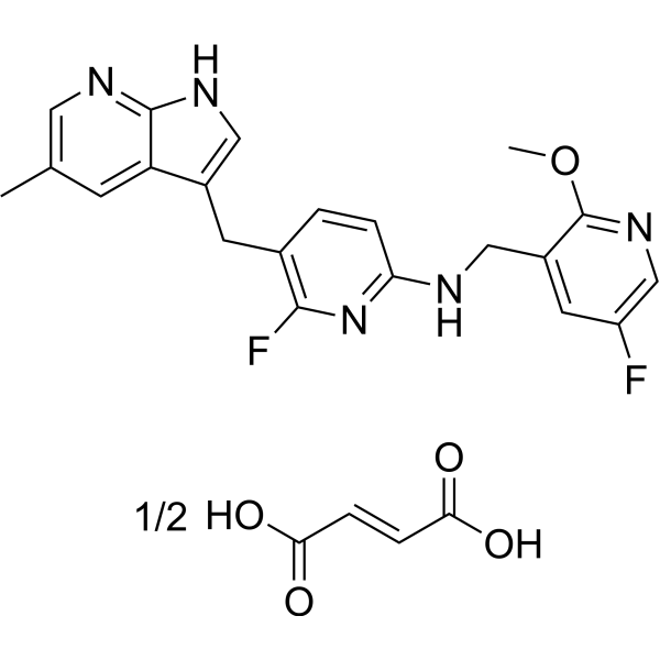
C23H21F2N5O3
|
|---|---|
| 分子量 |
453.45
|
| 精确质量 |
395.16
|
| 元素分析 |
C, 60.92; H, 4.67; F, 8.38; N, 15.44; O, 10.58
|
| 相关CAS号 |
PLX5622;1303420-67-8; 2743279-01-6 (HCl); 2749102-07-4 (fumarate)
|
| PubChem CID |
154731123
|
| 外观&性状 |
Off-white to light yellow solid powder
|
| tPSA |
226 Ų
|
| 氢键供体(HBD)数目 |
6
|
| 氢键受体(HBA)数目 |
18
|
| 可旋转键数目(RBC) |
14
|
| 重原子数目 |
66
|
| 分子复杂度/Complexity |
648
|
| 定义原子立体中心数目 |
0
|
| SMILES |
CC1=CC2=C(NC=C2CC3=C(N=C(C=C3)NCC4=C(N=CC(=C4)F)OC)F)N=C1.CC1=CC2=C(NC=C2CC3=C(N=C(C=C3)NCC4=C(N=CC(=C4)F)OC)F)N=C1.C(=C/C(=O)O)\C(=O)O
|
| InChi Key |
FMEISBYRQFIJPD-WXXKFALUSA-N
|
| InChi Code |
InChI=1S/2C21H19F2N5O.C4H4O4/c2*1-12-5-17-14(9-26-20(17)25-8-12)6-13-3-4-18(28-19(13)23)24-10-15-7-16(22)11-27-21(15)29-2;5-3(6)1-2-4(7)8/h2*3-5,7-9,11H,6,10H2,1-2H3,(H,24,28)(H,25,26);1-2H,(H,5,6)(H,7,8)/b;;2-1+
|
| 化学名 |
(E)-but-2-enedioic acid;6-fluoro-N-[(5-fluoro-2-methoxypyridin-3-yl)methyl]-5-[(5-methyl-1H-pyrrolo[2,3-b]pyridin-3-yl)methyl]pyridin-2-amine
|
| 别名 |
PLX5622; PLX-5622; PLX5622 (hemifumarate); PLX5622 hemifumarate; PLX 5622; PLX-5622 hemifumarate;
|
| HS Tariff Code |
2934.99.9001
|
| 存储方式 |
Powder -20°C 3 years 4°C 2 years In solvent -80°C 6 months -20°C 1 month 注意: 请将本产品存放在密封且受保护的环境中,避免吸湿/受潮。 |
| 运输条件 |
Room temperature (This product is stable at ambient temperature for a few days during ordinary shipping and time spent in Customs)
|
| 溶解度 (体外实验) |
DMSO :~100 mg/mL (~220.53 mM)
|
|---|---|
| 溶解度 (体内实验) |
配方 1 中的溶解度: ≥ 3.25 mg/mL (7.17 mM) (饱和度未知) in 10% DMSO + 40% PEG300 + 5% Tween80 + 45% Saline (这些助溶剂从左到右依次添加,逐一添加), 澄清溶液。
例如,若需制备1 mL的工作液,可将100 μL 32.5 mg/mL澄清DMSO储备液加入到400 μL PEG300中,混匀;然后向上述溶液中加入50 μL Tween-80,混匀;加入450 μL生理盐水定容至1 mL。 *生理盐水的制备:将 0.9 g 氯化钠溶解在 100 mL ddH₂O中,得到澄清溶液。 配方 2 中的溶解度: ≥ 3.25 mg/mL (7.17 mM) (饱和度未知) in 10% DMSO + 90% (20% SBE-β-CD in Saline) (这些助溶剂从左到右依次添加,逐一添加), 澄清溶液。 例如,若需制备1 mL的工作液,可将 100 μL 32.5 mg/mL 澄清 DMSO 储备液加入 900 μL 20% SBE-β-CD 生理盐水溶液中,混匀。 *20% SBE-β-CD 生理盐水溶液的制备(4°C,1 周):将 2 g SBE-β-CD 溶解于 10 mL 生理盐水中,得到澄清溶液。 View More
配方 3 中的溶解度: ≥ 3.25 mg/mL (7.17 mM) (饱和度未知) in 10% DMSO + 90% Corn Oil (这些助溶剂从左到右依次添加,逐一添加), 澄清溶液。 1、请先配制澄清的储备液(如:用DMSO配置50 或 100 mg/mL母液(储备液)); 2、取适量母液,按从左到右的顺序依次添加助溶剂,澄清后再加入下一助溶剂。以 下列配方为例说明 (注意此配方只用于说明,并不一定代表此产品 的实际溶解配方): 10% DMSO → 40% PEG300 → 5% Tween-80 → 45% ddH2O (或 saline); 假设最终工作液的体积为 1 mL, 浓度为5 mg/mL: 取 100 μL 50 mg/mL 的澄清 DMSO 储备液加到 400 μL PEG300 中,混合均匀/澄清;向上述体系中加入50 μL Tween-80,混合均匀/澄清;然后继续加入450 μL ddH2O (或 saline)定容至 1 mL; 3、溶剂前显示的百分比是指该溶剂在最终溶液/工作液中的体积所占比例; 4、 如产品在配制过程中出现沉淀/析出,可通过加热(≤50℃)或超声的方式助溶; 5、为保证最佳实验结果,工作液请现配现用! 6、如不确定怎么将母液配置成体内动物实验的工作液,请查看说明书或联系我们; 7、 以上所有助溶剂都可在 Invivochem.cn网站购买。 |
| 制备储备液 | 1 mg | 5 mg | 10 mg | |
| 1 mM | 2.2053 mL | 11.0266 mL | 22.0531 mL | |
| 5 mM | 0.4411 mL | 2.2053 mL | 4.4106 mL | |
| 10 mM | 0.2205 mL | 1.1027 mL | 2.2053 mL |
1、根据实验需要选择合适的溶剂配制储备液 (母液):对于大多数产品,InvivoChem推荐用DMSO配置母液 (比如:5、10、20mM或者10、20、50 mg/mL浓度),个别水溶性高的产品可直接溶于水。产品在DMSO 、水或其他溶剂中的具体溶解度详见上”溶解度 (体外)”部分;
2、如果您找不到您想要的溶解度信息,或者很难将产品溶解在溶液中,请联系我们;
3、建议使用下列计算器进行相关计算(摩尔浓度计算器、稀释计算器、分子量计算器、重组计算器等);
4、母液配好之后,将其分装到常规用量,并储存在-20°C或-80°C,尽量减少反复冻融循环。
计算结果:
工作液浓度: mg/mL;
DMSO母液配制方法: mg 药物溶于 μL DMSO溶液(母液浓度 mg/mL)。如该浓度超过该批次药物DMSO溶解度,请首先与我们联系。
体内配方配制方法:取 μL DMSO母液,加入 μL PEG300,混匀澄清后加入μL Tween 80,混匀澄清后加入 μL ddH2O,混匀澄清。
(1) 请确保溶液澄清之后,再加入下一种溶剂 (助溶剂) 。可利用涡旋、超声或水浴加热等方法助溶;
(2) 一定要按顺序加入溶剂 (助溶剂) 。
| NCT Number | Recruitment | interventions | Conditions | Sponsor/Collaborators | Start Date | Phases |
| NCT01282684 | Completed | Drug: PLX5622 Drug: Placebo |
Healthy | Plexxikon | January 2011 | Phase 1 |
| NCT01329991 | Completed | Drug: PLX5622 Drug: Placebo |
Rheumatoid Arthritis | Plexxikon | May 2011 | Phase 1 |
 CJC-1295 With DAC
 黄嘌呤钠盐
黄嘌呤钠盐
 四乙酰核糖
四乙酰核糖
 钨酸钠二水合物
InvivoChem的所有产品仅用于作科学研究,不面向患者销售
Copyright 2020 InvivoChem LLC | All Rights Reserved 粤ICP备20063088号-1


 463611831
