| 规格 | 价格 | 库存 | 数量 |
|---|---|---|---|
| 100mg |
|
||
| 500mg |
|
| 靶点 |
JAK1 (IC50 = 10 nM); JAK2 (IC50 = 18 nM); Tyk2 (IC50 = 84 nM); JAK3 (IC50 = 99 nM)
|
||
|---|---|---|---|
| 体外研究 (In Vitro) |
使用分离的酶系统和体外人类或犬细胞模型评估奥拉替尼针对 JAK 家族成员和导致细胞中 JAK 激活的细胞因子的效力和选择性。使用分离的酶系统评估 Oclacitinib 对 JAK 家族成员的抑制活性;浓度(IC50)分别为 10、18、99 和 84 nM 时,oclacitinib 可抑制 JAK1、JAK2、JAK3 和 TYK2 50%。 oclacitinib 对 JAK1 的选择性是 JAK2 的 1.8 倍,对 JAK1 的选择性是 JAK3 的 9.9 倍,因此对 JAK1 酶表现出最高的效力。 Oclacitinib 不会抑制 38 种非 JAK 激酶(IC50 > 1000 nM),但在 10 至 99 nM 的剂量(IC50)下可抑制 JAK 家族成员 50%。 oclacitinib 的 IC50 值范围为 36 至 249 nM,还降低与过敏、炎症和瘙痒相关的 JAK1 依赖性细胞因子(IL-2、IL-4、IL-6 和 IL-13)的活性。 Oclacitinib 对不激活细胞中 JAK1 酶的细胞因子(促红细胞生成素、粒细胞/巨噬细胞集落刺激因子、IL-12、IL-23;IC50 > 1000 nM)的影响可以忽略不计[1]。与用媒介物处理的耳朵相比,用托法替尼 (0.1%) 和奥拉替尼 (0.1%) 局部治疗显着减少了小鼠耳外植体的细胞迁移(所有 P < 0.05)。当将用 JAK 抑制剂处理的每个表皮与用媒介物处理的表皮进行比较时,MHC II 类阳性细胞或朗格汉斯细胞的数量显着减少(所有 P < 0.01)[2]。
|
||
| 体内研究 (In Vivo) |
与仅使用载体组相比,高剂量 Oclacitinib 组的抓挠次数明显减少 (P<0.01)。登记的客户拥有的狗(n = 436)可能被诊断为过敏性皮炎,并且主人评估患有中度至重度瘙痒。狗被随机接受赋形剂匹配的安慰剂或奥拉替尼,剂量为 0.4-0.6 mg/kg,每天口服两次。在第0天和第7天测量皮炎的强度,并使用改进的10cm视觉模拟量表(VAS)从第0天到第7天测量瘙痒程度。狗可以接受长达 28 天的试验。 Oclacitinib 24 小时快速起效[3]。
结果:两个治疗组的预处理所有者和兽医VAS评分相似Oclacitini在24小时内迅速起效。在每个评估日,平均Oclacitinib所有者Pruritus VAS评分明显优于安慰剂评分(P<0.0001)。经oclacitinib治疗后,瘙痒评分从7.58 cm降至2.59 cm。第7天的平均oclacitinib兽医皮炎VAS评分也明显优于安慰剂评分(P<0.0001)。两组报告的腹泻和呕吐频率相似。 结论和临床重要性:在这项研究中,奥拉西替尼对与过敏性皮炎相关的瘙痒提供了快速、有效和安全的控制,业主和兽医注意到瘙痒和皮炎VAS评分有了显著改善[3]。 |
||
| 酶活实验 |
Janus激酶活性测定和激酶选择性面板[1]
如前所述(Meyer等人,2010),使用Caliper微流体技术在分离的酶测定中使用JAK1(氨基酸852-1142;NP_002218)、JAK2(氨基酸808-1132;NP_004963)、JAK3(氨基酸781-1124;NP_000206)和TYK2(氨基酸870-1187;NP_003322)的重组人活性激酶结构域,以确定Oclacitinib对JAK家族成员的效力。与犬JAK酶中类似序列的序列同源性分别为98、98、100和90%(图S1)。使用SelectScreen™激酶分析服务进行Invitrogen激酶面板测试,以确定奥拉西替尼对38种不同非JAK激酶的效力奥拉西替尼的浓度为1μm。激酶特异性测定条件和数据分析在他们的网站上进行了描述http://www.lifetechnologies.com/us/en/home/products-and-services/services/custom-services/screening-and-profiling-services/selectscreen-profiling-service/selectscreen-kinase-profiling-service.html.该服务利用Z'-Lyte技术进行所有激酶筛查。所有测试都进行了两次。 |
||
| 细胞实验 |
白细胞介素-6细胞因子功能[1]
使用Invitrogen的CellSensor®STAT3-bla-HEK293T人上皮细胞系。将细胞在含有5%FBS的DMEM高糖培养基中以每孔1.875×10⁵个细胞的密度铺入384孔、黑壁、透明底部的测定板中,并在37°C、5%CO2下孵育Oclacitinib(0.0000954-25μm)或载体对照加入细胞1小时。然后将每毫升20纳克hIL-6加入细胞培养物5小时。通过用LiveBLAzer™-FRET B/G底物(CCF-4 AM)检测β-内酰胺酶活性来确定IL-6对STAT3β-内酰酶报告基因的激活。使用荧光板读数器获得460和530nm处的荧光发射值。460/530 nm比值表示为对照百分比,剂量反应数据使用4参数逻辑斯谛方程进行分析。 白细胞介素-13细胞因子功能[1] 使用美国典型培养物保藏中心的HT-29人结肠上皮细胞系。细胞在含有10%FBS、50 U/mL青霉素、50μg/mL链霉素和2 mm l-谷氨酰胺的McCoy 5A培养基中增殖。从培养瓶中胰蛋白酶消化细胞,在新鲜培养基中洗涤,并以每孔3×10⁵个细胞的密度重新悬浮在96孔的测定板上Oclacitini(0.0015-30μm))或载体对照加入细胞中,同时在冰上放置30分钟。然后加入每毫升1纳克hIL-13。细胞在37°C水浴中孵育30分钟,然后在1.75%甲醛的PBS中固定,在含有0.5%BSA的PBS中洗涤,并在-20°C的无水甲醇中孵育过夜。固定细胞用PE标记的人pSTAT6抗体染色。使用配备板式自动取样器的FACSCalibur对样品进行分析,并使用FlowJo软件7.6.1版进行分析。数据表示为平均荧光,然后表示为对照百分比。然后使用4参数逻辑斯谛方程分析剂量反应数据。 |
||
| 动物实验 |
|
||
| 药代性质 (ADME/PK) |
本研究旨在确定马来酸奥克拉替尼作为外用制剂在成年马匹中的药代动力学参数。六匹平均体重为528 kg的成年马匹单次接受0.5 mg/kg马来酸奥克拉替尼给药。分别于给药前及给药后15分钟、30分钟、45分钟、1小时、2小时、4小时、6小时、8小时、12小时、24小时、48小时和72小时采集血样。采用液相色谱-质谱联用技术测定马来酸奥克拉替尼的血浆浓度。结果表明,单室模型最符合其药代动力学参数。平均Cmax为486 ng/ml(范围423-549 ng/ml),Tmax估计为1.7 h(范围0.3-3.1 h)。估计T1/2为7.5-8 h。 https://pubmed.ncbi.nlm.nih.gov/35098559/
背景:奥克拉替尼是一种 Janus 激酶 (JK)1 抑制剂,已被证实对犬过敏性皮炎的治疗有效且安全。由于缺乏药代动力学数据,其在猫中的应用受到限制。 目的:测定猫口服和静脉注射奥克拉替尼后的药代动力学参数。 动物:6 只成年家养短毛猫。 方法和材料:采用两周期两处理设计,猫分别接受静脉注射和口服奥克拉替尼马来酸盐,剂量分别为 0.5 mg/kg 和 1 mg/kg。两次给药之间间隔一周的洗脱期。每只猫仅接受一次给药。采用高效液相色谱法测定奥克拉替尼静脉注射后0、5、15、30、1、4、6、10和24小时的血浆浓度,以及口服后0、15、30、1、2、4、6、10和24小时的血浆浓度。 结果:口服后,奥克拉替尼吸收迅速且几乎完全,绝对生物利用度为87%,达峰时间(Tmax)为35分钟。药物消除也非常迅速,半衰期为2.3小时,清除率为4.45 mL/min/kg(静脉注射后)。 结论和临床意义:猫体内奥克拉替尼的药代动力学参数与犬相似,但猫的吸收和消除速度略快,个体间差异也略大。建议猫使用更大剂量和/或更短给药间隔,以达到与犬相似的血药浓度。https://pubmed.ncbi.nlm.nih.gov/31769185/ 马来酸奥克拉替尼的药代动力学在四项独立研究中进行了评估。绝对生物利用度研究采用交叉设计,纳入10只犬。食物对生物利用度的影响在一项纳入18只犬的交叉研究中进行了研究。品种对药代动力学的影响在一项纳入比格犬和杂种犬的交叉研究中进行了评估。剂量比例和多次给药药代动力学在一项平行设计研究中进行了评估,每组8只犬。在所有四项研究中,均采集了系列血浆样本。口服后,马来酸奥克拉替尼吸收迅速且良好,达峰时间<1小时,绝对生物利用度为89%。空腹状态对口服马来酸奥克拉替尼的吸收速率和程度无显著影响,空腹组和餐后组的药代动力学参数无显著差异。在比格犬和混种犬的实验室群体中,奥克拉替尼的药代动力学也表现出相似性。口服后,马来酸奥克拉替尼的暴露量随剂量从0.6 mg/kg增加至3.0 mg/kg而呈比例增加。此外,在所有药代动力学研究中,未发现奥克拉替尼的药代动力学存在明显的性别差异。https://pubmed.ncbi.nlm.nih.gov/24330031/ |
||
| 参考文献 |
|
||
| 其他信息 |
Janus激酶(JAK)参与多种细胞信号通路,这些通路在过敏反应中由失调的细胞因子激活。本研究旨在确定新型JAK抑制剂oclacitinib是否能够降低与犬过敏性皮肤病相关的细胞因子的活性。利用分离的酶系统和体外人或犬细胞模型,我们测定了oclacitinib对JAK家族成员以及能够触发细胞内JAK激活的细胞因子的效力和选择性。Oclacitinib在10至99 nM的浓度范围内(IC50)可抑制JAK家族成员50%的活性,并且对38种非JAK激酶没有抑制作用(IC50 > 1000 nM)。Oclacitinib对JAK1的抑制作用最强(IC50 = 10 nM)。奥克拉替尼还能抑制参与过敏和炎症的JAK1依赖性细胞因子(IL-2、IL-4、IL-6和IL-13)以及瘙痒因子(IL-31)的功能,其IC50值范围为36至249 nM。奥克拉替尼对不激活细胞内JAK1酶的细胞因子(促红细胞生成素、粒细胞/巨噬细胞集落刺激因子、IL-12、IL-23;IC50值>1000 nM)的影响甚微。这些结果表明,奥克拉替尼是一种靶向治疗药物,可选择性地抑制参与过敏、炎症和瘙痒的JAK1依赖性细胞因子,并提示这些机制可能是奥克拉替尼有效控制犬过敏性皮肤病相关临床症状的机制。 [1]
过敏性皮肤病的患病率迅速增加,亟需开发缓解症状的治疗药物。本研究在小鼠皮炎模型中,分别口服或局部应用Janus激酶(JAK)抑制剂托法替尼和奥克拉替尼,并比较其减轻瘙痒和炎症反应的疗效。同时,分析了JAK抑制剂对骨髓来源树突状细胞(BMDC)的体外作用。在过敏性皮炎模型中,雌性BALB/c小鼠经甲苯-2,4-二异氰酸酯(TDI)致敏和激发。分别在TDI激发前30分钟和激发后4小时,口服或局部应用JAK抑制剂。测量小鼠的抓挠次数和耳廓厚度,检测激发皮肤中的细胞因子,并通过流式细胞术分析引流淋巴结中的细胞。体外实验表明,两种JAK抑制剂均显著抑制了骨髓来源树突状细胞(BMDC)的细胞因子生成、迁移和成熟。口服JAK抑制剂的小鼠抓挠行为显著减少,但耳廓厚度未见显著降低。相比之下,局部用药组的抓挠行为和耳廓厚度均显著低于对照组。然而,JAK抑制剂对细胞因子生成的调节作用存在差异,部分细胞因子显著降低,而部分细胞因子显著升高。总之,口服JAK抑制剂可显著减轻瘙痒行为,但对炎症反应的影响甚微;而局部用药则可同时改善瘙痒和炎症反应。尽管两种JAK抑制剂在体外和体内的JAK抑制谱有所不同,但它们的疗效却相当。[2] 背景:奥克拉替尼(Apoquel®)可抑制多种依赖于Janus激酶活性的促炎、促过敏和致痒细胞因子的功能。奥克拉替尼选择性抑制Janus激酶1。假设/目标:我们旨在通过一项随机、双盲、安慰剂对照试验,评估奥克拉替尼治疗过敏性皮炎相关瘙痒的安全性和有效性。方法:纳入436只主人评估为中度至重度瘙痒且初步诊断为过敏性皮炎的犬只。犬只被随机分配至奥克拉替尼组(0.4-0.6 mg/kg,每日两次口服)或安慰剂组(使用赋形剂匹配的安慰剂)。主人使用增强型 10 cm 视觉模拟评分量表 (VAS) 评估第 0 至 7 天的瘙痒严重程度,兽医则在第 0 天和第 7 天评估皮炎严重程度。犬只可参与研究 28 天。结果:治疗前,两组犬只的主人和兽医 VAS 评分相似。奥克拉替尼在 24 小时内迅速起效。在每个评估日,奥克拉替尼组的平均主人瘙痒 VAS 评分均显著优于安慰剂组(P < 0.0001)。奥克拉替尼治疗后,瘙痒评分从 7.58 cm 降至 2.59 cm。第 7 天,奥克拉替尼治疗组的兽医皮炎 VAS 评分平均值也显著优于安慰剂组(P < 0.0001)。两组腹泻和呕吐的发生频率相似。结论和临床意义:本研究表明,奥克拉替尼能够快速、有效且安全地控制过敏性皮炎相关的瘙痒,宠物主人和兽医均观察到瘙痒和皮炎 VAS 评分的显著改善。[3] |
| 精确质量 |
453.168
|
|---|---|
| CAS号 |
1208319-27-0
|
| 相关CAS号 |
1208319-27-0; 1640292-55-2; 1208319-26-9
|
| PubChem CID |
44631937
|
| 外观&性状 |
Typically exists as solid at room temperature
|
| tPSA |
174
|
| 氢键供体(HBD)数目 |
4
|
| 氢键受体(HBA)数目 |
10
|
| 可旋转键数目(RBC) |
7
|
| 重原子数目 |
31
|
| 分子复杂度/Complexity |
606
|
| 定义原子立体中心数目 |
0
|
| SMILES |
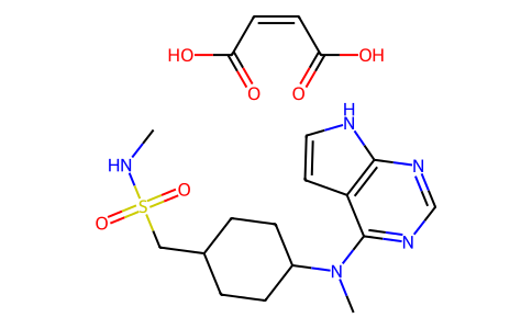
CNS(=O)(=O)CC1CCC(CC1)N(C)C2=NC=NC3=C2C=CN3.C(=C\C(=O)O)\C(=O)O
|
| InChi Key |
VQIGDTLRBSNOBV-BTJKTKAUSA-N
|
| InChi Code |
InChI=1S/C15H23N5O2S.C4H4O4/c1-16-23(21,22)9-11-3-5-12(6-4-11)20(2)15-13-7-8-17-14(13)18-10-19-15;5-3(6)1-2-4(7)8/h7-8,10-12,16H,3-6,9H2,1-2H3,(H,17,18,19);1-2H,(H,5,6)(H,7,8)/b;2-1-
|
| 化学名 |
(Z)-but-2-enedioic acid;N-methyl-1-[4-[methyl(7H-pyrrolo[2,3-d]pyrimidin-4-yl)amino]cyclohexyl]methanesulfonamide
|
| 别名 |
PF 03394197 maleate; Apoquel; OCLACITINIB MALEATE; 1208319-27-0; 1640292-55-2; Oclacitinib (maleate); Apoquel; PF-03394197 maleate; PF03394197 maleate;
|
| HS Tariff Code |
2934.99.9001
|
| 存储方式 |
Powder -20°C 3 years 4°C 2 years In solvent -80°C 6 months -20°C 1 month |
| 运输条件 |
Room temperature (This product is stable at ambient temperature for a few days during ordinary shipping and time spent in Customs)
|
| 溶解度 (体外实验) |
May dissolve in DMSO (in most cases), if not, try other solvents such as H2O, Ethanol, or DMF with a minute amount of products to avoid loss of samples
|
|---|
计算结果:
工作液浓度: mg/mL;
DMSO母液配制方法: mg 药物溶于 μL DMSO溶液(母液浓度 mg/mL)。如该浓度超过该批次药物DMSO溶解度,请首先与我们联系。
体内配方配制方法:取 μL DMSO母液,加入 μL PEG300,混匀澄清后加入μL Tween 80,混匀澄清后加入 μL ddH2O,混匀澄清。
(1) 请确保溶液澄清之后,再加入下一种溶剂 (助溶剂) 。可利用涡旋、超声或水浴加热等方法助溶;
(2) 一定要按顺序加入溶剂 (助溶剂) 。
 CJC-1295 With DAC
 黄嘌呤钠盐
黄嘌呤钠盐
 四乙酰核糖
四乙酰核糖
 钨酸钠二水合物
InvivoChem的所有产品仅用于作科学研究,不面向患者销售
Copyright 2020 InvivoChem LLC | All Rights Reserved 粤ICP备20063088号-1


 463611831
