
| 规格 | 价格 | 库存 | 数量 |
|---|---|---|---|
| 1mg |
|
||
| 5mg |
|
||
| 50mg |
|
||
| Other Sizes |
|

| 体外研究 (In Vitro) |
Prostratin 的 Ki 为 210 nM,并转录 [3H]PDBu 与 CEM 细胞结合 [1]。转录性急性髓系白血病 (AML) 细胞系(HL-60、NB4 和 U937 细胞)在前列腺素浓度 (125-1000 nM) 下无法增殖。在 HL-60 细胞中,前列腺素 (125–100 nM) 影响细胞周期相关分子(pRb 磷酸化、CDK 和 p21)并导致 AML 细胞中 G1 期停滞。 Prostratin 还会激活 PKC,从而导致 AML 细胞系变得活跃。此外,前列腺素产生的湿度对于 PKC 驱动的 MEK/ERK/MAP 信号负载激活是必要的 [2]。对于 HIV-1 诱导的激活,前列腺素诱导需要 PKD3 的活性形式。此外,前列腺素通过新 PKC 亚家族的 PKCε 激活 PKD3 [2]。
|
|---|---|
| 细胞实验 |
细胞活力测定 [2]
细胞类型: HL-60、NB4 和 U937 细胞 测试浓度: 125nM、250nM、500nM、1000nM 孵化持续时间:24小时、48小时、72小时 实验结果:剂量依赖性抑制3]。急性髓系白血病 (AML) 细胞系的生长。 细胞周期分析[2] 细胞类型: HL-60、NB4 和 U937 细胞 测试浓度: 125 nM、250 nM、500 nM、1000 nM 孵育时间:24 小时 实验结果:在一定浓度下诱导 G0/G1 期积累依赖方式。 蛋白质印迹分析[2] 细胞类型: HL-60 细胞 测试浓度: 125 nM、250 nM、500 nM , 1000 nM 孵育时间:24小时 实验结果:细胞周期相关分子(pRb磷酸化、CDK和p21)的影响)在 60 个细胞中的 HL- 中。 |
| 参考文献 |
|
| 其他信息 |
前列腺素是一种佛波醇酯,它作为一种代谢产物发挥作用。
据报道,前列腺素存在于三角大戟、费氏大戟以及其他有相关数据的生物体中。 |
| 分子式 |
C22H30O6
|
|---|---|
| 分子量 |
390.4700
|
| 精确质量 |
390.204
|
| CAS号 |
60857-08-1
|
| PubChem CID |
454217
|
| 外观&性状 |
White to off-white solid powder
|
| 密度 |
1.3±0.1 g/cm3
|
| 沸点 |
550.5±50.0 °C at 760 mmHg
|
| 熔点 |
216-219℃
|
| 闪点 |
188.7±23.6 °C
|
| 蒸汽压 |
0.0±3.4 mmHg at 25°C
|
| 折射率 |
1.600
|
| LogP |
1.84
|
| tPSA |
104.06
|
| 氢键供体(HBD)数目 |
3
|
| 氢键受体(HBA)数目 |
6
|
| 可旋转键数目(RBC) |
3
|
| 重原子数目 |
28
|
| 分子复杂度/Complexity |
825
|
| 定义原子立体中心数目 |
7
|
| SMILES |
C[C@@H]1C[C@@]2([C@@H](C2(C)C)[C@H]3[C@]1([C@@H]4C=C(C(=O)[C@]4(CC(=C3)CO)O)C)O)OC(=O)C
|
| InChi Key |
BOJKFRKNLSCGHY-HXGSDTCMSA-N
|
| InChi Code |
InChI=1S/C22H30O6/c1-11-6-16-20(26,18(11)25)9-14(10-23)7-15-17-19(4,5)21(17,28-13(3)24)8-12(2)22(15,16)27/h6-7,12,15-17,23,26-27H,8-10H2,1-5H3/t12-,15+,16-,17-,20-,21+,22-/m1/s1
|
| 化学名 |
[(1R,2S,6R,10S,11R,13S,15R)-1,6-dihydroxy-8-(hydroxymethyl)-4,12,12,15-tetramethyl-5-oxo-13-tetracyclo[8.5.0.02,6.011,13]pentadeca-3,8-dienyl] acetate
|
| HS Tariff Code |
2934.99.9001
|
| 存储方式 |
Powder -20°C 3 years 4°C 2 years In solvent -80°C 6 months -20°C 1 month 注意: 本产品在运输和储存过程中需避光。 |
| 运输条件 |
Room temperature (This product is stable at ambient temperature for a few days during ordinary shipping and time spent in Customs)
|
| 溶解度 (体外实验) |
DMSO : ~50 mg/mL (~128.05 mM)
|
|---|---|
| 溶解度 (体内实验) |
配方 1 中的溶解度: ≥ 1.25 mg/mL (3.20 mM) (饱和度未知) in 10% DMSO + 40% PEG300 + 5% Tween80 + 45% Saline (这些助溶剂从左到右依次添加,逐一添加), 澄清溶液。
例如,若需制备1 mL的工作液,可将100 μL 12.5 mg/mL澄清的DMSO储备液加入到400 μL PEG300中,混匀;再向上述溶液中加入50 μL Tween-80,混匀;然后加入450 μL生理盐水定容至1 mL。 *生理盐水的制备:将 0.9 g 氯化钠溶解在 100 mL ddH₂O中,得到澄清溶液。 配方 2 中的溶解度: ≥ 1.25 mg/mL (3.20 mM) (饱和度未知) in 10% DMSO + 90% (20% SBE-β-CD in Saline) (这些助溶剂从左到右依次添加,逐一添加), 澄清溶液。 例如,若需制备1 mL的工作液,可将 100 μL 12.5 mg/mL澄清DMSO储备液加入900 μL 20% SBE-β-CD生理盐水溶液中,混匀。 *20% SBE-β-CD 生理盐水溶液的制备(4°C,1 周):将 2 g SBE-β-CD 溶解于 10 mL 生理盐水中,得到澄清溶液。 View More
配方 3 中的溶解度: ≥ 1.25 mg/mL (3.20 mM) (饱和度未知) in 10% DMSO + 90% Corn Oil (这些助溶剂从左到右依次添加,逐一添加), 澄清溶液。 1、请先配制澄清的储备液(如:用DMSO配置50 或 100 mg/mL母液(储备液)); 2、取适量母液,按从左到右的顺序依次添加助溶剂,澄清后再加入下一助溶剂。以 下列配方为例说明 (注意此配方只用于说明,并不一定代表此产品 的实际溶解配方): 10% DMSO → 40% PEG300 → 5% Tween-80 → 45% ddH2O (或 saline); 假设最终工作液的体积为 1 mL, 浓度为5 mg/mL: 取 100 μL 50 mg/mL 的澄清 DMSO 储备液加到 400 μL PEG300 中,混合均匀/澄清;向上述体系中加入50 μL Tween-80,混合均匀/澄清;然后继续加入450 μL ddH2O (或 saline)定容至 1 mL; 3、溶剂前显示的百分比是指该溶剂在最终溶液/工作液中的体积所占比例; 4、 如产品在配制过程中出现沉淀/析出,可通过加热(≤50℃)或超声的方式助溶; 5、为保证最佳实验结果,工作液请现配现用! 6、如不确定怎么将母液配置成体内动物实验的工作液,请查看说明书或联系我们; 7、 以上所有助溶剂都可在 Invivochem.cn网站购买。 |
| 制备储备液 | 1 mg | 5 mg | 10 mg | |
| 1 mM | 2.5610 mL | 12.8051 mL | 25.6102 mL | |
| 5 mM | 0.5122 mL | 2.5610 mL | 5.1220 mL | |
| 10 mM | 0.2561 mL | 1.2805 mL | 2.5610 mL |
1、根据实验需要选择合适的溶剂配制储备液 (母液):对于大多数产品,InvivoChem推荐用DMSO配置母液 (比如:5、10、20mM或者10、20、50 mg/mL浓度),个别水溶性高的产品可直接溶于水。产品在DMSO 、水或其他溶剂中的具体溶解度详见上”溶解度 (体外)”部分;
2、如果您找不到您想要的溶解度信息,或者很难将产品溶解在溶液中,请联系我们;
3、建议使用下列计算器进行相关计算(摩尔浓度计算器、稀释计算器、分子量计算器、重组计算器等);
4、母液配好之后,将其分装到常规用量,并储存在-20°C或-80°C,尽量减少反复冻融循环。
计算结果:
工作液浓度: mg/mL;
DMSO母液配制方法: mg 药物溶于 μL DMSO溶液(母液浓度 mg/mL)。如该浓度超过该批次药物DMSO溶解度,请首先与我们联系。
体内配方配制方法:取 μL DMSO母液,加入 μL PEG300,混匀澄清后加入μL Tween 80,混匀澄清后加入 μL ddH2O,混匀澄清。
(1) 请确保溶液澄清之后,再加入下一种溶剂 (助溶剂) 。可利用涡旋、超声或水浴加热等方法助溶;
(2) 一定要按顺序加入溶剂 (助溶剂) 。
|
|
|
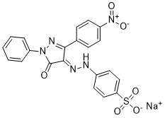 PHPS1 Sodium
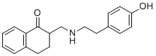 BE 2254
 环胞啶硫酸盐
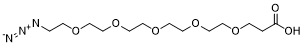 Azido-PEG5-acid
InvivoChem的所有产品仅用于作科学研究,不面向患者销售
Copyright 2020 InvivoChem LLC | All Rights Reserved 粤ICP备20063088号-1


 463611831
