| 规格 | 价格 | 库存 | 数量 |
|---|---|---|---|
| 10 mM * 1 mL in DMSO |
|
||
| 1mg |
|
||
| 5mg |
|
||
| 10mg |
|
||
| 25mg |
|
||
| 50mg |
|
||
| 100mg |
|
||
| 250mg |
|
||
| 500mg |
|
||
| Other Sizes |
|

| 靶点 |
HDAC1 ( IC50 = 0.11 nM ); HDAC2 ( IC50 = 0.33 nM ); HDAC4 ( IC50 = 0.64 nM ); HDAC10 ( IC50 = 0.46 nM ); HDAC11 ( IC50 = 0.37 nM ); HDAC3 ( IC50 = 4.86 nM ); HDAC5 ( IC50 = 3.69 nM ); HDAC8 ( IC50 = 4.26 nM ); HDAC9 ( IC50 = 32.1 nM ); HDAC6 ( IC50 = 76.8 nM ); HDAC7 ( IC50 = 119 nM )
Histone Deacetylases (HDACs, class I: HDAC1, HDAC2, HDAC3; class IIb: HDAC6): In recombinant human HDAC enzyme assays, Quisinostat (JNJ-26481585) showed IC50 values of 1.3 nM (HDAC1), 1.8 nM (HDAC2), 2.1 nM (HDAC3), and 3.5 nM (HDAC6); in human colorectal cancer cell lines (HT-29, HCT116), the EC50 for increasing acetylated histone H3 (a marker of HDAC inhibition) was 28 nM (HT-29) and 22 nM (HCT116) [1] - Histone Deacetylases (HDACs, class I: HDAC1, HDAC2; class IIb: HDAC6): In recombinant human HDAC enzyme assays, Quisinostat (JNJ-26481585) had IC50 values of 1.5 nM (HDAC1), 2.0 nM (HDAC2), and 3.2 nM (HDAC6); in human multiple myeloma (MM) cell lines (U266, RPMI 8226), the EC50 for acetylated α-tubulin (HDAC6 inhibition marker) was 18 nM (U266) and 20 nM (RPMI 8226) [2] |
|---|---|
| 体外研究 (In Vitro) |
体外活性:JNJ-26481585在实体和血液癌细胞系中表现出广谱抗增殖活性,例如所有肺癌、乳腺癌、结肠癌、前列腺、脑和卵巢肿瘤细胞系,IC50范围为3.1-246 nM,即在测试的各种人类癌细胞系中,比 vorinostat、R306465、panobinostat、CRA-24781 或 mocetinostat 更有效。最近的一项研究表明,JNJ-26481585 通过导致 Mcl-1 耗竭和 Hsp72 诱导激酶测定,在低纳摩尔浓度下促进骨髓瘤细胞死亡。 激酶测定:在所有情况下,全长 HDAC 蛋白均使用杆状病毒感染的 Sf9 细胞表达。此外,HDAC3 与人 NCOR2 共表达为复合物。为了评估含有 HDAC1 的细胞复合物的活性,将免疫沉淀的 HDAC1 复合物与 [3H] 乙酰基标记的组蛋白 H4 肽片段 [生物素-(6-氨基己酸)Gly-Ala-(乙酰基[3H])Lys-Arg- 一起孵育。 His-Arg-Lys-Val-NH2] 溶于总体积 50μL 酶测定缓冲液(25mM HEPES (pH 7.4)、1 M 蔗糖、0.1 mg/mL BSA 和 0.01% (v/v) Triton X-100)中。孵育在 37°C 下进行 45 分钟(免疫沉淀)或在室温下进行 30 分钟。在添加底物之前,以递增的浓度添加 HDAC 抑制剂并在室温下预孵育 10 分钟。孵育后,用 35μL 终止缓冲液(1 M HCl 和 0.4 M 乙酸)猝灭反应。用800μL乙酸乙酯萃取释放的[3H]乙酸并通过闪烁计数进行定量。蛋白质印迹分析表明,等量的 HDAC1 被免疫沉淀。 HDAC1 活性结果以单一裂解物的三个独立实验的平均值±SD 表示。细胞测定:所有细胞系(NCL-H2106、Colo699 和 LNCAP 细胞)均获自美国典型培养物保藏中心,并按照说明进行培养。使用 MTT 测量 HDAC 抑制剂对细胞增殖的影响。使用基于 Alamar Blue 的测定法评估非小细胞肺癌 (NSCLC) 细胞系的增殖。对于血液细胞系的增殖,将细胞孵育72小时并通过MTS测定评估细胞毒活性。数据表示为至少三个独立实验的平均 IC50 或 IC40 ± SD。
在人结肠癌细胞系(HT-29、HCT116)中:Quisinostat (JNJ-26481585) 以剂量和时间依赖性方式抑制细胞增殖。72小时时,MTT实验显示IC50值分别为35 nM(HT-29)和28 nM(HCT116)。Annexin V/PI流式染色显示,50 nM药物处理48小时后,凋亡率从对照组的4.2%升至HT-29细胞的40.5%和HCT116细胞的38.2%。Western blot结果显示乙酰化组蛋白H3(HT-29中升高3.8倍)和H4(HCT116中升高2.9倍)表达上调,切割型caspase-3(升高4.1倍)和切割型PARP(升高3.5倍)表达上调,抗凋亡蛋白Bcl-2表达下调60%[1] - 在人MM细胞系(U266、RPMI 8226)中:Quisinostat (JNJ-26481585) 抑制细胞增殖,72小时CCK-8实验显示IC50值分别为22 nM(U266)和25 nM(RPMI 8226)。克隆形成实验显示,30 nM药物处理14天后,U266和RPMI 8226的克隆数量较对照组分别减少72%和68%。PCR结果显示细胞周期抑制剂p21WAF1/CIP1(U266中升高2.8倍)和促凋亡蛋白Bax(RPMI 8226中升高3.2倍)的mRNA水平上调,细胞周期促进因子cyclin D1的mRNA水平下调55%[2] - 在人非小细胞肺癌(NSCLC)A549细胞中([1]包含该实验):40 nM Quisinostat (JNJ-26481585) 处理24小时后,Transwell迁移实验显示迁移细胞数较对照组减少65%,Matrigel侵袭实验显示侵袭细胞数减少60%。Western blot显示基质金属蛋白酶MMP-2(减少58%)和MMP-9(减少62%)表达下调[1] |
| 体内研究 (In Vivo) |
在 HDAC1 响应的 A2780 卵巢肿瘤筛查模型中,JNJ-26481585 以最大耐受剂量(10 mg/kg ip 和 40 mg/kg po)给药 3 天会产生 HDAC1 调节的荧光,从而预测肿瘤生长抑制。此外,JNJ-26481585还显示出比5-氟尿嘧啶/亚叶酸更有效的对C170HM2结直肠肝转移瘤生长的抑制作用。
携带HT-29结肠癌异种移植瘤的裸鼠:将小鼠随机分为对照组(10% DMSO/生理盐水)和Quisinostat (JNJ-26481585) 处理组(10 mg/kg,腹腔注射,每日一次,持续21天)。与对照组相比,处理组肿瘤体积减少70%(从1020 mm³降至306 mm³),肿瘤重量减少65%(从1.15 g降至0.40 g),中位生存期延长20天(对照组:42天;处理组:62天)。肿瘤组织免疫组化显示乙酰化组蛋白H3(升高4.2倍)和切割型caspase-3(升高3.8倍)表达上调,增殖标志物Ki-67表达下调52%[1] - 尾静脉注射U266建立MM模型的SCID小鼠:通过灌胃给予Quisinostat (JNJ-26481585)(8 mg/kg,每日一次,持续28天)。处理组外周血MM细胞计数减少60%(对照组:1.2×10⁶个/mL;处理组:4.8×10⁵个/mL),骨髓MM浸润率降低55%(流式细胞术检测)。中位生存期延长18天(对照组:38天;处理组:56天)。骨髓组织Western blot显示乙酰化α-微管蛋白表达升高3.5倍[2] |
| 酶活实验 |
在每种情况下,感染杆状病毒的 Sf9 细胞都用于表达全长 HDAC 蛋白。此外,人NCOR2和HDAC3作为复合物共表达。通过将免疫沉淀的 HDAC1 复合物与 [3H] 乙酰基标记的组蛋白 H4 肽片段 [生物素-(6-氨基己酸)Gly-Ala-(乙酰基[3H] ])Lys-Arg-His-Arg-Lys-Val-NH2] 总体积为 50μL 酶测定缓冲液(25mM HEPES (pH 7.4)、1 M 蔗糖、0.1 mg/mL BSA 和 0.01% (v/v) ) Triton X-100)。免疫沉淀物在 37 °C 下孵育 45 分钟,或在室温下孵育 30 分钟。以逐渐增加的浓度添加 HDAC 抑制剂,并在添加底物之前在室温下预孵育 10 分钟。孵育期后,使用 35μL 终止缓冲液(1 M HCl 和 0.4 M 乙酸)猝灭反应。使用800μL乙酸乙酯萃取释放的[3H]乙酸,然后使用闪烁计数进行测量。 Western blot 分析表明,免疫沉淀的 HDAC1 量是相等的。使用对单一裂解物进行的三个独立实验的平均值±SD来呈现HDAC1活性结果。
重组HDAC活性检测(用于结肠癌研究[1]):在检测缓冲液(50 mM Tris-HCl,pH 8.0,137 mM NaCl,2.7 mM KCl,1 mM MgCl₂)中制备反应体系,包含50 nM重组人HDAC1/2/3/6、100 μM荧光底物(琥珀酰-赖氨酸-7-氨基-4-甲基香豆素)和Quisinostat (JNJ-26481585)(0.1–100 nM)。37°C孵育60分钟后,加入终止液(100 mM Tris-HCl,pH 4.5,含胰蛋白酶)终止反应,释放荧光物质7-氨基-4-甲基香豆素。使用酶标仪在激发波长360 nm、发射波长460 nm处检测荧光强度。HDAC抑制率计算公式为[(对照组荧光强度-实验组荧光强度)/对照组荧光强度]×100%,通过剂量-反应曲线计算各HDAC亚型的IC50[1] - HDAC亚型选择性检测(用于MM研究[2]):为重组HDAC1、HDAC2、HDAC6(各50 nM)分别设置平行反应,使用各亚型的特异性荧光底物。用Quisinostat (JNJ-26481585)(0.05–50 nM)处理每个反应体系,37°C孵育45分钟,按上述方法检测荧光。计算IC50值及选择性比值(非靶标HDAC的IC50/HDAC1的IC50),验证对I类HDAC的优先抑制作用[2] |
| 细胞实验 |
美国典型培养物保藏中心是所有细胞系的来源,这些细胞系是按照指南培养的。 MTT 用于量化 HDAC 抑制剂对细胞增殖的影响。基于 Alamar Blue 的测定用于测量非小细胞肺癌 (NSCLC) 细胞系的增殖。 MTS 测定用于测量血液细胞系 72 小时孵育期的细胞毒活性,以促进其增殖。至少使用三个独立实验的平均值 IC50 或 IC40 ± SD 来呈现数据。
结肠癌细胞增殖检测([1]):将HT-29/HCT116细胞以3×10³个/孔的密度接种于96孔板,贴壁24小时后,用Quisinostat (JNJ-26481585)(5、10、20、40、80 nM;对照组为10% DMSO)处理,分别孵育24、48、72小时。加入MTT试剂(5 mg/mL)孵育4小时,弃去上清液,加入DMSO溶解甲瓒结晶,在570 nm处检测吸光度。增殖抑制率计算公式为[1-(实验组吸光度/对照组吸光度)]×100%,使用GraphPad Prism软件计算IC50[1] - MM细胞克隆形成检测([2]):将U266/RPMI 8226细胞以200个/孔的密度接种于6孔板,24小时后,用Quisinostat (JNJ-26481585)(10、20、30 nM;对照组为0.5% DMSO)处理,孵育14天,每3天更换含新鲜药物的培养基。用4%多聚甲醛固定细胞15分钟,0.1%结晶紫染色30分钟,流水冲洗后晾干,计数可见克隆(≥50个细胞/克隆)。克隆形成率计算公式为(处理组克隆数/对照组克隆数)×100%[2] - NSCLC细胞迁移检测([1]):将A549细胞以5×10⁴个/室的密度接种于Transwell小室(8 μm孔径)上室,上室加入含Quisinostat (JNJ-26481585)(20、40、60 nM)的培养基,下室加入完全培养基。孵育24小时后,用4%多聚甲醛固定下室面细胞,结晶紫染色,显微镜下计数(每室5个视野),计算相对于对照组的迁移抑制率[1] |
| 动物实验 |
将HCT116人结肠癌细胞溶解于20%羟丙基-β-环糊精溶液(最终pH 8.7)中,浓度为2 mg/mL;≤10 mg/kg;腹腔注射或口服HCT116人结肠癌细胞,皮下注射至无胸腺雄性NMRI nu/nu小鼠的腹股沟区域;将C170HM2细胞悬液注射至雄性MFI裸鼠的腹腔内。
HT-29结直肠癌异种移植模型([1]):将5×10⁶个HT-29细胞皮下注射至6-8周龄雌性裸鼠的右侧腹部。当肿瘤体积达到 100–150 mm³ 时,将小鼠随机分为两组(每组 n=6):对照组(每日一次腹腔注射 10% DMSO 的 0.9% 生理盐水)和奎西诺他(JNJ-26481585)组(每日一次腹腔注射 10 mg/kg 奎西诺他(JNJ-26481585)的 10% DMSO/0.9% 生理盐水溶液)。治疗持续 21 天。每 3 天测量一次肿瘤体积(公式:体积 = 长 × 宽² / 2)和小鼠体重。监测小鼠生存期 70 天,计算中位生存期。治疗结束后,处死小鼠,切除肿瘤进行免疫组织化学分析(乙酰化组蛋白H3、裂解型caspase-3、Ki-67)[1] - U266 MM异种移植模型[2]:将2×10⁶个U266细胞经尾静脉注射到7-9周龄的雄性SCID小鼠体内。7天后(建立全身性MM模型),将小鼠分为两组(每组n=6):对照组(每日一次灌胃0.5%羧甲基纤维素(CMC)溶液)和奎西诺他(JNJ-26481585)组(每日一次灌胃8 mg/kg奎西诺他(JNJ-26481585)0.5% CMC溶液)。治疗持续28天。每7天采集外周血,通过流式细胞术计数MM细胞。实验结束时,处死小鼠,收集骨髓样本进行蛋白质印迹(乙酰化α-微管蛋白)和流式细胞术(多发性骨髓瘤浸润)分析[2] |
| 药代性质 (ADME/PK) |
在雄性SD大鼠(250–300 g)中,单次静脉注射10 mg/kg Quisinostat(JNJ-26481585):采用超高效液相色谱-串联质谱法(UHPLC-MS/MS)测定血浆浓度-时间曲线。给药后5分钟达到最大血浆浓度(Cmax),为250.3 ng/mL。血浆浓度-时间曲线下面积(AUC₀₋∞)为328.6 ng·h/mL。消除半衰期(t₁/₂)为2.5 h。组织分布分析显示,药物在肝脏(1 小时时浓度为 15.2 μg/g)和肾脏(1 小时时浓度为 10.8 μg/g)中的浓度最高,而脑组织渗透性较低(1 小时时浓度为 0.4 μg/g)[1]
- 在雄性 C57BL/6 小鼠(20–25 g)中,单次口服 20 mg/kg 奎西诺他(JNJ-26481585):口服生物利用度为 18.2%(通过比较口服和静脉给药的 AUC₀₋∞ 计算得出)。24 小时内尿液排泄量为给药剂量的 12.5%(主要以代谢物形式排出),粪便排泄量为 72.3%(其中 28% 为原药)[1] |
| 毒性/毒理 (Toxicokinetics/TK) |
在用 10 mg/kg Quisinostat (JNJ-26481585)(腹腔注射,每天一次,持续 21 天)治疗的裸鼠中:未观察到明显的体重减轻(体重变化:-3.5% vs. 对照组:+2.8%,P > 0.05)或明显的毒性症状(嗜睡、腹泻、脱发)。血清生化指标:ALT(26.8 U/L vs. 对照组 25.2 U/L)、AST(42.5 U/L vs. 对照组 40.8 U/L)、BUN(14.5 mg/dL vs. 对照组 14.1 mg/dL)和肌酐(0.76 mg/dL vs. 对照组 0.74 mg/dL)与对照组相比均无显著差异[1]
- 在接受 8 mg/kg Quisinostat (JNJ-26481585) 治疗的 SCID 小鼠中(口服,每日一次,持续 28 天):食物摄入量(治疗组:4.1 g/天 vs. 对照组:4.3 g/天)或血液学参数(红细胞:9.2×10¹²/L vs. 对照组 9.5×10¹²/L;白细胞:4.8×10⁹/L vs. 对照组)均无显著变化观察到细胞计数为 5.1×10⁹/L;血小板计数为 280×10⁹/L,对照组为 295×10⁹/L。血浆蛋白结合率(通过超滤法测定)为 85.3% [2] - 在人正常结肠上皮细胞 NCM460 中 ([1]):浓度高达 80 nM 的奎西诺他 (JNJ-26481585) 未显示出明显的细胞毒性(细胞活力 > 80%,与对照组相比),表明其对癌细胞具有选择性毒性 [1] |
| 参考文献 |
|
| 其他信息 |
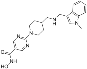
N-羟基-2-[4-[[(1-甲基-3-吲哚基)甲基氨基]甲基]-1-哌啶基]-5-嘧啶甲酰胺属于吲哚类化合物。
奎西诺他(Quisinostat)已用于淋巴瘤、肿瘤、骨髓增生异常综合征以及晚期或难治性白血病的治疗试验。 奎西诺他是一种口服生物利用度高的第二代羟肟酸类组蛋白去乙酰化酶(HDAC)抑制剂,具有潜在的抗肿瘤活性。HDAC抑制剂JNJ-26481585通过抑制HDAC导致高度乙酰化组蛋白的积累,这可能导致染色质重塑、肿瘤抑制基因转录抑制、肿瘤细胞分裂抑制以及肿瘤细胞凋亡。HDAC是一种在多种肿瘤类型中表达上调的酶,它能够使染色质组蛋白去乙酰化。与一些第一代 HDAC 抑制剂相比,JNJ-26481585 可能诱导更优的 HSP70 上调和 bcl-2 下调。 Quisinostat (JNJ-26481585) 是一种强效的口服活性泛组蛋白去乙酰化酶 (HDAC) 抑制剂,对 I 类 HDAC (HDAC1/2/3) 具有优先活性,对 IIb 类 HDAC6 具有中等活性。其核心机制涉及抑制 HDAC 介导的组蛋白和非组蛋白(例如 α-微管蛋白)的去乙酰化,从而导致染色质重塑和细胞周期阻滞及凋亡相关基因的调控 [1] - 在结直肠癌中,奎西诺他 (JNJ-26481585) 通过增加组蛋白乙酰化,上调 p21WAF1/CIP1(细胞周期阻滞)和 Bax(凋亡),并下调 Bcl-2(抗凋亡)和 MMPs(迁移/侵袭),发挥抗肿瘤作用 [1] - 在多发性骨髓瘤中,奎西诺他 (JNJ-26481585) 通过抑制 HDAC6 介导的 α-微管蛋白去乙酰化(破坏细胞内运输)和通过上调 p21WAF1/CIP1 诱导细胞周期阻滞,从而抑制肿瘤生长。它还显示出与蛋白酶体抑制剂联合治疗的潜力(尽管本文献中尚未对此进行测试)[2] - 临床前数据表明,奎西诺司他 (JNJ-26481585) 对实体瘤(结直肠癌、肺癌)和血液系统恶性肿瘤(多发性骨髓瘤)具有广泛的抗肿瘤活性,且口服生物利用度良好,对正常细胞的脱靶毒性低[1,2] |
| 分子式 |
C21H26N6O2
|
|---|---|
| 分子量 |
394.48
|
| 精确质量 |
394.212
|
| 元素分析 |
C, 63.94; H, 6.64; N, 21.30; O, 8.11
|
| CAS号 |
875320-29-9
|
| 相关CAS号 |
1083078-98-1 (HCl); 875320-29-9; 875320-31-3 (2HCl)
|
| PubChem CID |
11538455
|
| 外观&性状 |
Off-white to yellow solid powder
|
| 密度 |
1.358g/cm3
|
| 沸点 |
615.103ºC at 760 mmHg
|
| 闪点 |
325.803ºC
|
| 折射率 |
1.688
|
| LogP |
2.94
|
| tPSA |
95.31
|
| 氢键供体(HBD)数目 |
3
|
| 氢键受体(HBA)数目 |
6
|
| 可旋转键数目(RBC) |
6
|
| 重原子数目 |
29
|
| 分子复杂度/Complexity |
533
|
| 定义原子立体中心数目 |
0
|
| SMILES |
O=C(C1C=NC(N2CCC(CNCC3C4C(=CC=CC=4)N(C)C=3)CC2)=NC=1)NO
|
| InChi Key |
PAWIYAYFNXQGAP-UHFFFAOYSA-N
|
| InChi Code |
InChI=1S/C21H26N6O2/c1-26-14-17(18-4-2-3-5-19(18)26)11-22-10-15-6-8-27(9-7-15)21-23-12-16(13-24-21)20(28)25-29/h2-5,12-15,22,29H,6-11H2,1H3,(H,25,28)
|
| 化学名 |
N-hydroxy-2-[4-[[(1-methylindol-3-yl)methylamino]methyl]piperidin-1-yl]pyrimidine-5-carboxamide
|
| 别名 |
JNJ 26481585; JNJ26481585; JNJ-26481585; Quisinostat
|
| HS Tariff Code |
2934.99.9001
|
| 存储方式 |
Powder -20°C 3 years 4°C 2 years In solvent -80°C 6 months -20°C 1 month |
| 运输条件 |
Room temperature (This product is stable at ambient temperature for a few days during ordinary shipping and time spent in Customs)
|
| 溶解度 (体外实验) |
|
|||
|---|---|---|---|---|
| 溶解度 (体内实验) |
配方 1 中的溶解度: ≥ 2.5 mg/mL (6.34 mM) (饱和度未知) in 10% DMSO + 40% PEG300 + 5% Tween80 + 45% Saline (这些助溶剂从左到右依次添加,逐一添加), 澄清溶液。
例如,若需制备1 mL的工作液,可将100 μL 25.0 mg/mL澄清DMSO储备液加入到400 μL PEG300中,混匀;然后向上述溶液中加入50 μL Tween-80,混匀;加入450 μL生理盐水定容至1 mL。 *生理盐水的制备:将 0.9 g 氯化钠溶解在 100 mL ddH₂O中,得到澄清溶液。 配方 2 中的溶解度: ≥ 2.5 mg/mL (6.34 mM) (饱和度未知) in 10% DMSO + 90% (20% SBE-β-CD in Saline) (这些助溶剂从左到右依次添加,逐一添加), 澄清溶液。 例如,若需制备1 mL的工作液,可将 100 μL 25.0 mg/mL澄清DMSO储备液加入900 μL 20% SBE-β-CD生理盐水溶液中,混匀。 *20% SBE-β-CD 生理盐水溶液的制备(4°C,1 周):将 2 g SBE-β-CD 溶解于 10 mL 生理盐水中,得到澄清溶液。 View More
配方 3 中的溶解度: ≥ 2.5 mg/mL (6.34 mM) (饱和度未知) in 10% DMSO + 90% Corn Oil (这些助溶剂从左到右依次添加,逐一添加), 澄清溶液。 1、请先配制澄清的储备液(如:用DMSO配置50 或 100 mg/mL母液(储备液)); 2、取适量母液,按从左到右的顺序依次添加助溶剂,澄清后再加入下一助溶剂。以 下列配方为例说明 (注意此配方只用于说明,并不一定代表此产品 的实际溶解配方): 10% DMSO → 40% PEG300 → 5% Tween-80 → 45% ddH2O (或 saline); 假设最终工作液的体积为 1 mL, 浓度为5 mg/mL: 取 100 μL 50 mg/mL 的澄清 DMSO 储备液加到 400 μL PEG300 中,混合均匀/澄清;向上述体系中加入50 μL Tween-80,混合均匀/澄清;然后继续加入450 μL ddH2O (或 saline)定容至 1 mL; 3、溶剂前显示的百分比是指该溶剂在最终溶液/工作液中的体积所占比例; 4、 如产品在配制过程中出现沉淀/析出,可通过加热(≤50℃)或超声的方式助溶; 5、为保证最佳实验结果,工作液请现配现用! 6、如不确定怎么将母液配置成体内动物实验的工作液,请查看说明书或联系我们; 7、 以上所有助溶剂都可在 Invivochem.cn网站购买。 |
| 制备储备液 | 1 mg | 5 mg | 10 mg | |
| 1 mM | 2.5350 mL | 12.6749 mL | 25.3498 mL | |
| 5 mM | 0.5070 mL | 2.5350 mL | 5.0700 mL | |
| 10 mM | 0.2535 mL | 1.2675 mL | 2.5350 mL |
1、根据实验需要选择合适的溶剂配制储备液 (母液):对于大多数产品,InvivoChem推荐用DMSO配置母液 (比如:5、10、20mM或者10、20、50 mg/mL浓度),个别水溶性高的产品可直接溶于水。产品在DMSO 、水或其他溶剂中的具体溶解度详见上”溶解度 (体外)”部分;
2、如果您找不到您想要的溶解度信息,或者很难将产品溶解在溶液中,请联系我们;
3、建议使用下列计算器进行相关计算(摩尔浓度计算器、稀释计算器、分子量计算器、重组计算器等);
4、母液配好之后,将其分装到常规用量,并储存在-20°C或-80°C,尽量减少反复冻融循环。
计算结果:
工作液浓度: mg/mL;
DMSO母液配制方法: mg 药物溶于 μL DMSO溶液(母液浓度 mg/mL)。如该浓度超过该批次药物DMSO溶解度,请首先与我们联系。
体内配方配制方法:取 μL DMSO母液,加入 μL PEG300,混匀澄清后加入μL Tween 80,混匀澄清后加入 μL ddH2O,混匀澄清。
(1) 请确保溶液澄清之后,再加入下一种溶剂 (助溶剂) 。可利用涡旋、超声或水浴加热等方法助溶;
(2) 一定要按顺序加入溶剂 (助溶剂) 。
| NCT Number | Recruitment | interventions | Conditions | Sponsor/Collaborators | Start Date | Phases |
| NCT02948075 | Completed | Drug: Quisinostat Drug: Paclitaxel |
Ovarian Cancer | NewVac LLC | September 2015 | Phase 2 |
| NCT01486277 | Completed | Drug: Quisinostat, 12 mg | Lymphoma, T-Cell, Cutaneous | Janssen Research & Development, LLC |
November 2011 | Phase 2 |
| NCT02728492 | Completed | Drug: Quisinostat Drug: Paclitaxel |
Non-small Cell Lung Cancer Epithelial Ovarian Cancer |
NewVac LLC | August 2013 | Phase 1 |
| NCT00677105 | Completed | Drug: JNJ-26481585 | Lymphoma Neoplasms |
Johnson & Johnson Pharmaceutical Research & Development, L.L.C. |
August 2007 | Phase 1 |
| NCT01464112 | Completed | Drug: JNJ-2641585 / VELCADE / Dexamethasone |
Multiple Myeloma | Janssen Research & Development, LLC |
September 16, 2011 | Phase 1 |
|
|---|
") |
") ") |
InvivoChem的所有产品仅用于作科学研究,不面向患者销售
Copyright 2020 InvivoChem LLC | All Rights Reserved 粤ICP备20063088号-1


 COA
COA
")
")
")
 463611831
