| 规格 | 价格 | 库存 | 数量 |
|---|---|---|---|
| 10 mM * 1 mL in DMSO |
|
||
| 1mg |
|
||
| 5mg |
|
||
| 10mg |
|
||
| 25mg |
|
||
| 50mg |
|
||
| 100mg |
|
||
| 250mg |
|
||
| Other Sizes |
|

| 靶点 |
cdc2/cyclin B (IC50 = 0.65 μM); cdk2/cyclin A (IC50 = 0.7 μM); Cdk2/cyclin E2 (IC50 = 0.7 μM); CDK5/p35 (IC50 = 0.16 μM); GST-erk1 (IC50 = 30 μM); erk1 (IC50 = 34 μM); erk2 (IC50 = 14 μM); IR tyrosine kinase (IC50 = 70 μM)
|
|---|---|
| 体外研究 (In Vitro) |
体外活性:Roscovitine 对一些细胞周期蛋白依赖性激酶表现出高效和高选择性,对于 cdc2/cyclin B、cdk2/cyclin A、cdk2/cyclin E 和 cdk5/p53 的 IC50 分别为 0.65、0.7、0.7 和 0.16 μM。 Roscovitine 可逆性抑制海星卵母细胞和海胆胚胎微摩尔范围内的前相/中期转变,抑制非洲爪蟾卵提取物中的体外 M 相促进因子活性和体外 DNA 合成,并抑制哺乳动物细胞系的增殖,平均 IC50 16μM。在系膜细胞中,Roscovitine 导致 CDK2 活性呈剂量依赖性降低,在 7.5、12.5 和 25 mM 浓度下,Roscovitine 分别导致 CDK2 活性降低 25、50% 和 100%。最近的一项研究表明,Roscovitine 可抑制盘基网柄菌中的 cdk5 激酶活性、细胞增殖、多细胞发育和 cdk5 核转位,而不影响无菌生长期间 cdk5 蛋白的表达。激酶测定:在 30°C 的缓冲液 C 中测定激酶活性。从数据中减去空白值,并将活性计算为在 10 分钟孵育期间掺入蛋白质受体中的磷酸盐的摩尔量。使用适当稀释的 DMSO 进行对照。在少数情况下,底物的磷酸化通过 SDS/PAGE 后的放射自显影进行评估。 p34cdc2/cyclin B 通过亲和层析从 M 期海星 (M. glacialis) 卵母细胞中纯化。在终体积为 30 μL 的 15 μM [γ-32P]ATP (3000 Ci/mmol;1 mCi/mL) 存在下,用 1 mg 组蛋白 H1/mL 进行测定。在 30 °C 下孵育 10 分钟后,将 25 μL 等份上清液点样到 Whatman P81 磷酸纤维素纸片上,20 秒后,将滤膜清洗 5 次(每次至少 5 分钟)。 10mL磷酸/L水的溶液。将湿滤器转移到 6 mL 塑料闪烁瓶中,添加 5 mL ACS 闪烁液,并在 Packard 计数器中测量放射性。激酶活性表示为 10 分钟孵育期间掺入组蛋白 H1 中的磷酸盐的摩尔量或最大活性的百分比。 p33cdk2/cyclin A 和 p33cdk2/cyclinE 由感染各种杆状病毒的 sf9 昆虫细胞提取物重建。细胞周期蛋白 A 和 E 是与谷胱甘肽 S-转移酶的融合蛋白,复合物在谷胱甘肽-琼脂糖珠上纯化。如 p34cdc2/细胞周期蛋白 B 激酶所述,在 15 μM [γ-32P]ATP 存在下,用 1 mg/mL 组蛋白 H1 测定激酶活性,持续 10 分钟,最终体积为 30 μL。 p33cdk5/p35 从牛脑中纯化,不包括 Mono S 色谱步骤。将来自 Superose 12 柱的活性级分合并并浓缩至终浓度约为 25 μg 酶/mL。在 15 μM [γ-32P]ATP 存在下,用 1 mg/mL 组蛋白 HI 检测激酶,检测时间为 10 分钟,最终体积为 30 μL,如 p34cdc2/细胞周期蛋白 B 激酶所述。 p33cdk5/cyclin D1 从昆虫细胞裂解物中获得。 Cdk4 是一种与谷胱甘肽-S-转移酶的融合蛋白,活性复合物在谷胱甘肽-琼脂糖珠上纯化。在 15 μM [γ-32P]ATP 存在下,用纯化的视网膜母细胞瘤蛋白(与谷胱甘肽-S-转移酶复合)测定其激酶活性,最终体积为 30 μL。孵育 15 分钟后,添加 30 μL Laemmli 样品缓冲液。磷酸化底物通过 10% SDS/PAGE 解析,并通过 Hyperfilm MP 过夜暴露的放射自显影和光密度测定法进行分析。 p33cdk4/cyclinD 2 从昆虫细胞裂解物中获得。在终体积为 30 μL 的 15 μM [γ-32P]ATP 存在下,使用纯化的视网膜母细胞瘤蛋白(与谷胱甘肽-S-转移酶复合)进行测定。孵育 30 分钟后,添加 30 μL Laemmli 样品缓冲液。磷酸化底物通过 10% SDS/PAGE 解析,并通过 Hyperfilm MP 过夜暴露的放射自显影和光密度测定进行分析。 MAP 激酶 erkl(用谷胱甘肽-S-转移酶标记)在细菌中表达,在谷胱甘肽-琼脂糖珠上纯化,并在 15 μM [γ-32P]ATP 存在下用 1 mg 髓磷脂碱性蛋白/ml 进行测定,如上所述p34cdc2cyclin B 激酶。 His-标记的erk1和erk2在体外被丝裂原激活的蛋白激酶激酶激活、纯化(Ni亲和力和Mono Q)并如上所述在10分钟内以30μL的终体积进行测定。从杆状病毒感染的 sf9 昆虫细胞中纯化蛋白激酶 C 亚型,并在 15 μM [γ-32P]ATP 存在下用 1 mg/mL 硫酸鱼精蛋白进行测定,在 30 °C 下进行 10 分钟,最终体积为 30 μL。如 cdc2 激酶所述,在 Whatman P81 磷酸纤维素纸上回收磷酸化的硫酸鱼精蛋白。如对p34cdc2/细胞周期蛋白B激酶所述,在15μM[γ-32P]ATP存在下用1mg组蛋白H1/ml测定从牛心脏纯化的cAMP依赖性蛋白激酶的催化亚基。在 15 μM [γ-32P]ATP 存在下,如针对 p34cdc2/细胞周期蛋白 B 激酶所述,用 1 mg 组蛋白 H1/mL 测定从牛气管平滑肌纯化至同质的 cGMP 依赖性蛋白激酶。从大鼠肝细胞质中分离酪蛋白激酶 2,并用 1 mg 酪蛋白/mL 和 15 μM [γ-32P]ATP 进行测定。将底物点样在 Whatmann 3MM 过滤器上并用 10%(质量/体积)三氯乙酸洗涤。从鸡砂中纯化的肌球蛋白轻链激酶在 100 nM 钙调蛋白、100 μM CaCl2、50 mM Hepes、5 mM MgCl、1 mM 二硫苏糖醇和 0.1 mg BSA/ml 存在下在 pH 7.5 下使用基于以下的合成肽进行测定平滑肌肌球蛋白轻链磷酸化位点,并在 15 μM [γ-32P]ATP 存在下,最终体积为 50 μL。如上所述,在磷酸纤维素过滤器上监测放射性磷酸盐的掺入。 ASK-γ 是 GSK-3 的植物同源物,在大肠杆菌中表达为谷胱甘肽-S-转移酶融合蛋白,并在谷胱甘肽-琼脂糖上纯化。使用 5 μg 髓磷脂碱性蛋白,在 15 μM [γ-32P]ATP 存在下,在 30 °C 下检测 ASK-γ 激酶 10 分钟,终体积为 30 μL。如 p34cdc2/细胞周期蛋白 B 激酶所述,在 Whatman P81 磷酸纤维素纸上回收磷酸化髓磷脂碱性蛋白。胰岛素受体酪氨酸激酶结构域 (CIRK-41) 在杆状病毒系统中过表达并纯化至均质。在 15 μM [γ-32P]ATP 存在下,最终体积为 30 μL,使用 5 μg Raytide 在 30 °C 下测定其激酶活性 10 分钟。按照 p34cdc2/细胞周期蛋白 B 激酶的描述,在 Whatman P81 磷酸纤维素纸上回收磷酸化的 Raytide。 c-src 激酶从受感染的 Sf9 细胞中纯化。 v-abl 激酶在大肠杆菌中表达,并在 IgG Affigel 10 上进行亲和纯化。两种激酶均在 30 °C 下使用 5 μg Raytide、在 15 μM [γ-32P]ATP 存在下、在最终体积为 30 μL。按照 p34cdc2/细胞周期蛋白 B 激酶的描述,在 Whatman P81 磷酸纤维素纸上回收磷酸化的 Raytide。细胞测定:包含九种肿瘤类型的 60 个人类肿瘤细胞系(白血病、非小细胞肺癌、结肠癌、中枢神经系统癌症、黑色素瘤、卵巢癌、肾癌、前列腺癌、乳腺癌)提前培养 24 小时连续暴露于 0.01-100 μM roscovitine 48 小时。使用磺基罗丹明 B 蛋白测定来评估细胞毒性。
|
| 体内研究 (In Vivo) |
Roscovitine,剂量为 50 mg/kg,可显着抑制尤文氏肉瘤家族肿瘤 (ESFT) 异种移植物的生长。 Roscovitine 增强阿霉素的抗肿瘤作用,而不增加毒性,其机制涉及细胞周期停滞而不是荷有已建立的 MCF7 异种移植物的裸鼠的细胞凋亡。
研究人员随后通过评估药物治疗对肿瘤生长的影响,使用材料和方法中所述的A4573 ESFT细胞裸鼠异种移植物,研究了Seliciclib (Roscovitine)在体内的作用。当肿瘤体积达到约130 mm3时,按照不同的时间表,给动物腹腔注射roscovitine或单独的载体溶液,并在长达几周的时间内测量肿瘤生长。如图5A所示,与对照组动物相比,接受罗索维汀治疗的小鼠的肿瘤生长明显较慢,这反映了切除后观察到的单个肿瘤的大小明显较小(图5A,插图)。在完成第一个为期5天的治疗系列后的一天,接受roscovitine治疗的动物的肿瘤相对于治疗开始时的大小仅增长了约1.25倍,而未接受治疗的小鼠的肿瘤体积已经达到其原始大小的约14.5倍。这些值代表了肿瘤体积的约11.5倍的差异,尽管接受罗斯科维汀治疗的动物的肿瘤继续缓慢生长,但在对照动物(肿瘤已生长到其初始大小的约15倍)必须按照机构动物护理和使用指南处死时(第13天;图5A),肿瘤大小的显著差异(约7.5倍)仍然很明显。从roscovitine治疗的第1天开始计算,对照组动物的肿瘤在2天内达到原始体积的三倍,而接受治疗的动物的肿瘤需要10天才能将其初始体积增加三倍(图5A)。总的来说,这一差异表明,罗斯科维汀治疗使肿瘤生长减少了约5倍。[4] 此外,最重要的是,与未经治疗的对照组动物相比,连续5天仅以50mg/kg/d的剂量腹腔注射一次(总剂量为250mg/kg)的Seliciclib (Roscovitine)将肿瘤大小减小了约85%(第5-8天;图5A),而据报道,包括每天以100mg/kg/d的剂量腹腔内注射三次,持续5天(总剂量1500 mg/kg)的治疗计划分别仅将裸鼠体内人结肠(LoVo)和子宫(MESSA-DX5)肿瘤细胞系诱导的肿瘤生长减少了45%和62%(19)。我们的治疗在总剂量降低6倍的情况下取得了更好的抗肿瘤反应,这一事实强烈表明,与其他人类肿瘤细胞相比,罗斯科维汀对ESFT的疗效要高得多。这些结果表明,roscovitine有效地抑制了ESFT细胞在体内和培养中的生长。为了进一步阐明roscovitine在体内的作用机制,我们检查了肿瘤组织是否显示出任何凋亡的迹象。如图5B所示,TUNEL检测(图5B,,中)和切割的半胱氨酸天冬氨酸蛋白酶-3的免疫组织化学检测(图5B,右)的结果表明,罗斯科维汀还通过半胱氨酸天冬氨酰蛋白酶依赖机制诱导体内ESFT肿瘤的凋亡。相比之下,在仅注射载体溶液的对照动物(图5B,上图)的肿瘤中可检测到可忽略的凋亡迹象。[4] 在MCF7异种移植物模型中,与单一药物阿霉素相比,Seliciclib(Roscovitine)+阿霉素的疗效[5] 图3显示了MCF7对照肿瘤(未经治疗或仅用载体治疗)、用阿霉素或单一药物Seliciclib(Roscovitine)治疗的肿瘤以及用Seliciclib+阿霉素治疗的肿瘤的生长情况。在治疗结束时,与seliciclib+阿霉素相比,用单一药物(阿霉素或seliciclib)治疗的肿瘤的平均相对大小分别为304 mm3和180 mm3。与溶媒对照组相比,这些分别对应于48%和70%的肿瘤生长抑制,具有统计学意义(Student t检验p<0.05)。在治疗结束时,seliciclib+阿霉素治疗组的肿瘤体积明显低于载体+阿霉素处理组(p<0.05)。未治疗组和载体治疗组的肿瘤大小倍增时间为7天,阿霉素或seliciclib治疗组为11天,seliciclib+阿霉素治疗组为23天。治疗组没有体重减轻或行为改变。 |
| 酶活实验 |
缓冲液 C 中的激酶活性在 30 °C 下测量。数据去除空白值,活性计算为在 10 分钟孵育过程中掺入蛋白质受体中的磷酸盐的摩尔量。适当的 DMSO 稀释液用于对照。 SDS/PAGE 后,有时使用放射自显影来评估底物的磷酸化。通过使用亲和层析,从 M 期海星 (M. glacialis) 卵母细胞中分离出 p34cdc2/cyclin B。在最终体积为 30 μL 的情况下,测定中使用 1 mg 组蛋白 H1/mL 以及 15 μM [γ-32P]ATP (3000 Ci/mmol;1 mCi/mL)。 30 °C 孵育 10 分钟后,将 25 μL 等份上清液点样到 Whatman P81 磷酸纤维素纸上。 20 秒后,将过滤器在 10mL 磷酸/L 水的溶液中洗涤五次(每次至少五分钟)。将湿滤器转移至 6 mL 塑料闪烁瓶中后,添加 5 mL ACS 闪烁液,并使用 Packard 计数器测量放射性。激酶活性报告为最大活性的百分比或孵育 10 分钟后掺入组蛋白 H1 中的磷酸盐的摩尔量。重组的 p33cdk2/cyclin A 和 p33cdk2/cyclin E 由杆状病毒感染的 sf9 昆虫细胞提取物制成。谷胱甘肽 S-转移酶融合蛋白、细胞周期蛋白 A 和 E 在谷胱甘肽-琼脂糖珠上纯化。与 p34cdk2/细胞周期蛋白 B 激酶一样,使用 1 mg/mL 组蛋白 H1 和 15 μM [γ-32P]ATP 在 10 分钟内测量激酶活性最终体积为 30 μL。采用牛脑纯化p33cdk5/p35;不包括 Mono S 色谱步骤。将 Superose 12 柱的活性级分合并并浓缩,直至达到约 25 μg 酶/mL 的最终浓度。与 p34cdk2/cyclin B 激酶一样,在 15 μM [γ-32P]ATP 存在下,使用 1 mg/mL 组蛋白 HI 检测该激酶,超过过程 10 分钟,最终体积为 30 μL。 p33cdk5/cyclin D1 的来源是昆虫细胞裂解物。谷胱甘肽-S-转移酶和 Cdk4 形成融合蛋白,并使用谷胱甘肽-琼脂糖珠纯化活性复合物。在最终体积为 30 μL 的情况下,在 15 μM [γ-32P]ATP 存在下,使用纯化的视网膜母细胞瘤蛋白(与谷胱甘肽-S-转移酶复合)测量其激酶活性。孵育 15 分钟后,添加 30 μL Laemmli 样品缓冲液。使用 10% SDS/PAGE 分离已磷酸化的底物,并使用放射自显影、光密度测定法和 Hyperfilm MP 过夜暴露进行检查。 p33cdk4/cyclinD 2 的来源是昆虫细胞裂解物。在终体积 30 μL 中,使用纯化的视网膜母细胞瘤蛋白(与谷胱甘肽-S-转移酶复合)和 15 μM [γ-32P]ATP 进行测试。孵育 30 分钟后,添加 30 μL Laemmli 样品缓冲液。使用 10% SDS/PAGE 分离磷酸化底物,并在暴露于 Hyperfilm MP 整夜后使用光密度测定法和放射自显影术进行检查。在谷胱甘肽-琼脂糖珠上纯化,并在 15 μM [γ-32P]ATP 存在下用 1 mg 髓磷脂碱性蛋白/ml 进行测定,MAP 激酶 erkl(用谷胱甘肽-S-转移酶标记)是在细菌中产生,如前面提到的 p34cdc2cyclin B 激酶。在体外,丝裂原激活的蛋白激酶激酶激活His标记的erk1和erk2,然后使用Ni亲和力和Mono Q进行纯化。按照前面提到的方案,在最终体积为30微升的情况下进行测定十分钟。使用受感染的 sf9 昆虫细胞分离蛋白激酶 C 亚型,然后使用 1 mg/mL 硫酸鱼精蛋白和 15 μM [γ-32P]ATP。 Whatman P81 磷酸纤维素纸用于回收磷酸化的硫酸鱼精蛋白,就像用于 CDC2 激酶一样。 cAMP 依赖性蛋白激酶的催化亚基从牛心脏中纯化出来,使用 1 mg 组蛋白 Hl/ml 和 15 μM [γ-32P]ATP 进行测量,就像 p34 cdc2/细胞周期蛋白 B 激酶。从牛气管平滑肌中匀浆和纯化后,使用 1 mg 组蛋白 Hl/mL 和 15 μM [γ-32P]ATP 测量 cGMP 依赖性蛋白激酶,就像 p34 cdc2/细胞周期蛋白 B 激酶。使用大鼠肝细胞质分离酪蛋白激酶 2,然后使用 1 mg 酪蛋白/mL 和 15 μM [γ-32P]ATP 进行测试。在 Whatmann 3MM 过滤器上点样后,用 10%(质量/体积)三氯乙酸清洁基材。基于平滑肌肌球蛋白轻链磷酸化位点的合成肽用于分析从鸡砂中纯化的肌球蛋白轻链激酶。测定的最终体积为 50 μL,条件包括 100 nM 钙调蛋白、100 μM CaCl2、50 mM Hepes、5 mM MgCl、1 mM 二硫苏糖醇和 0.1 mg BSA/ml,pH 值7.5。如前所述,在磷酸纤维素过滤器上跟踪放射性磷酸盐的掺入。 GSK-3 的植物同源物 ASK-γ 在大肠杆菌中表达为谷胱甘肽-S-转移酶融合蛋白后,在谷胱甘肽-琼脂糖上纯化。 30°C 10 分钟,将 5 μg 髓磷脂碱性蛋白添加到终体积 30 μL 的 15 μM [γ-32P]ATP 中,以测试 ASK-γ 激酶。在 Whatman P81 磷酸纤维素纸上,以与 p34cdc2/cyclin B 激酶相同的方式回收磷酸化髓磷脂碱性蛋白。在杆状病毒系统中,胰岛素受体酪氨酸激酶结构域 (CIRK-41) 过度表达并均质纯化。使用 5 μg Raytide 和 15 μM [γ-32P]ATP 在最终体积 30 μL 中在 30 °C 下测量 10 分钟,测量其激酶活性。如 p34cdc2/cyclin B 激酶所述,磷酸化的 Raytide 在 Whatman P81 磷酸纤维素纸上回收。从被感染的 Sf9 细胞中分离出 c-src 激酶。在大肠杆菌中表达后,使用 IgG Affigel 10 亲和纯化 v-abl 激酶。该测定在 30°C 下进行 10 分钟,使用 5 μg Raytide,15 μM [γ-32P]ATP,最终体积为 30 μL。如 p34cdc2/cyclin B 激酶所述,磷酸化的 Raytide 在 Whatman P81 磷酸纤维素纸上回收。
|
| 细胞实验 |
使用的细胞是大鼠肾小管上皮细胞(NRK52E)。 NRK52E 细胞的治疗涉及使用 CDK5 抑制剂 (R)-Seliciclib (Roscovitine) (Ros.;10 μM) 和激活剂 p35 (15 μM)、PPARγ 激动剂 BRL 49653 (Rosi.;50 nM) 和 ERK1/2 抑制剂U0126 (50 nm)。 72 小时治疗期后,从每组中提取细胞进行进一步分析。
细胞凋亡和细胞周期测定。[4] 通过活细胞计数和/或末端脱氧核苷酸转移酶介导的缺口末端标记(TUNEL)检测来评估细胞凋亡。细胞活力通过台盼蓝排斥法测定:将细胞悬浮在0.04%台盼蓝PBS中,置于血细胞计数器上,在显微镜下计数。使用基于红色的TMR原位死亡检测试剂盒进行TUNEL检测,以原位检测凋亡细胞。细胞在室载玻片中培养至5×104个细胞的群体密度。在Seliciclib(Roscovitine)暴露16小时后,用PBS洗涤细胞,在室温下用新鲜制备的多聚甲醛(PBS中4%)固定30分钟,在PBS中漂洗三次,用PBS中0.2%的Triton X-100渗透30分钟,并在37°C下在黑暗中的加湿气氛中与TUNEL反应混合物一起孵育1小时。用尼康E600荧光显微镜观察TUNEL阳性细胞。对于细胞周期分析,在暴露于Seliciclib(Roscovitine)后24小时收获细胞,在PBS中洗涤一次,在柠檬酸盐缓冲液(pH 7.6)中固定,在含有20μg/mL碘化丙啶的PBS中重悬,并在37°C下孵育30分钟,然后在FACScan仪器上进行流式细胞术分析,在Vincent T.Lombardi综合癌症中心的流式细胞仪/细胞分选共享资源进行。相同的原位死亡检测试剂盒用于对脱链5μm肿瘤切片进行TUNEL检测。 半胱天冬酶测定。[4] TC-71和A4573细胞的培养是通过在96孔组织培养板上每孔放置2×10⁴个细胞(用于胱天蛋白酶活性测定)或在6孔板上每孔放置2×10⁵个细胞(进行凋亡测定)来建立的。孵育过夜后,细胞用10μmol/L的Seliciclib (Roscovitine)、5μg/mL的顺铂(作为半胱氨酸天冬氨酸蛋白酶-3依赖性凋亡的阳性诱导剂)或DMSO载体(作为阴性对照)处理24小时,每种药物都有或没有终浓度为20μmol/L的Ac-DEVD-CHO半胱氨酸天冬氨酰蛋白酶-3/7抑制剂。所有治疗均一式三份。治疗后,如上所述确定凋亡诱导的程度,并按照制造商的方案使用Apo ONE均质caspase-3/7测定法进行caspase-3/7活性测定。简而言之,一旦试剂和细胞培养板平衡到室温,将等体积的试剂直接加入细胞培养物中,以500rpm摇动培养板,在带有485/535激发/发射滤光片和增益设置为25的荧光板读数器中加入试剂8小时后测定荧光输出。 细胞毒性试验[5] 将亚流细胞胰蛋白酶消化,接种到100μl培养基中的96孔组织培养板中。孵育过夜后,从贴壁细胞中吸出培养基,并加入从储备溶液中新鲜制备的具有预定药物浓度的新鲜培养基。阿霉素储备溶液在10 mM的无菌蒸馏水中。在DMSO中制备Seliciclib (Roscovitine)。500 nM的阿霉素和20μM的seliciclib作为单一药物使用,或以24小时间隔顺序给药或联合给药。细胞暴露于药物72小时。使用MTT法在四孔中测定每种药物浓度的细胞存活率,如下所示:向每孔中加入50μl 2 mg/ml的MTT PBS溶液。将平板置于37°C、5%CO2下4小时。小心地从每孔中取出培养基,加入50μl DMSO,并使用微孔板读数器测定OD540。用培养基处理的细胞仅作为100%细胞存活的对照。 细胞周期分析[5] 对于细胞周期谱分析,将细胞接种到150毫米的平板上,并在标准条件下生长。将亚融合培养物暴露于500 nM的阿霉素和20μM的Seliciclib (Roscovitine)作为单一药物或以24小时的间隔依次给予(Seliciclib (Roscovitine),然后是阿霉素)。在48、72或96小时处理后收获细胞,并通过掺入BrdUrd(溴脱氧尿苷)然后碘化丙啶染色进行分析。在每个时间点,用稀释在DMEM(10%FCS,1%P/S)培养基中的30uM BrdUrd在37°C下标记细胞15-20分钟。保留提取的培养基,洗涤细胞,也保留上清液。将细胞胰蛋白酶化(5%胰蛋白酶,2%EDTA),与保留的培养基一起加入并洗涤。加入PBS,以1200 rpm的速度将细胞沉淀5分钟。将这些MCF7重新悬浮在1ml PBS和3ml ETOH中,在涡旋的同时逐滴加入,并在4°C下孵育过夜。通过以2500 rpm离心5分钟使细胞沉淀,加入2 ml新鲜制备的胃蛋白酶溶液(1 mg/ml,30 mM HCl pH 1.5),并在37°C下混合细胞30分钟。通过离心再次将细胞造粒,向每个样品中加入1ml 2M HCl,并在室温下孵育20分钟。将MCF7重新悬浮在200ul Becton Dickinson抗BrdUrd抗体中,该抗体在抗体缓冲液(PBS,0.5%BSA,0.5%吐温20)中稀释1:50,并在室温下孵育1小时。在PBS中洗涤后,将细胞在室温下在黑暗中在200ul FITC偶联的抗小鼠抗体中孵育30分钟,该抗体在抗体缓冲液中稀释至20ug/ml。最后,用PBS洗涤细胞,将其重新悬浮在含有25 ug/ml碘化丙啶反染剂的500 ul PBS中,并在黑暗中置于冰上,直至分析。 |
| 动物实验 |
大鼠:雄性Sprague Dawley大鼠(6-8周龄)接受单次腹腔注射,注射液为柠檬酸缓冲液(非糖尿病组)或0.1 M pH 4.5的柠檬酸缓冲液(糖尿病组),均用链脲佐菌素(65 mg/kg)稀释。注射三天后,使用葡萄糖分析仪,采用葡萄糖氧化酶法测定血浆葡萄糖浓度。本研究纳入的大鼠均被归类为糖尿病大鼠,其血糖水平高于16.7 mM。每周测定一次血浆葡萄糖水平。Seliciclib(Roscovitine)(25 mg/kg)每日一次腹腔注射至糖尿病大鼠体内,直至处死,以研究CDK5抑制剂对肾小管间质纤维化的影响。以DMSO作为对照。
小鼠:将处于指数生长期的UMSCC47细胞皮下注射到雌性裸鼠的骶骨区域。每只小鼠接种100 μL含有2×10⁵个细胞的50% Matrigel和50% PBS混合液。待肿瘤生长至可检测大小后,小鼠接受腹腔注射载体或Seliciclib(Roscovitine),剂量为16.5 mg/kg。记录小鼠的一般行为、肿瘤生长情况和体重。每三天测量一次肿瘤体积。一旦肿瘤体积大于0.5 cm³,处死小鼠。 体内研究。 [4]将4 × 10⁶个A4573细胞悬浮于100 μL Matrigel基底膜基质中,皮下注射到小鼠右侧后腹部。异种移植瘤生长至平均肿瘤体积为129 ± 30 mm³。首先将Seliciclib(Roscovitine)溶解于无水甲醇或DMSO(1体积)。使用含有10% Tween 80、20% N-二甲基乙酰胺和70%聚乙二醇400的稀释剂制备载体溶液。将小鼠随机分为两组(每组6只),并开始治疗。一组小鼠接受Seliciclib(Roscovitine)治疗,每日一次腹腔注射,剂量为50 mg/kg,连续给药5天或分两个5天疗程给药,中间间隔2天。对照组按照相同的方案接受腹腔注射载体溶液。所有小鼠均采用二氧化碳窒息法处死。接受 Seliciclib(Roscovitine)治疗的小鼠在首次注射后 7 天或治疗结束后 4 周内处死。在这些时间点,取出肿瘤,测量其大小,并制备用于 TUNEL 检测的样本。原发肿瘤体积采用公式 V = (1/2)a × b² 计算,其中 a 为肿瘤最长轴,b 为肿瘤最短轴。定量实验数据以平均值 ± 标准误 (SE) 表示。组间差异的统计分析采用单因素方差分析 (ANOVA) 和非配对 Student's t 检验。[4] MCF7 异种移植瘤 [5] MCF7 异种移植瘤研究已获得英国癌症研究中心 (UKCCCR) 的许可(许可证号 60/3045),并遵循其指导原则。在注射雌激素受体阳性MCF7细胞前至少2天,将17β-雌二醇缓释片(0.72 mg/片)植入雌性裸鼠(nu/nu)体内。将1 × 10⁸个MCF7细胞悬浮于DMEM和Matrigel基质胶中,皮下注射至小鼠两侧。小鼠饲养于无菌条件下,置于独立通风笼中,温度控制在24°C,光照时间控制在12小时/天。 用于异种移植研究的阿霉素和Seliciclib(Roscovitine)制剂[5] 阿霉素溶于水中,于4°C保存,最长可达1个月。将塞利西利(罗斯科维汀)溶于 90:10 的 PEG400:DMSO 混合溶液中,超声处理 30 分钟,并置于 4°C 保存。每周制备新鲜的塞利西利(罗斯科维汀)制剂。 治疗方案 [5] 根据之前的试验,本研究选择浓度为 400 mg/kg 的塞利西利(罗斯科维汀)。当肿瘤体积在 50–150 mm³ 范围内时,将小鼠分为 4 组,每组 10 只,分别测试 400 mg/kg 塞利西利(Roscovitine)(经口胃管给药)联合 1.5 mg/kg 多柔比星(相当于临床剂量;腹腔注射)的治疗方案,每组采用一种给药方案(表 I),每周重复一次,持续 3 周。 |
| 药代性质 (ADME/PK) |
药代动力学[6]
所有受试者均在第1天不同时间点(最长12小时)以及第5天(120小时)采集血浆样本,并储存于-80°C直至采用LC-MS/MS法测定roscovitine及其M3代谢物的浓度。roscovitine和M3的整体药代动力学数据分别见图4和图5。即使接受相同剂量的roscovitine治疗,受试者之间也观察到显著差异。一项考虑细胞色素P450多态性及其他因素的更详细分析已发表[20]。药代动力学分析采用WinNonlin®软件进行非房室模型分析。测定了罗斯科维汀(表S19)及其代谢物M3(表S20)的以下参数:曲线下面积(AUCt和AUCInf)、最大浓度(Cmax)、达峰时间(Tmax)和半衰期(t1/2)。药代动力学参数汇总如下:各治疗组的观察例数、均值、标准差(SD)、均值标准差(SEM)、中位数、最小值和最大值(表S21)。 200 mg、400 mg和800 mg组的罗斯科维汀最大浓度分别为10.6-344 ng/mL、54.2-1,533 ng/mL和307-3,783 ng/mL。前两组的峰浓度出现在1至4小时之间,而800 mg组的峰浓度出现在2至6小时之间。三个组的暴露量分别为 43.5-3,385 ng·h/mL、344-20,210 ng·h/mL 和 1,767-60,437 ng·h/mL(图 4,表 S19)。200 mg、400 mg 和 800 mg 组的 M3 最大浓度分别为 87.6-1,600 ng·h/mL、62.2-2,811 ng·h/mL 和 754-4,190 ng/mL。前两组的峰值浓度出现在 1 至 2 小时之间,而 400 mg 和 800 mg 组的峰值浓度出现在 1 至 4 小时之间。三个组的暴露量分别为 270-8,294 ng·h/mL、262-35,114 ng·h/mL 和 5,881-116,512 ng·h/mL(图 4,表 S20)。对于罗斯科维汀和 M3,AUCt 和 Cmax 的增加均与给药剂量呈显著正相关(图 4,表 S19 和 S20)。随着剂量的增加,M3/罗斯科维汀的中位 AUCt 和 Cmax 比值接近 1(图 4,表 S21)。 |
| 毒性/毒理 (Toxicokinetics/TK) |
安全性评估[6]
主要终点为安全性评估(表1、2、S6-S8)。所有受试者在ROSCO-CF研究期间均至少出现过一次不良事件(AE)。11名接受安慰剂的受试者共报告了60例AE,23名接受roscovitine治疗的受试者共报告了132例AE(表1-3、S6-S11)。各组AE的中位数分别为:安慰剂组5例,200 mg组2例,400 mg组8例,800 mg组5例。安慰剂组的总体AE发生率为5.46例/受试者,roscovitine组为5.74例/受试者。每位受试者最高级别AE的分布情况见表S9A。两组试验组间AE的发生率无显著差异。在34名受试者中,5名出现严重不良事件(SAE):安慰剂组0/11,200 mg组1/8,400 mg组1/8,800 mg组3/7(表S9B)。 不良事件(严重和非严重)[6] 不良事件根据临床反应类型,使用MedDRA 21.1分类词典进行分组(表1)。胃肠道疾病、感染和侵染以及呼吸系统、胸部和纵隔疾病是报告最多的三种不良事件。与安慰剂组相比,接受罗斯科维汀治疗的受试者更常报告心脏、眼部、肝胆和肌肉骨骼疾病。安慰剂组未报告“心脏事件”,罗斯科维汀组2报告1例,罗斯科维汀组3报告2例。其中一例“窦性心动过速”被报告为严重不良事件。心动过速和窦性心动过速是许多临床疾病中常见的临床反应。在“眼部疾病”方面,罗斯科维汀组2仅报告了一例“非严重不良事件”。罗斯科维汀组2报告了6例“肝胆疾病”事件,罗斯科维汀组3报告了3例。观察到的“肌肉骨骼疾病”反应为非特异性反应,如肌痛、疼痛和关节痛。仅在罗斯科维汀组3报告了一例“肾脏疾病”和一例痛经(生殖系统疾病)。 不良事件严重程度[6] 所有不良事件的严重程度均由研究者使用特定的严重程度分级标准进行评估(表S6-S8)。安慰剂组报告了46例1级不良事件和14例2级不良事件。在罗斯科维汀组中,报告了95例1级不良事件、35例2级不良事件和2例3级不良事件。三个罗斯科维汀组中不良事件的严重程度分布详见表S6-S8,并在表S9A中进行了总结。 不良事件的预期性[6] 使用最新的塞利西利研究者手册来评估不良事件的预期性。罗斯科维汀组中报告了一些未列出的不良事件:1例畏光和1例视力障碍,均被报告为非严重不良事件。囊性纤维化(CF)状态可被视为感染性疾病(如胃肠炎、CF感染性肺部急性加重、鼻咽炎、口腔疱疹、咽炎、鼻炎、扁桃体炎和病毒感染)的混杂因素。心电图T波倒置数据作为疑似非预期严重不良反应(SUSAR)报告给卫生部门。对该受试者进行给药前后的心电图评估,发现其曾出现过其他T波倒置。 “血肌酸磷酸激酶升高”的病例可能与肌痛和关节痛等非特异性肌肉骨骼疾病有关。 严重不良反应[6] 所有出现至少一次严重不良反应的受试者均接受了罗斯科维汀治疗。总体而言,5名受试者共出现8次严重不良反应(表2)。其中3名受试者在同一天出现2次严重不良反应。严重不良反应的临床类型分布显示,肝胆事件和感染性疾病的发生率高于其他受试者(表2)。罗斯科维汀剂量递增组的分布显示,高剂量组受试者出现严重不良反应的次数更多(表2)。独立数据安全监测委员会认为,3 例肝胆疾病严重不良事件可能与罗斯科维汀治疗有关(2 例为 400 毫克,1 例为 800 毫克),1 例心脏疾病和 1 例肾脏和泌尿系统疾病也与罗斯科维汀治疗(800 毫克)有关(表 S10)。 |
| 参考文献 | |
| 其他信息 |
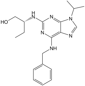
Seliciclib 是一种 2,6-二氨基嘌呤,其 C-6、C-2-N 和 N-9 位分别带有苄氨基、(2R)-1-羟基丁-2-基和异丙基取代基。它是细胞周期蛋白依赖性激酶 (CDK) 抑制剂家族中的一种实验性候选药物。它既是 EC 2.7.11.22(细胞周期蛋白依赖性激酶)抑制剂,也是一种抗病毒药物。
R-roscovitine(Seliciclib 或 CYC202)是一种细胞周期蛋白依赖性激酶 (CDK) 抑制剂,可优先抑制包括 CDK2、CDK7 和 CDK9 在内的多种酶靶点,从而改变受试细胞的生长周期。由Cyclacel公司开发的seliciclib正被研究用于治疗非小细胞肺癌(NSCLC)、白血病、HIV感染、单纯疱疹病毒感染以及慢性炎症性疾病的机制。 据报道,seliciclib存在于蛇尾兰(Ophioparma ventosa)中,并有相关数据。 seliciclib是一种口服小分子细胞周期蛋白依赖性激酶(CDK)抑制剂,具有潜在的促凋亡和抗肿瘤活性。CDK是丝氨酸/苏氨酸激酶,在细胞周期调控中发挥重要作用,并在多种恶性肿瘤中过度表达。Seliciclib主要通过与CDK 2、7和9竞争ATP结合位点来抑制这些激酶,从而阻断细胞周期进程。此外,该药物似乎还能干扰CDK介导的RNA聚合酶II羧基末端结构域的磷酸化,从而抑制RNA聚合酶II依赖的转录。这可能导致抗凋亡因子(例如髓系细胞白血病序列1 (Mcl-1))的下调,Mcl-1是一种对多种肿瘤细胞类型存活至关重要的蛋白质。抗凋亡因子的下调可能导致细胞凋亡的诱导,从而进一步增强seliciclib的抗增殖作用。 一种嘌呤衍生物,是细胞周期蛋白依赖性激酶的竞争性抑制剂,具有作为抗肿瘤和抗病毒药物的治疗潜力。 药物适应症 已研究用于治疗乳腺癌、肺癌、淋巴瘤(未具体说明)、多发性骨髓瘤、淋巴细胞白血病和癌症/肿瘤(未具体说明)。 细胞周期蛋白依赖性激酶 (CDK) 在细胞分裂周期 (CDC) 的细胞内调控中发挥着至关重要的作用。这些激酶及其调节因子在人类肿瘤中经常失调。酶筛选近期发现了一些细胞周期蛋白依赖性激酶的特异性抑制剂,例如丁内酯I、黄酮吡啶醇和嘌呤类化合物奥洛莫辛。在一系列针对纯化的cdc2/细胞周期蛋白B的C2、N6、N9取代腺嘌呤化合物中,2-(1-乙基-2-羟乙基氨基)-6-苄基氨基-9-异丙基嘌呤(罗斯科维汀)对某些细胞周期蛋白依赖性激酶表现出高效性和高选择性。罗斯科维汀的激酶特异性通过25种高纯度激酶(包括蛋白激酶A、G和C亚型、肌球蛋白轻链激酶、酪蛋白激酶2、胰岛素受体酪氨酸激酶、c-src和v-abl)进行了研究。结果表明,罗斯科维汀对大多数激酶的抑制作用并不显著。 cdc2/cyclin B、cdk2/cyclin A、cdk2/cyclin E 和 cdk5/p35 仅受到显著抑制(IC50 值分别为 0.65、0.7、0.7 和 0.2 μM)。cdk4/cyclin D1 和 cdk6/cyclin D2 受罗斯科维汀抑制作用极弱(IC50 > 100 μM)。细胞外信号调节激酶 erk1 和 erk2 分别受到抑制,IC50 值分别为 34 μM 和 14 μM。罗斯科维汀可逆地使海星卵母细胞和海胆胚胎停滞在晚期前期。罗斯科维汀抑制体外 M 期促进因子活性和非洲爪蟾卵提取物中的体外 DNA 合成。它能阻断孕酮诱导的非洲爪蟾卵母细胞成熟以及体内延伸因子eEF-1的磷酸化。罗斯科维汀抑制哺乳动物细胞系的增殖,平均IC50值为16 μM。在罗斯科维汀存在的情况下,L1210细胞停滞于G1期并积累于G2期。罗斯科维汀抑制cdc2/细胞周期蛋白B对波形蛋白Ser55位点的体内磷酸化。由于其对某些细胞周期蛋白依赖性激酶的独特选择性,罗斯科维汀可作为一种有用的抗有丝分裂试剂用于细胞周期研究,并可能对控制cdc2、cdk2或cdk5激酶活性失调的细胞具有潜在价值。[1] 肾小球损伤的特征是系膜细胞(MC)增殖和基质形成。我们旨在确定使用嘌呤类似物罗斯科维汀降低细胞周期蛋白依赖性激酶2 (CDK2) 的活性是否能减少体外和体内系膜细胞的增殖。罗斯科维汀 (25 μM) 可抑制胎牛血清 (FCS) 诱导的培养系膜细胞增殖 (P < 0.0001)。实验性系膜增生性肾小球肾炎 (Thy1 模型) 大鼠被分为两组。预防组从第 1 天开始,每天腹腔注射溶于二甲基亚砜 (DMSO) 的罗斯科维汀 (2.8 mg/kg)。治疗组从第 3 天开始,即系膜细胞增殖建立之时,每天接受罗斯科维汀注射。对照组 Thy1 大鼠仅接受 DMSO 注射。在Roscovitine预防组中,第5天和第10天MC增殖(PCNA+/OX7+双重免疫染色)减少了50%以上;在治疗组中,第5天MC增殖也减少了50%以上(P < 0.0001)。早期给予Roscovitine可降低第5天和第10天IV型胶原、层粘连蛋白和纤连蛋白的免疫染色强度(r = 0.984;P < 0.001),这与肾功能改善(尿蛋白/肌酐比值、血尿素氮,P < 0.05)相关。我们得出结论,在实验性肾小球肾炎中,使用Roscovitine降低CDK2活性可减少细胞增殖和基质生成,从而改善肾功能,这可能是一种治疗以增殖为特征的疾病的有效方法。 [2] 罗斯科维汀是一种细胞周期蛋白依赖性激酶 (Cdk) 抑制剂,在微摩尔浓度下可抑制激酶活性和盘基网柄菌的无菌培养生长。在 50 µM 罗斯科维汀处理的培养物中,通过过表达 Cdk5-GFP,细胞生长几乎完全恢复;在 100 µM 罗斯科维汀处理的培养物中,细胞生长恢复约 50%。这支持了 Cdk5 在盘基网柄菌细胞增殖过程中发挥重要作用的观点,并表明 Cdk5 是该药物的主要靶点。罗斯科维汀不影响无菌培养过程中 Cdk5 蛋白的表达,但抑制其核转位。这一新结果提示,罗斯科维汀的作用可能部分归因于其在其他系统中对 Cdk5 核转位的改变。在利用AX3全细胞裂解液进行的实验中,roscovitine抑制了激酶活性,但在利用Cdk5-GFP过表达细胞裂解液进行的实验中则未观察到此现象。在高浓度下,roscovitine会抑制子实体和子实体的形成。形成的子实体较小,产生的孢子相对较少,且许多孢子呈圆形。然而,roscovitine并不影响柄细胞的分化。结合之前的研究结果,这些数据表明roscovitine在生长过程中抑制Cdk5,并在发育中后期抑制尚未明确的Cdks。[3] 尤文氏肉瘤家族肿瘤(ESFT)包含几种特征明确的恶性肿瘤,这些肿瘤具有特别强的侵袭性。尽管近年来在多模式治疗方法和积极的局部控制措施方面取得了进展,但仍有相当一部分患者因疾病进展而死亡。此外,在过去的15到20年中,这一结果并没有显著改善。因此,迫切需要新的、更有效的治疗方案来治疗尤文氏肉瘤/原发性骨肉瘤(ESFT)。由于ESFT细胞过度表达多种细胞周期蛋白依赖性激酶(CDK),我们探索了罗斯科维汀(roscovitine)对ESFT的疗效。罗斯科维汀是一种CDK抑制剂,在抗癌活性临床试验中显示出对人体出乎意料的安全性。结果表明,ESFT细胞系对罗斯科维汀均敏感。罗斯科维汀治疗除了对细胞周期产生相对较小的影响外,还同时导致促凋亡蛋白BAX表达上调以及survivin和XIAP表达下调,从而诱导caspase依赖性细胞凋亡。此外,体内实验表明,腹腔注射罗斯科维汀也能显著减缓ESFT异种移植瘤的皮下生长。这些结果强烈提示罗斯科维汀可能是一种有效的ESFT治疗药物,建议在临床试验中评估其对ESFT的疗效,并将其纳入未来的治疗方案。[4] 我们旨在确定在MCF7乳腺癌异种移植模型中,塞利西利(CYC202,R-罗斯科维汀)是否能在不增加毒性的情况下增强阿霉素的抗肿瘤作用。我们比较了塞利西利联合阿霉素与单药阿霉素或塞利西利治疗MCF7细胞和携带已建立的MCF7异种移植瘤的裸鼠的疗效。采用细胞周期分析、免疫组织化学和实时PCR检测治疗后的细胞和肿瘤。塞利西利显著增强了阿霉素的抗肿瘤作用,且未增加小鼠毒性。MIB1(Ki67)免疫组织化学染色显示,治疗后细胞增殖减少。与未治疗的对照组相比,单独使用阿霉素或seliciclib治疗或联合治疗后,p21和p27的水平升高。然而,与对照组相比,治疗组肿瘤中p53蛋白(DO1、CM1)、survivin或p53磷酸化(SER15)均未发生变化。总之,CDK抑制剂seliciclib(R-roscovitine)通过细胞周期阻滞而非细胞凋亡机制,增强阿霉素在MCF7肿瘤中的抗肿瘤作用,且不增加毒性。[5] |
| 分子式 |
C19H26N6O
|
|
|---|---|---|
| 分子量 |
354.45
|
|
| 精确质量 |
354.216
|
|
| 元素分析 |
C, 64.38; H, 7.39; N, 23.71; O, 4.51
|
|
| CAS号 |
186692-46-6
|
|
| 相关CAS号 |
|
|
| PubChem CID |
160355
|
|
| 外观&性状 |
White to off-white solid powder
|
|
| 密度 |
1.3±0.1 g/cm3
|
|
| 沸点 |
577.5±60.0 °C at 760 mmHg
|
|
| 闪点 |
303.1±32.9 °C
|
|
| 蒸汽压 |
0.0±1.7 mmHg at 25°C
|
|
| 折射率 |
1.643
|
|
| LogP |
1.68
|
|
| tPSA |
87.89
|
|
| 氢键供体(HBD)数目 |
3
|
|
| 氢键受体(HBA)数目 |
6
|
|
| 可旋转键数目(RBC) |
8
|
|
| 重原子数目 |
26
|
|
| 分子复杂度/Complexity |
417
|
|
| 定义原子立体中心数目 |
1
|
|
| SMILES |
O([H])C([H])([H])[C@@]([H])(C([H])([H])C([H])([H])[H])N([H])C1=NC(=C2C(=N1)N(C([H])=N2)C([H])(C([H])([H])[H])C([H])([H])[H])N([H])C([H])([H])C1C([H])=C([H])C([H])=C([H])C=1[H]
|
|
| InChi Key |
BTIHMVBBUGXLCJ-OAHLLOKOSA-N
|
|
| InChi Code |
InChI=1S/C19H26N6O/c1-4-15(11-26)22-19-23-17(20-10-14-8-6-5-7-9-14)16-18(24-19)25(12-21-16)13(2)3/h5-9,12-13,15,26H,4,10-11H2,1-3H3,(H2,20,22,23,24)/t15-/m1/s1
|
|
| 化学名 |
(2R)-2-[[6-(benzylamino)-9-propan-2-ylpurin-2-yl]amino]butan-1-ol
|
|
| 别名 |
Seliciclib; R-Roscovitine; CYC-202; roscovitine; Seliciclib; 186692-46-6; R-Roscovitine; (R)-roscovitine; Roscovitin; CYC202; Roscovitin; Roscovitine; CYC202; CYC 202
|
|
| HS Tariff Code |
2934.99.9001
|
|
| 存储方式 |
Powder -20°C 3 years 4°C 2 years In solvent -80°C 6 months -20°C 1 month |
|
| 运输条件 |
Room temperature (This product is stable at ambient temperature for a few days during ordinary shipping and time spent in Customs)
|
| 溶解度 (体外实验) |
|
|||
|---|---|---|---|---|
| 溶解度 (体内实验) |
配方 1 中的溶解度: ≥ 2.5 mg/mL (7.05 mM) (饱和度未知) in 10% DMSO + 40% PEG300 + 5% Tween80 + 45% Saline (这些助溶剂从左到右依次添加,逐一添加), 澄清溶液。
例如,若需制备1 mL的工作液,可将100 μL 25.0 mg/mL澄清DMSO储备液加入到400 μL PEG300中,混匀;然后向上述溶液中加入50 μL Tween-80,混匀;加入450 μL生理盐水定容至1 mL。 *生理盐水的制备:将 0.9 g 氯化钠溶解在 100 mL ddH₂O中,得到澄清溶液。 配方 2 中的溶解度: ≥ 2.5 mg/mL (7.05 mM) (饱和度未知) in 10% DMSO + 90% (20% SBE-β-CD in Saline) (这些助溶剂从左到右依次添加,逐一添加), 澄清溶液。 例如,若需制备1 mL的工作液,可将 100 μL 25.0 mg/mL澄清DMSO储备液加入900 μL 20% SBE-β-CD生理盐水溶液中,混匀。 *20% SBE-β-CD 生理盐水溶液的制备(4°C,1 周):将 2 g SBE-β-CD 溶解于 10 mL 生理盐水中,得到澄清溶液。 View More
配方 3 中的溶解度: ≥ 2.5 mg/mL (7.05 mM) (饱和度未知) in 10% DMSO + 90% Corn Oil (这些助溶剂从左到右依次添加,逐一添加), 澄清溶液。 配方 4 中的溶解度: ≥ 2.5 mg/mL (7.05 mM) (饱和度未知) in 5% DMSO + 40% PEG300 + 5% Tween80 + 50% Saline (这些助溶剂从左到右依次添加,逐一添加), 澄清溶液。 *生理盐水的制备:将 0.9 g 氯化钠溶解在 100 mL ddH₂O中,得到澄清溶液。 配方 5 中的溶解度: ≥ 2.5 mg/mL (7.05 mM) (饱和度未知) in 5% DMSO + 95% (20% SBE-β-CD in Saline) (这些助溶剂从左到右依次添加,逐一添加), 澄清溶液。 *20% SBE-β-CD 生理盐水溶液的制备(4°C,1 周):将 2 g SBE-β-CD 溶解于 10 mL 生理盐水中,得到澄清溶液。 配方 6 中的溶解度: 1% DMSO +30% polyethylene glycol+1% Tween 80 : 30 mg/mL 1、请先配制澄清的储备液(如:用DMSO配置50 或 100 mg/mL母液(储备液)); 2、取适量母液,按从左到右的顺序依次添加助溶剂,澄清后再加入下一助溶剂。以 下列配方为例说明 (注意此配方只用于说明,并不一定代表此产品 的实际溶解配方): 10% DMSO → 40% PEG300 → 5% Tween-80 → 45% ddH2O (或 saline); 假设最终工作液的体积为 1 mL, 浓度为5 mg/mL: 取 100 μL 50 mg/mL 的澄清 DMSO 储备液加到 400 μL PEG300 中,混合均匀/澄清;向上述体系中加入50 μL Tween-80,混合均匀/澄清;然后继续加入450 μL ddH2O (或 saline)定容至 1 mL; 3、溶剂前显示的百分比是指该溶剂在最终溶液/工作液中的体积所占比例; 4、 如产品在配制过程中出现沉淀/析出,可通过加热(≤50℃)或超声的方式助溶; 5、为保证最佳实验结果,工作液请现配现用! 6、如不确定怎么将母液配置成体内动物实验的工作液,请查看说明书或联系我们; 7、 以上所有助溶剂都可在 Invivochem.cn网站购买。 |
| 制备储备液 | 1 mg | 5 mg | 10 mg | |
| 1 mM | 2.8213 mL | 14.1064 mL | 28.2127 mL | |
| 5 mM | 0.5643 mL | 2.8213 mL | 5.6425 mL | |
| 10 mM | 0.2821 mL | 1.4106 mL | 2.8213 mL |
1、根据实验需要选择合适的溶剂配制储备液 (母液):对于大多数产品,InvivoChem推荐用DMSO配置母液 (比如:5、10、20mM或者10、20、50 mg/mL浓度),个别水溶性高的产品可直接溶于水。产品在DMSO 、水或其他溶剂中的具体溶解度详见上”溶解度 (体外)”部分;
2、如果您找不到您想要的溶解度信息,或者很难将产品溶解在溶液中,请联系我们;
3、建议使用下列计算器进行相关计算(摩尔浓度计算器、稀释计算器、分子量计算器、重组计算器等);
4、母液配好之后,将其分装到常规用量,并储存在-20°C或-80°C,尽量减少反复冻融循环。
计算结果:
工作液浓度: mg/mL;
DMSO母液配制方法: mg 药物溶于 μL DMSO溶液(母液浓度 mg/mL)。如该浓度超过该批次药物DMSO溶解度,请首先与我们联系。
体内配方配制方法:取 μL DMSO母液,加入 μL PEG300,混匀澄清后加入μL Tween 80,混匀澄清后加入 μL ddH2O,混匀澄清。
(1) 请确保溶液澄清之后,再加入下一种溶剂 (助溶剂) 。可利用涡旋、超声或水浴加热等方法助溶;
(2) 一定要按顺序加入溶剂 (助溶剂) 。
| NCT Number | Recruitment | interventions | Conditions | Sponsor/Collaborators | Start Date | Phases |
| NCT03774446 | Recruiting | Drug: Seliciclib | Cushing Disease | Cedars-Sinai Medical Center | August 2025 | Phase 2 |
| NCT00999401 | Completed | Drug: sapacitabine and seliciclib |
Advanced Solid Tumors | Cyclacel Pharmaceuticals, Inc. | April 2009 | Phase 1 |
| NCT02649751 | Terminated | Drug: Roscovitine Drug: Placebo |
Cystic Fibrosis | University Hospital, Brest | February 22, 2016 | Phase 2 |
| NCT02160730 | Terminated | Drug: R-roscovitine | Cushings Disease | Shlomo Melmed, MD | May 2014 | Phase 2 |
| NCT00372073 | Terminated | Drug: seliciclib Drug: Placebo |
Non-small Cell Lung Cancer | Cyclacel Pharmaceuticals, Inc. | August 9, 2006 | Phase 2 |
|
|---|
 M1-20
M1-20
 CDK12-Cyclin K Ligand-Linker Conjugates 1
 CGP-74514 dihydrochloride
 (S)-CDK2 degrader 6
InvivoChem的所有产品仅用于作科学研究,不面向患者销售
Copyright 2020 InvivoChem LLC | All Rights Reserved 粤ICP备20063088号-1


 COA
COA

 463611831
