| 规格 | 价格 | 库存 | 数量 |
|---|---|---|---|
| 5mg |
|
||
| 10mg |
|
||
| 25mg |
|
||
| 50mg |
|
||
| 100mg |
|
||
| 250mg |
|
||
| Other Sizes |
|

| 靶点 |
C3a receptor (IC50 = 200 nM)
|
|---|---|
| 体外研究 (In Vitro) |
SB 290157 的 IC50 为 200 nM,可作为 125I-C3a 放射性配体的竞争性拮抗剂,与表达人 C3aR (RBL-C3aR) 的大鼠嗜碱性白血病-2H3 细胞结合。 SB290157抑制 RBL-C3aR 细胞和人中性粒细胞中 C3a 诱导的 Ca2+ 动员和 C3a 诱导的 C3aR 内化,IC50 值分别为 27.7 和 28 nM。由于 SB 290157 不会与 C5aR 或其他六种趋化性 G 蛋白偶联受体发生激动性相互作用,因此它对 C3aR 表现出选择性。此外,在 C3a 诱导的 Ca2+ 动员方面,表达小鼠和豚鼠 C3aR 的 RBL-2H3 细胞会受到 SB 290157 的抑制。它能有效抑制 C3a 诱导的对灌注大鼠尾动脉场刺激的收缩反应的增强,以及 C3a 介导的豚鼠血小板 ATP 释放[1]。
过敏毒素C3a是一种强效的趋化肽和炎症介质,在补体激活过程中释放,与G蛋白偶联受体结合并激活。C3aR的分子克隆促进了鉴定C3aR非肽拮抗剂的研究。鉴定并化学优化了一种在高通量筛选中选择性抑制C3aR的化学先导物。产生的拮抗剂N(2)-[(2,2-二苯乙氧基)乙酰基]-L-精氨酸(SB290157)作为(125)I-C3a放射性配体与表达人C3aR(RBL-C3aR)的大鼠嗜碱性白血病(RBL)-2H3细胞结合的竞争性拮抗剂,IC(50)为200 nMSB290157是一种功能拮抗剂,以浓度依赖的方式阻断C3a诱导的C3aR内化,并阻断C3a在RBL-C3aR细胞和人中性粒细胞中诱导的Ca(2+)动员,IC(50)分别为27.7和28 nM。SB 290157对C3aR具有选择性,因为它不拮抗C5aR或其他六种趋化性G蛋白偶联受体。功能拮抗不仅限于人类C3aR;SB 290157还抑制了C3a诱导的表达小鼠和豚鼠C3aRS的RBL-2H3细胞的Ca(2+)动员:它能有效抑制C3a介导的豚鼠血小板ATP释放,并抑制C3a诱导对灌注大鼠尾动脉场刺激的收缩反应增强[1]。 为了鉴定非肽C3aR拮抗剂,使用由RBL-C3aR细胞和125I-C3a制备的膜配置了高通量放射配体结合分析。在高通量筛选中测试了来自SmithKline Beecham化合物集合的约240000种化合物,得到64种确认的活性化合物。其中一种化合物SKF 63649作为选择性C3aR拮抗剂得到了进一步的研究进展(图1,a)。随后对该化合物的化学优化导致了SB290157的发现(图1,B)。在125I-C3a竞争性结合实验中评估了这两种化合物对C3aR的亲和力。在该测定中,SB 290157对C3aR的亲和力比SKF 63649高一个数量级;IC50值分别为200和3000 nM(图2,A)。相关结构SB 280936(图1C)在浓度高达10μM的竞争性结合试验中对该受体没有亲和力,并用作阴性对照。 为了确定这些化合物是否是功能性拮抗剂,使用了基于FLIPR的C3a诱导的RBL-C3aR细胞中Ca2+动员试验。SKF 63649和SB290157分别表现出对1 nM C3a诱导的Ca2+动员的浓度依赖性抑制,IC50分别为350 nM(n=2)和27.7±2.9 nM(=3)(图2B)。在高达20μM的浓度下,SB 280936对C3a诱导的RBL-C3aR细胞中Ca2+动员没有影响。通过测试天然表达C3aR的细胞的活性,我们研究了拮抗剂抑制新鲜分离的外周血中性粒细胞中C3a诱导的Ca2+动员的能力。这两种化合物都是拮抗剂,对SKF 63649和SB290157的IC50分别为388和30 nM。SB 290157对C3aR具有选择性,因为它不会拮抗C5a诱导的人中性粒细胞或RBL-C5aR细胞中的Ca2+动员,也不会抑制中性粒细胞上其他五种GPCR的Ca2+调动反应,即白三烯B4、fMLP、血小板活化因子、CXCR1和CXCR2。 对SB290157抑制C3a诱导的HMC-1细胞趋化性的能力进行了评估,HMC-1细胞是一种天然表达C3aR的人肥大细胞系,C3a对其具有趋化性。浓度为5μM的SB 290157显著抑制了C3a介导的HMC-1细胞的趋化性(图2C)。SB 290157对HMC-1细胞的C5a介导的趋化性没有影响(数据未显示)。 测试拮抗剂对C3a诱导的C3aR内化的抑制作用。中性粒细胞与10 nM C3a孵育3分钟足以刺激90%的C3aR内化。SKF 63649和SB290157均以浓度依赖的方式抑制了10 nM C3a诱导的C3aR内化(图3)。在拮抗剂浓度大于1μM的情况下,C3a诱导的C3aR内化减少了约50%(图3)。在该试验中,SB 280936对C3aR内化没有影响(图3)。 除了对人C3aR的功能拮抗作用外,SB290157还是C3a诱导的稳定表达小鼠和豚鼠C3aRs的RBL 2H3细胞Ca2+动员的强效抑制剂(表I)。SB290157抑制C3a诱导的小鼠和豚鼠C3aRs的Ca2+动员的IC50分别为7和12.5 nM。SB 280936在小鼠和豚鼠C3aRs上均无活性。 为了评估拮抗剂对人类以外物种内源性C3aRs的功能活性,评估了它们对1 nM(本试验中的EC80浓度)C3a诱导的豚鼠血小板ATP释放的抑制作用,豚鼠血小板是天然表达C3aR的细胞。SKF 63649和SB290157均以浓度依赖的方式抑制,IC50值分别为385±185和30±14 nM[1]。 |
| 体内研究 (In Vivo) |
在豚鼠 LPS 诱导的气道中性粒细胞增多和大鼠佐剂诱导的关节炎模型中,SB290157分别减轻爪水肿并抑制中性粒细胞募集[1]。只有三小时后,拮抗剂才能有效消除关节肿胀;在 30 mg/kg 剂量下,观察到关节肿胀可抑制 50%。三小时后,C3 水平远低于表现出补体消耗的初始小鼠。此外,值得注意的是,C3 激活随着抗 OVA pAb 的分级浓度而增加 [2]。
在豚鼠LPS诱导的气道中性粒细胞减少模型中评估了SB290157。如图6所示,以气溶胶形式给药的LPS(10μg/ml)在LPS暴露后48小时产生白细胞浸润(比未暴露的动物高5倍),尤其是中性粒细胞(>1000倍)。通过给予SB290157,30mg/kg i.p.b.i.d.,气道中性粒细胞减少(39%)(LPS+载体=33.2±300万中性粒细胞或50.4%的总白细胞恢复;LPS+SB 290157=20.3±170万中性粒血球或31.5%的总白血球;p=0.02,Fisher保护最小二乘差)。治疗动物的白细胞总数与载体治疗动物没有显著差异(LPS+载体=65.1±1130万;LPS+SB 290157=62.0±1010万)。 还使用预防性给药方案在佐剂诱导的关节炎模型中评估了SB290157。从佐剂注射当天开始,将化合物施用于雄性Lewis大鼠。SB 290157经腹膜内给药,每日两次,并在第20天测量爪子炎症。在连续20天接受30mg/kg b.i.d剂量的动物中,第20天爪水肿抑制率为41%(p<0.001)。接受3或10 mg/kg SB 290157 b.i.d.的大鼠足爪水肿没有显著影响[1]。 研究了C3a受体拮抗剂(C3aRA)是否参与抑制抗OVA pAb诱导的关节炎,因为众所周知,过敏毒素C3a在补体激活期间有效炎症反应的发展中起着至关重要的作用。在DBA/1J小鼠中,通过在关节内(i.a.)注射OVA(0小时)前0.5小时给予抗OVA pAb诱导关节炎。在0.5和3小时观察到关节肿胀的两个峰值。通过在0和2小时注射浓度为10和30 mg/kg的SB290157,研究了C3aRA在关节炎中的作用。拮抗剂仅能在3小时减少关节肿胀,30 mg/kg的浓度对关节肿胀的抑制率约为50%。与显示补体消耗的幼稚小鼠相比,C3水平在3小时显著降低。此外,观察到C3活化,并相应于抗OVA pAb的分级浓度而增加。结果还表明,C3aRA能够降低滑膜组织中IL-1β的表达。综上所述,结果表明C3aRA可能有效抑制关节炎[2]。 |
| 酶活实验 |
受体内化试验[1]
如所述,使用C3aR特异性兔多克隆抗血清进行流式细胞术内化测定。测试了100 nM至10μM范围内的SB290157、1-萘氧基乙酰精氨酸(SKF 63649)和N-[(3,5-二氯苯基)甲基]-N-(3-吡啶羰基)甘油-1-精氨酸](SB 280936)在用10 nM C3a刺激人类中性粒细胞后抑制C3aR内化的能力,该浓度诱导C3aR从细胞表面几乎完全消失(>90%的受体内化)。中性粒细胞在37°C下与化合物和10 nM C3a共孵育3分钟,然后评估受体内化情况。 结合分析[1] C3a拮抗剂的高通量闪烁邻近试验(SPA)。[1] 使用RBL-C3aR细胞膜建立了一种主要的高通量放射性配体结合测定法。125I-C3a以高亲和力(Kd=8 pM)结合RBL-C3aR细胞,并且是可饱和的。 所有结合分析均在96孔微量滴定板中进行。Bolton Hunter定制碘化由NEN Research Products使用sp.act进行。2200 Ci/mmol。结合缓冲液由20 mM双三丙烷(pH 8.0)、25 mM NaCl、1 mM MgSO4和0.1 mM EDTA组成。每个孔含有:125I-C3a(16 pM)、70μg麦胚凝集素SPA珠、0.20μg RBL-C3aR膜、23μg/ml BSA和0.03%的3-[(3-甲酰氨基丙基)二甲基氨基]-1-丙磺酸结合缓冲液。此外,非特异性结合的对照孔包括超过15nM的未标记C3a。 在冰上摇动的同时,将膜与SPA珠预结合30分钟。将膜和珠粒的混合物在2000rpm下离心3分钟。去除上清液,将沉淀物重新悬浮在含有50μg/ml BSA的结合缓冲液中至原始体积,然后分配到微量滴定板中。将拮抗剂溶解在纯DMSO中,得到20倍的溶液,然后与H2O按1:1的比例混合,得到10倍、50%的DMSO工作溶液。添加顺序为10μl样品、45μl膜结合SPA珠,然后是45μl放射性标记配体,在含有0.06%3-[(3-氨基丙基)二甲基铵]-1-丙磺酸酯的结合缓冲液中。用Dynex Technologies的板密封剂覆盖板,摇动20分钟,并在室温下再孵育40分钟。然后将平板以2000rpm离心3分钟,然后在Wallac 1450 MicroβPlus液体闪烁计数器上计数。 对后续研究具有约束力。[1] 结合测定基本上如前所述进行(26)。简而言之,在室温下,将2-5×10⁵个RBL-C3aR细胞与100 pM 125I-C3a和不同浓度的拮抗剂在20 mM HEPES(pH 7.4)、125 mM NaCl、5 mM KCl、1 mM CaCl2、1 mM MgCl2、0.25%BSA和0.5 mM葡萄糖(HAG-CM)中孵育45分钟。使用HV Millipore MultiScreen测定板通过真空过滤去除未结合的配体,该板具有用HAG-CM平衡的Durapore 0.45-μm孔径膜。用100μl/孔的HAG-CM洗涤过滤器两次并干燥。在贝克曼伽马计数器5500B上对板进行计数。使用KaleidaGraph v3.09进行数据分析。 |
| 细胞实验 |
HMC-1趋化性测定[1]
使用Neuro Probe 96孔一次性趋化板(5μm孔径)评估HMC-1细胞的C3a介导的趋化性。膜的顶面预先涂有100ng层粘连蛋白或纤维连接蛋白。在28μl RPMI 1640中向较低的孔中加入不同浓度的C3a(有和没有拮抗剂)。组装过滤器,将2至5×10⁵个细胞加入25μl的顶部孔中。将板在37°C和5%CO2下孵育60分钟。移除过滤器,用PBS冲洗膜的顶面;然后用Diff-Quik(Baxter,Dade Division,Miami,FL)对细胞进行染色。通过在三个连续的高功率场中计数细胞,在显微镜下定量迁移的细胞数量。 荧光成像板读数仪(FLIPR)Ca2+动员测定[1] 使用Fluo 3负载的RBL-C3aR细胞和使用FLIPR的微量滴定板进行C3aR Ca2+动员研究。简而言之,收获细胞(约80%融合),以约40000个细胞/孔的速度将其铺在96孔黑壁透明底板(Packard观察板)上,并在培养箱中生长18-24小时。在测定当天,吸出培养基,用100μl Eagle's MEM替换,该MEM含有l-谷氨酰胺、0.1%BSA、4μM氟-3-乙酰氧基甲酯和1.5 mM亚磺吡喃酮的Earle盐。将板在37°C下孵育60分钟;吸出培养基,用不含氟-3-乙酰氧基甲酯的相同培养基替换,并在37°C下孵育10分钟。将细胞洗涤三次,并在37°C下在100μl测定缓冲液(120 mM NaCl、4.6 mM KCl、1.03 mM KH2 PO4、25 mM NaHCO3、1.0 mM CaCl2、1.1 mM MgCl2、11 mM葡萄糖、20 mM HEPES(pH 7.4)和1.5 mM亚磺吡喃酮)中孵育。如前所述,将板放入FLIPR中进行分析。定量了加入激动剂后荧光的最大变化。测定每种拮抗剂浓度下C3a诱导的最大Ca2+动员百分比。IC50定义为抑制1nM C3a诱导的最大反应的50%的测试化合物的浓度,从浓度-反应曲线中获得。对于激动剂效力,EC50定义为产生50%最大C3a诱导反应的浓度。 |
| 动物实验 |
大鼠尾动脉收缩[1]
将体重400~600 g的雄性Sprague Dawley正常血压大鼠处死,取尾部并置于生理缓冲液中。将尾部固定于解剖板上,暴露尾动脉,切取一段30~40 mm长的动脉段并置于缓冲液中。将该动脉段切成两段等长的动脉段,每段动脉段两端均用PE50导管插管,并用4-0手术丝线固定导管。将插管后的动脉段置于管状玻璃腔室中,同时用38℃的氧合Krebs缓冲液进行管腔内灌注和管腔外灌注。管腔内灌注速率为1 ml/min,管腔外灌注速率为2 ml/min。在这些条件下,基线灌注压稳定在 25 至 50 mmHg 之间。经过 20 至 30 分钟的稳定期后,通过位于腔室两端的铂电极,每隔 30 秒对动脉周围交感神经进行电刺激,以获得短暂的、尖峰状的灌注压升高。刺激由持续 1 秒、电压 70 V、脉宽 0.7 ms、频率 15 Hz 的方波脉冲串组成。这些刺激参数导致灌注压较基线升高 50 至 100 mmHg。当反应稳定后,将其中一段动脉暴露于通过灌注液输送的 SB290157 中,而另一段动脉则不进行任何处理。在暴露于SB290157(10 nM、100 nM 和 1 μM)15 分钟后,将 C3a(100 nM)加入到灌注流中,分别注入到两个动脉段,并监测其对灌注压的影响。通常情况下,C3a 会提高灌注压。C3a 介导的灌注压升高作用迅速减弱(1-2 分钟)。 豚鼠气道中性粒细胞增多模型[1] 雄性 Hartley 豚鼠购自 Charles River Breeding Laboratories,饲养于屏障设施中。每次将四只豚鼠放入一个经过改造的塑料箱(20 升)中,该塑料箱设有进气口和排气口;箱盖上的小型风扇可增加气溶胶循环。将溶于生理盐水(30 μg/ml)的LPS气溶胶通过改良的DeVilbiss Pulmosonic雾化器产生,并以250 ml/min的流速经吸入口输送至实验箱内,持续15分钟。在LPS刺激前1小时和刺激后4小时,腹腔注射SB290157(30 mg/kg)或赋形剂(20%聚乙二醇400 (PEG) 生理盐水),并在次日间隔6小时每日两次(bid)给药。第三组动物未接触LPS,仅接受赋形剂。在LPS刺激后48小时进行支气管肺泡灌洗(BAL)。豚鼠经戊巴比妥过量麻醉处死后,用50 ml Dulbecco氏PBS(5 × 10 ml)灌洗肺部,并在轻柔按摩胸部后吸出灌洗液。将支气管肺泡灌洗液离心,沉淀物重悬于 0.25% NaCl 溶液中以裂解残留红细胞;再次离心后,沉淀物重悬于 1 ml 0.9% NaCl 溶液中。计数总细胞数后,制备玻片,染色,并通过计数至少 200 个细胞,将嗜酸性粒细胞、中性粒细胞和单核细胞进行区分,并将结果表示为占总细胞数的百分比以及每种细胞的实际数量。该测量和表达技术此前已通过组织学方法验证,能够准确反映内皮和内皮下气道白细胞增多。细胞数量和百分比采用 ANOVA 方差分析,然后进行 Fisher 最小二乘法检验进行统计学比较。 佐剂诱导性关节炎[1] 雄性近交系 Lewis 大鼠购自 Charles River Breeding Laboratories。在同一实验中,仅使用同龄动物。佐剂诱导性关节炎 (AIA) 的诱导方法如前所述。简而言之,将 0.75 mg 悬浮于石蜡油中的丁酸分枝杆菌注射到 6-8 周龄(160-180 g)雄性 Lewis 大鼠尾根部。于第 20 天采用排水法测量后爪体积。将 SB290157 悬浮于由 5% 乙醇、10% Cremaphor-El 和 85% 生理盐水组成的溶剂中,于佐剂注射当天开始,每日两次腹腔注射,剂量分别为 30、10 和 3 mg/kg,最终注射体积为 0.5 ml。对笼子进行改造,使患病动物能够自由获取食物和水。对照组动物仅注射溶剂。爪体积变化以每组10-12只动物的平均值和标准误(SEM)表示,后爪水肿抑制率的计算方法如前所述。统计分析中,采用Student's t检验比较SB290157治疗组大鼠与未治疗对照组的爪体积。 豚鼠药代动力学研究[1] 本药代动力学研究采用三只雄性Hartley豚鼠进行。在无菌条件下,每只豚鼠在研究日至少5天前接受手术植入股静脉和动脉静脉导管。研究日,喂食后的动物接受单次腹腔注射SB290157(30 mg/kg)(总注射量为3 ml/kg)。给药溶液用含20%聚乙二醇(PEG)的生理盐水配制。在给予SB290157后的不同时间间隔,通过动脉导管采集血样;离心分离血浆。采用液相色谱/质谱联用(LC/MS/MS)法定量血浆中SB290157的浓度(定量下限为10 ng/ml)。采用非房室模型分析血浆浓度-时间数据。DBA/1J小鼠关节炎的诱导[2] 在给予OVA前0.5 h(-0.5 h),通过静脉注射(iv)给予小鼠0.2 ml大鼠抗OVA多克隆抗体(10 mg/ml)。将10 µg OVA溶解于25 µl PBS中,并通过关节内注射(ia)(0 h)给予小鼠。单独注射OVA作为基线。抗OVA多克隆抗体注射引起的关节厚度净增加值,是通过从抗OVA多克隆抗体注射小鼠的关节厚度中减去未免疫的OVA注射小鼠的关节厚度计算得出的。C3aR拮抗剂SB290157(10或30 mg/kg)腹腔注射两次,分别在OVA注射后0小时(即注射后立即注射)和2小时注射。以5%乙醇PBS溶液作为溶剂对照。在注射前、注射后0.5小时以及之后每小时测量一次关节肿胀程度,直至OVA注射后5小时。 |
| 药代性质 (ADME/PK) |
本研究评估了SB 290157在豚鼠和小鼠腹腔注射给药后的药代动力学特征。豚鼠研究结果总结于图5。当以30 mg/kg的剂量腹腔注射SB 290157时,在长达8小时内均检测到高浓度且持续的血浆药物浓度(>100 ng/ml,0.25 μM)(图5)。达到的Cmax为7000 ng/ml,表观半衰期(t1/2)为0.89 ± 0.26 h。小鼠腹腔注射SB 290157后也获得了类似的药代动力学数据(t1/2 = 1.47 ± 0.10 h;数据未显示)。[1]
|
| 参考文献 |
|
| 其他信息 |
通过高通量筛选鉴定出的小分子非肽类C3aR拮抗剂SKF 63649,能以低微摩尔级的亲和力抑制C3aR的结合。经化学优化得到化合物SB 290157后,其对C3aR的亲和力提高了一个数量级。SB 290157是一种功能性拮抗剂,对新鲜分离的中性粒细胞上表达的天然受体以及稳定表达于RBL-2H3细胞上的重组C3aR均表现出同等效力的C3a诱导Ca2+动员反应抑制作用。SB 290157不仅是人C3aR的功能性拮抗剂,也是小鼠、大鼠和豚鼠C3aR的功能性拮抗剂。 SB 290157 对小鼠、豚鼠和人 C3a 受体诱导的 Ca2+ 动员的抑制效力相似(IC50 = 7–30 nM)。考虑到这些不同物种 C3aR 的序列同一性相对较低(总体同一性为 60–65%),这一结果令人有些意外。SB 290157 对 C3aR 具有选择性,不拮抗 C5aR 或人中性粒细胞上的其他 5 种趋化性 GPCR。
在人中性粒细胞 Ca2+ 动员试验和豚鼠 ATP 释放试验中,SB 290157 的拮抗效力具有良好的相关性。该结果支持重组受体拮抗剂的数据,表明其对两种物种内源性 C3aR 的效力相似。然而,功能性测定中测得的拮抗剂效价比全细胞结合测定中估计的亲和力高约7倍。这可能是由于测定方案本身的差异造成的,包括:功能性测定的孵育时间(秒)与结合测定的平衡条件(30-60分钟)不同;测定温度不同(结合测定在室温下进行,而功能性测定在37°C下进行);或者可能是碘化对C3a与其受体亲和力的影响。C3a碘化对其与C3aR相互作用的影响似乎很小,因为在竞争性结合测定中测得的C3a与C3aR的亲和力与已发表的C3aR解离常数(Kd)(0.1-1.0 nM)非常吻合。在结合和功能测定中,SB 290157 作为 C3aR 拮抗剂的效力始终比最初的高通量筛选结果 SKF 63649 高 10 倍。 C3aR 拮抗剂化合物对 C3a 诱导的 C3aR 内化有显著影响,在 10 nM C3a 刺激下,抑制了近 50% 的受体内化。在浓度低于 10 μM 时,SB 290157 对 C3a 诱导的受体内吞作用的拮抗作用似乎比 SKF 63649 更强,这与该化合物在结合和功能测定中获得的效力一致。 SB 290157 还显著抑制了 HMC-1 细胞的 C3a 介导的趋化作用以及灌注大鼠尾动脉对电场刺激的 C3a 诱导的收缩反应。在这些测定中难以进行浓度-反应研究,但 SB 290157 对小鼠、大鼠和豚鼠 C3a 受体的拮抗效力与对人 C3aR 的效力相当。这些数据,结合小鼠和豚鼠腹腔注射SB 290157后血浆浓度高且持续的测定结果,表明该化合物适合用于动物模型研究,以帮助阐明C3a和C3aR的生理和病理生理作用。 我们利用两种炎症动物模型研究了C3aR拮抗剂SB 290157。在第一种模型中,SB 290157抑制了豚鼠LPS诱导的气道中性粒细胞增多模型中的中性粒细胞募集和聚集。这种抑制活性似乎具有中性粒细胞特异性,因为在受刺激肺组织中回收的中性粒细胞数量减少,但回收的细胞总数没有显著抑制。这有些出乎意料,因为C3a对中性粒细胞没有趋化作用,尽管中性粒细胞表达C3aR,表现出特异性结合,并且对C3a有短暂的钙反应。 SB 290157 的作用可能是对中性粒细胞募集的间接影响,而非直接影响。 SB 290157 也已在 AIA 的疾病修饰大鼠模型中进行了测试。在接受 SB 290157(30 mg/kg,腹腔注射)的 Lewis 大鼠中观察到了抗炎活性。与未治疗的对照组动物相比,爪肿胀显著减轻(41%)。对于 C3aR 拮抗剂而言,这在侵袭性关节炎模型中具有显著活性,并可能表明 C3a 参与了该疾病的发病机制。 我们的数据表明,SB 290157 是一种高亲和力、选择性和竞争性的 C3aR 拮抗剂。它在两种体内炎症模型中均具有活性;因此,它有望成为一种工具化合物,用于进一步研究以阐明 C3aR 激活的生理和病理生理作用。 [1]综上所述,现有数据表明,SB 290157可通过降低关节肿胀程度、中性粒细胞迁移和IL-1β生成来抑制关节炎的发生。目前的研究结果提示,C3a可能参与了SB 290157加重关节炎的过程。因此,SB 290157可能对抑制关节炎有效。[2] |
| 分子式 |
C24H29F3N4O6
|
|---|---|
| 分子量 |
526.505476713181
|
| 精确质量 |
526.203
|
| 元素分析 |
C, 54.75; H, 5.55; F, 10.83; N, 10.64; O, 18.23
|
| CAS号 |
1140525-25-2
|
| 相关CAS号 |
1140525-25-2 (TFA);259218-28-5;
|
| PubChem CID |
16760645
|
| 外观&性状 |
White to yellow solid powder
|
| tPSA |
177
|
| 氢键供体(HBD)数目 |
5
|
| 氢键受体(HBA)数目 |
10
|
| 可旋转键数目(RBC) |
12
|
| 重原子数目 |
37
|
| 分子复杂度/Complexity |
619
|
| 定义原子立体中心数目 |
1
|
| SMILES |
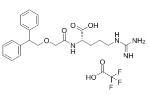
C1=CC=C(C=C1)C(COCC(=O)N[C@@H](CCCN=C(N)N)C(=O)O)C2=CC=CC=C2.C(=O)(C(F)(F)F)O
|
| InChi Key |
ZJRMPPVJAQWGEG-FYZYNONXSA-N
|
| InChi Code |
InChI=1S/C22H28N4O4.C2HF3O2/c23-22(24)25-13-7-12-19(21(28)29)26-20(27)15-30-14-18(16-8-3-1-4-9-16)17-10-5-2-6-11-17;3-2(4,5)1(6)7/h1-6,8-11,18-19H,7,12-15H2,(H,26,27)(H,28,29)(H4,23,24,25);(H,6,7)/t19-;/m0./s1
|
| 化学名 |
(2S)-5-(diaminomethylideneamino)-2-[[2-(2,2-diphenylethoxy)acetyl]amino]pentanoic acid;2,2,2-trifluoroacetic acid
|
| 别名 |
1140525-25-2; SB290157 trifluoroacetate; SB 290157 trifluoroacetate salt; SB290157 (trifluoroacetate); (2S)-5-(diaminomethylideneamino)-2-[[2-(2,2-diphenylethoxy)acetyl]amino]pentanoic acid;2,2,2-trifluoroacetic acid; SB 290157 trifluoroacetate; N2-[2-(2,2-Diphenylethoxy)acetyl]-L-arginine 2,2,2-Trifluoroacetate;; SB290157 trifluoroacetate salt;
|
| HS Tariff Code |
2934.99.9001
|
| 存储方式 |
Powder -20°C 3 years 4°C 2 years In solvent -80°C 6 months -20°C 1 month 注意: 请将本产品存放在密封且受保护的环境中,避免吸湿/受潮。 |
| 运输条件 |
Room temperature (This product is stable at ambient temperature for a few days during ordinary shipping and time spent in Customs)
|
| 溶解度 (体外实验) |
DMSO : ~100 mg/mL (~189.93 mM)
Ethanol : ~100 mg/mL (~189.93 mM) |
|---|---|
| 溶解度 (体内实验) |
配方 1 中的溶解度: ≥ 5 mg/mL (9.50 mM) (饱和度未知) in 10% EtOH + 40% PEG300 + 5% Tween80 + 45% Saline (这些助溶剂从左到右依次添加,逐一添加), 澄清溶液。
例如,若需制备1 mL的工作液,将 100 μL 50.0 mg/mL 澄清乙醇储备液加入到 400 μL PEG300 中,混匀;然后向上述溶液中加入50 μL Tween-80,混匀;加入450 μL生理盐水定容至1 mL。 *生理盐水的制备:将 0.9 g 氯化钠溶解在 100 mL ddH₂O中,得到澄清溶液。 配方 2 中的溶解度: ≥ 5 mg/mL (9.50 mM) (饱和度未知) in 10% EtOH + 90% (20% SBE-β-CD in Saline) (这些助溶剂从左到右依次添加,逐一添加), 澄清溶液。 例如,若需制备1 mL的工作液,可将 100 μL 50.0 mg/mL 澄清乙醇储备液加入到 900 μL 20% SBE-β-CD 生理盐水溶液中,混匀。 *20% SBE-β-CD 生理盐水溶液的制备(4°C,1 周):将 2 g SBE-β-CD 溶解于 10 mL 生理盐水中,得到澄清溶液。 View More
配方 3 中的溶解度: ≥ 5 mg/mL (9.50 mM) (饱和度未知) in 10% EtOH + 90% Corn Oil (这些助溶剂从左到右依次添加,逐一添加), 澄清溶液。 配方 4 中的溶解度: ≥ 2.08 mg/mL (3.95 mM) (饱和度未知) in 10% DMSO + 40% PEG300 + 5% Tween80 + 45% Saline (这些助溶剂从左到右依次添加,逐一添加), 澄清溶液。 例如,若需制备1 mL的工作液,可将100 μL 20.8 mg/mL澄清的DMSO储备液加入400 μL PEG300中,混匀;再向上述溶液中加入50 μL Tween-80,混匀;然后加入450 μL生理盐水定容至1 mL。 *生理盐水的制备:将 0.9 g 氯化钠溶解在 100 mL ddH₂O中,得到澄清溶液。 配方 5 中的溶解度: ≥ 2.08 mg/mL (3.95 mM) (饱和度未知) in 10% DMSO + 90% (20% SBE-β-CD in Saline) (这些助溶剂从左到右依次添加,逐一添加), 澄清溶液。 例如,若需制备1 mL的工作液,可将100μL 20.8mg/mL澄清的DMSO储备液加入到900μL 20%SBE-β-CD生理盐水中,混匀。 *20% SBE-β-CD 生理盐水溶液的制备(4°C,1 周):将 2 g SBE-β-CD 溶解于 10 mL 生理盐水中,得到澄清溶液。 配方 6 中的溶解度: ≥ 2.08 mg/mL (3.95 mM) (饱和度未知) in 10% DMSO + 90% Corn Oil (这些助溶剂从左到右依次添加,逐一添加), 澄清溶液。 例如,若需制备1 mL的工作液,可将 100 μL 20.8 mg/mL 澄清 DMSO 储备液加入到 900 μL 玉米油中并混合均匀。 1、请先配制澄清的储备液(如:用DMSO配置50 或 100 mg/mL母液(储备液)); 2、取适量母液,按从左到右的顺序依次添加助溶剂,澄清后再加入下一助溶剂。以 下列配方为例说明 (注意此配方只用于说明,并不一定代表此产品 的实际溶解配方): 10% DMSO → 40% PEG300 → 5% Tween-80 → 45% ddH2O (或 saline); 假设最终工作液的体积为 1 mL, 浓度为5 mg/mL: 取 100 μL 50 mg/mL 的澄清 DMSO 储备液加到 400 μL PEG300 中,混合均匀/澄清;向上述体系中加入50 μL Tween-80,混合均匀/澄清;然后继续加入450 μL ddH2O (或 saline)定容至 1 mL; 3、溶剂前显示的百分比是指该溶剂在最终溶液/工作液中的体积所占比例; 4、 如产品在配制过程中出现沉淀/析出,可通过加热(≤50℃)或超声的方式助溶; 5、为保证最佳实验结果,工作液请现配现用! 6、如不确定怎么将母液配置成体内动物实验的工作液,请查看说明书或联系我们; 7、 以上所有助溶剂都可在 Invivochem.cn网站购买。 |
| 制备储备液 | 1 mg | 5 mg | 10 mg | |
| 1 mM | 1.8993 mL | 9.4965 mL | 18.9930 mL | |
| 5 mM | 0.3799 mL | 1.8993 mL | 3.7986 mL | |
| 10 mM | 0.1899 mL | 0.9496 mL | 1.8993 mL |
1、根据实验需要选择合适的溶剂配制储备液 (母液):对于大多数产品,InvivoChem推荐用DMSO配置母液 (比如:5、10、20mM或者10、20、50 mg/mL浓度),个别水溶性高的产品可直接溶于水。产品在DMSO 、水或其他溶剂中的具体溶解度详见上”溶解度 (体外)”部分;
2、如果您找不到您想要的溶解度信息,或者很难将产品溶解在溶液中,请联系我们;
3、建议使用下列计算器进行相关计算(摩尔浓度计算器、稀释计算器、分子量计算器、重组计算器等);
4、母液配好之后,将其分装到常规用量,并储存在-20°C或-80°C,尽量减少反复冻融循环。
计算结果:
工作液浓度: mg/mL;
DMSO母液配制方法: mg 药物溶于 μL DMSO溶液(母液浓度 mg/mL)。如该浓度超过该批次药物DMSO溶解度,请首先与我们联系。
体内配方配制方法:取 μL DMSO母液,加入 μL PEG300,混匀澄清后加入μL Tween 80,混匀澄清后加入 μL ddH2O,混匀澄清。
(1) 请确保溶液澄清之后,再加入下一种溶剂 (助溶剂) 。可利用涡旋、超声或水浴加热等方法助溶;
(2) 一定要按顺序加入溶剂 (助溶剂) 。
 MEDI7814
 Bisphenol A bissulfate disodium
 Factor B-IN-6
 Factor B-IN-7
InvivoChem的所有产品仅用于作科学研究,不面向患者销售
Copyright 2020 InvivoChem LLC | All Rights Reserved 粤ICP备20063088号-1


 COA
COA
 463611831
