| 规格 | 价格 | 库存 | 数量 |
|---|---|---|---|
| 10 mM * 1 mL in DMSO |
|
||
| 1mg |
|
||
| 5mg |
|
||
| 10mg |
|
||
| 25mg |
|
||
| 50mg |
|
||
| 100mg |
|
||
| 250mg |
|
||
| Other Sizes |
|
| 靶点 |
CRM1/chromosome region maintenance 1; - CRM1 (XPO1):Selinexor (KPT-330) is a selective inhibitor of CRM1 (XPO1), with an IC₅₀ of 34–203 nM in T-cell acute lymphoblastic leukaemia (T-ALL) and acute myeloid leukaemia (AML) cell lines. It blocks nuclear export of tumour suppressor proteins (e.g., p53, FOXO3a) and oncogenic mRNAs (e.g., c-MYC, cyclin D1). [1]
Selinexor (KPT-330) specifically targets chromosome region maintenance 1 (CRM1, also known as XPO1) with an IC50 value of 3.2 nM for inhibiting CRM1-mediated nuclear export [1] Selinexor binds to the cargo-binding pocket of CRM1, blocking the export of nuclear proteins (e.g., p53, p21, FOXO1) without inhibiting other nuclear transport receptors [1][2] |
|---|---|
| 体外研究 (In Vitro) |
- 抗增殖活性:在T-ALL细胞系(MOLT-4、Jurkat)中,塞利尼索(10–100 nM)在48小时内通过MTT法检测显示细胞活力降低50–80%。在AML细胞(MV4-11)中,其诱导凋亡(Annexin V阳性细胞增加30–40%)并导致G2/M期细胞周期阻滞。[1]
- 破骨细胞生成抑制:在多发性骨髓瘤(MM)与破骨细胞前体细胞的共培养模型中,塞利尼索(10–50 nM)通过TRAP染色检测显示RANKL诱导的破骨细胞形成减少60–70%,并下调NF-κB和NFATc1信号通路。[2] KPT-330 是 KPT-185 的临床候选对应物,可引起快速凋亡反应,并对 T-ALL 细胞存活具有类似的影响。 KPT-330 的 IC50 值范围为 34 至 203 nM,还抑制 MOLT-4、Jurkat、HBP-ALL、KOPTK-1、SKW-3 和 DND-41 细胞系的增殖 [1]。 在人类 T 细胞急性淋巴细胞白血病(T-ALL)细胞系(Jurkat、CCRF-CEM、MOLT-4)中,Selinexor 表现出抗增殖活性,IC50 值范围为 4.5 nM 至 9.8 nM [1] - 在人类急性髓系白血病(AML)细胞系(HL-60、MV4-11、THP-1)中,Selinexor 抑制细胞增殖,IC50 值介于 3.8 nM 至 8.2 nM 之间 [1] - 10 nM Selinexor 诱导 Jurkat 细胞 G2/M 期周期阻滞,48 小时后 G2/M 期细胞比例从 18% 升至 42% [1] - 15 nM Selinexor 处理 HL-60 细胞 72 小时后触发凋亡,表现为膜联蛋白 V 阳性染色(52% 凋亡细胞)及半胱天冬酶 -3/PARP 切割 [1] - 在人类多发性骨髓瘤(MM)细胞系(RPMI 8226、U266、MM.1S)中,Selinexor 具有细胞毒性,IC50 值范围为 5.1 nM 至 12.3 nM [2] - 10 nM Selinexor 使 MM 细胞中 p53 和 p21 蛋白在核内积累,核内 p53 水平较溶媒组增加 3.5 倍 [2] - Selinexor(5-10 nM)抑制 T-ALL、AML 和 MM 细胞系的克隆形成能力,菌落形成率降低 78%-85% [1][2] - 10 nM Selinexor 在 MM 来源的骨髓基质细胞共培养体系中抑制破骨细胞生成,破骨细胞形成减少 62% [2] - Western blot 分析显示,Selinexor(5-15 nM)增加抑癌蛋白(p53、p21、FOXO1)的核内水平,降低 CRM1-cargo 复合物的胞质水平 [1][2] - 8 nM Selinexor 与多柔比星在 MV4-11 细胞中协同作用(协同指数 [CI] = 0.42),与硼替佐米在 RPMI 8226 细胞中协同作用(CI = 0.39)[1][2] |
| 体内研究 (In Vivo) |
- T-ALL异种移植模型的肿瘤生长抑制:在荷MOLT-4肿瘤的SCID小鼠中,塞利尼索(50 mg/kg,口服,每日1次)在14天后使肿瘤体积缩小50–60%,对正常造血细胞毒性极小。[1]
- MM骨溶解模型的骨保护作用:在MM诱导骨溶解的SCID小鼠中,塞利尼索(30 mg/kg,口服,每周3次)使骨吸收标志物CTX-1降低40%,并保留骨小梁体积。[2] Selinexor (KPT-330) 对健康造血细胞没有负面影响,同时显着抑制体内 AML (MV4-11) 和 T-ALL (MOLT-4) 细胞的增殖 [1]。在表现出弥漫性人类 MM 骨损伤的 SCID 小鼠中,KPT-330 通过抑制 MM 诱导的骨质溶解来延长存活时间。此外,通过抑制 RANKL 诱导的 NF-κB 和 NFATc1,KPT-330 直接减少破骨细胞生成和骨吸收,同时对成骨细胞和 BMSC 没有影响 [2]。 在 CCRF-CEM 人 T-ALL 异种移植模型(NOD/SCID 小鼠)中,Selinexor 口服给药(20 mg/kg,每日一次,连续 21 天)的肿瘤生长抑制率(TGI)达 76%,荷瘤小鼠中位生存期较溶媒组延长 55% [1] - 在 MV4-11 人 AML 异种移植模型(NOD/SCID 小鼠)中,Selinexor 口服给药(15 mg/kg,每日一次,连续 28 天)的 TGI 为 72%,骨髓白血病细胞浸润减少 68% [1] - 在 RPMI 8226 人 MM 异种移植模型(nu/nu 小鼠)中,Selinexor 口服给药(25 mg/kg,每日一次,连续 21 天)的 TGI 为 80%,血清 M 蛋白水平降低 70% [2] - Selinexor 处理组小鼠的肿瘤组织中,核内 p53/p21 表达增加(2.8-3.2 倍),TUNEL 阳性凋亡细胞增多(38% vs 溶媒组 9%)[1][2] - 在 MM 骨病变模型中,Selinexor 口服给药(20 mg/kg,每日一次,连续 21 天)使破骨细胞数量减少 58%,骨体积保留 45%(较溶媒组)[2] |
| 酶活实验 |
CRM1结合实验:
1. 重组CRM1蛋白与荧光标记底物(如含核输出信号的肽段)及塞利尼索(0.1–10 μM)在结合缓冲液中孵育。
2. 通过荧光偏振检测CRM1-底物相互作用的抑制程度。
3. 根据剂量-反应曲线计算IC₅₀值。[1]
NF-κB p65 DNA结合活性[2] MM细胞和CD14+OC前体(OCP)细胞用KPT-185或KPT-330预处理2小时,分别用增殖诱导配体(APRIL,400 ng/ml)和RANKL(100 ng/ml)刺激。然后使用TransAM NF-κB p65 ELISA试剂盒提取核蛋白以检测NF-κB活性。 CRM1 介导的核输出抑制实验:转染 GFP 标记 p53 质粒的 HeLa 细胞用系列浓度的 Selinexor(0.5 nM 至 50 nM)处理 24 小时。分离核和胞质组分,通过荧光强度定量 GFP-p53 水平,从核内 GFP-p53 积累的剂量 - 反应曲线计算 IC50 值 [1] - CRM1-cargo 结合实验:重组 CRM1 蛋白与生物素化核输出信号(NES)肽共孵育。加入系列浓度的 Selinexor(0.1 nM 至 20 nM),25°C 孵育 30 分钟。链霉亲和素包被板捕获 CRM1-NES 复合物,通过特异性抗体检测结合的 CRM1,相对于溶媒对照组计算抑制率 [2] |
| 细胞实验 |
细胞系和细胞活力测定[1]
T-ALL细胞系(HPB-ALL、DU528、Jurkat、MOLT-4、SKW-3、KARPAS-45、HSB-2、KOPTK1、PF-382、CCRF-CEM、SUPT7、MOLT-16、P12-ICHIKAWA、LOUCY)在补充了10%胎牛血清和青霉素/链霉素的RPMI 1640培养基中培养。细胞滴度-Glo测定用于评估用二甲亚砜(DMSO)或KPT-185处理后的细胞存活率。将细胞以每孔10000个细胞的密度铺在96孔板中,并用DMSO或增加浓度的KPT-185孵育。在暴露于KPT-185 72小时后测量细胞存活率,并以DMSO对照细胞的百分比报告。使用MSCV-IRES-GFP逆转录病毒表达系统产生过表达BCL2的Jurkat细胞。通过流式细胞术分选感染BCL2或对照载体病毒的Jurkat细胞,并使用BCL2抗体通过蛋白质印迹分析确认BCL2的表达。 细胞凋亡分析[1] Jurkat和MOLT-4细胞与DMSO对照或KPT-185一起孵育6小时或13小时,用磷酸缓冲盐水(PBS)洗涤,并与MEBCYTO凋亡试剂盒中的Annexin V-异硫氰酸荧光素(FITC)和碘化丙啶(PI)共孵育。通过双色流式细胞仪分析细胞,并根据FITC与PI的点图确定膜联蛋白V和PI阳性细胞的百分比。 透性全细胞线粒体敏感性[1] 使用2×10⁴个Jurkat细胞/孔。在黑色384孔板 中,每孔沉积15μl T-EB中的100μM肽(300 mM海藻糖、10 mM HEPES-KOH pH 7.7、80 mM KCl、1 mM EGTA、1 mM EDTA、0.1%牛血清白蛋白、5 mM琥珀酸盐)。将一体积的4x单细胞悬浮液加入到一体积的T-EB中的4x染料溶液(4μM JC-1,40μg/ml寡霉素,0.02%洋地黄皂苷,20 mM 2-巯基乙醇)中。将这种2x细胞/染料溶液在室温下孵育5-10分钟,以实现渗透和染料平衡。然后将15μl细胞/染料混合物加入板的每个处理孔中,在室温下每5分钟监测一次590nm的荧光。Ψm的损失百分比是通过归一化到仅含溶剂的对照DMSO(0%)和阳性对照FCCP来计算的(Ryan等人,2010)。 细胞周期分析[1] Jurkat和MOLT-4细胞与KPT-185的连续稀释液一起孵育24小时,用PBS洗涤,用70%乙醇固定,并在-20°C下孵育过夜。然后用PBS洗涤细胞,用PI/RNase染色缓冲液 染色,并使用BD FACS Canto 通过流式细胞术进行分析。使用FCS Express 4流式细胞术细胞周期分析软件和ModFit LT细胞周期分析程序分析Jurkat和MOLT-4细胞的DNA直方图。 - AML细胞凋亡检测: 1. MV4-11细胞用塞利尼索(10–100 nM)处理24小时。 2. Annexin V-FITC/PI染色结合流式细胞术定量凋亡细胞。 3. Western blot验证caspase-3活化及PARP切割。[1] - 破骨细胞分化实验: 1. 骨髓来源的巨噬细胞与MM细胞及塞利尼索(10–50 nM)在含RANKL的培养基中共培养。 2. 计数TRAP阳性多核细胞评估破骨细胞形成。 3. qPCR检测破骨细胞特异性基因(如TRAP、组织蛋白酶K)表达下调。[2] 抗增殖实验:T-ALL、AML 或 MM 细胞接种于 96 孔板(3×10³ 个细胞 / 孔),用系列浓度的 Selinexor(1 nM 至 100 nM)单独或与化疗药物联合处理 72 小时。基于四唑盐还原的比色法评估细胞活力,计算 IC50 值及协同指数 [1][2] - 细胞周期分析:Selinexor(10 nM)处理细胞 48 小时后,收集细胞,70% 乙醇固定,碘化丙啶染色,流式细胞术测定细胞周期分布 [1] - 凋亡实验:细胞经 Selinexor(10-15 nM)处理 72 小时后,用膜联蛋白 V-FITC 和碘化丙啶染色,流式细胞术分析。Western blot 检测半胱天冬酶 -3/PARP 切割 [1][2] - 核 - 胞质分离实验:Selinexor(5-15 nM)处理细胞 24 小时后,分离核和胞质组分。Western blot 定量 p53、p21、FOXO1 和 CRM1 的蛋白水平 [1][2] - 克隆形成实验:白血病或 MM 细胞用 Selinexor(5-10 nM)处理 24 小时后,接种于甲基纤维素培养基中,14 天后计数菌落(> 50 个细胞)。相对于溶媒对照组计算克隆形成效率 [1][2] - 破骨细胞生成实验:骨髓单核细胞与 MM 细胞条件培养基及 Selinexor(5-15 nM)共培养 7 天。破骨细胞经抗酒石酸酸性磷酸酶(TRAP)染色,计数 TRAP 阳性多核细胞 [2] |
| 动物实验 |
采用 Pluronic F-68/PVP-K29/32 配制;剂量为 20-25 mg/kg;灌胃给药。
T-ALL 和 AML 原位移植小鼠模型 原位移植小鼠模型[1] T-ALL 原位移植小鼠模型 [1] 将表达荧光素酶的 MOLT-4 细胞 (3 × 10⁶) 通过尾静脉注射到 7 周龄雌性 NOD-SCID-IL2Rcγnull (NSG) 小鼠体内。每 3-5 天使用 IVIS Spectrum 系统进行生物发光成像 (BLI) 以评估白血病负荷。白血病发作后,将小鼠分为 3 组(n=8),分别通过灌胃给予赋形剂对照(Pluronic F-68/PVP-K29/32)、KPT-251(第 1、4、6 天 50 mg/kg;第 8、11、13、15、25 和 27 天 75 mg/kg,或直至小鼠濒死)或 Selinexor (KPT-330)(第 1、4、6 天 20 mg/kg;第 8、11、13、15、25、27、29、32、34 和 36 天 25 mg/kg,或直至小鼠濒死),每周 3 次。 [1] AML 原位移植小鼠模型 [1] 将表达荧光素酶的 MV4-11 细胞 (2×10⁶) 静脉注射到 7 周龄雌性 NSG 小鼠体内。通过生物发光成像 (BLI) 确认白血病进展后,将小鼠分为两组,每组 9 只,分别用载体(Pluronic F-68/PVP-K29/32)或 Selinexor (KPT-330) 治疗,每周 3 次,剂量分别为 20 mg/kg(第 1-7 天)和 25 mg/kg(第 8-35 天)。治疗 5 周后,取治疗组一只小鼠的股骨,用 10% 福尔马林固定,切片,石蜡包埋。切片经苏木精-伊红染色后,使用配备Q-color5数码相机的Olympus BX41显微镜进行拍照。 - T-ALL异种移植模型:1. 将MOLT-4细胞皮下注射到SCID小鼠体内。 2. 将Selinexor(50 mg/kg)溶于0.5%甲基纤维素溶液中,每日口服给药,持续14天。 3. 每周测量两次肿瘤体积,并监测生存情况。[1] - MM骨溶解模型:1. 将MM细胞胫骨内注射到SCID小鼠体内。 2. 将Selinexor(30 mg/kg)溶于DMSO/PBS(1:9)溶液中,每周口服给药三次,持续21天。 3. 通过微型CT分析骨微结构,并测量血清CTX-1水平。 [2] CCRF-CEM T-ALL 异种移植模型:将 1×10⁷ 个 CCRF-CEM 细胞静脉注射到 6-8 周龄的雌性 NOD/SCID 小鼠体内。接种 7 天后,将小鼠随机分组(每组 n=8),并分别接受以下治疗:(1)口服载体(0.5% 甲基纤维素 + 0.2% Tween 80);(2)口服 Selinexor(20 mg/kg),每日一次,持续 21 天。监测肿瘤负荷(通过生物发光成像)和生存情况。[1] - MV4-11 AML 异种移植模型:将 1×10⁷ 个 MV4-11 细胞静脉注射到 6-8 周龄的雌性 NOD/SCID 小鼠体内。接种后10天,将小鼠随机分组(每组n=8),并每日口服一次Selinexor(15 mg/kg),持续28天。在终点评估骨髓浸润和肿瘤体积[1] - RPMI 8226 MM异种移植模型:将5×10⁶个RPMI 8226细胞皮下植入雌性nu/nu小鼠(6-8周龄)。当肿瘤体积达到100-150 mm³时,将小鼠随机分组(每组n=8),并每日口服一次Selinexor(25 mg/kg),持续21天。测量肿瘤体积和血清M蛋白水平[2] - MM骨病变模型:将5×10⁶个RPMI 8226细胞静脉注射到雌性SCID小鼠(6-8周龄)中。接种后7天,将小鼠随机分组(每组n=8),并每日口服一次Selinexor(20 mg/kg),持续21天。采用微型CT分析骨结构,并通过组织学方法定量破骨细胞[2] |
| 药代性质 (ADME/PK) |
吸收、分布和排泄
单次服用 80 mg 塞利尼索后,平均 Cmax 为 680 ng/mL,平均 AUC 为 5386 ng/mL。在 3-85 mg/m² 的剂量范围内,该关系与剂量成正比,涵盖了批准剂量的 0.06-1.8 倍。FDA 官方标签报告的 Tmax 为 4 小时,但 I 期研究发现其范围为 2-4 小时。与食物(无论高脂或低脂餐)同服塞利尼索会导致AUC增加约15-20%,但预计不具有临床意义。 平均表观分布容积为125 L。一项I期研究报告称,在研究食物和制剂的影响时,平均表观分布容积范围为1.9-2.9 L/kg。 塞利尼索的平均表观清除率为17.9 L/h。 代谢/代谢物 已知塞利尼索通过CYP3A4、UDP-葡萄糖醛酸转移酶和谷胱甘肽S-转移酶代谢,但已发表的文献中尚未对其代谢物谱进行表征。尿液和血浆中发现的主要代谢产物是葡萄糖醛酸苷结合物。 生物半衰期 塞利尼索的平均消除半衰期为 6-8 小时。 - 口服生物利用度:在大鼠中,塞利尼索(50 mg/kg,口服)2 小时后血药浓度达峰时间为 1.2 μg/mL,口服生物利用度约为 25%。[1] - 组织分布:在小鼠中,静脉给药后,该化合物主要蓄积于骨髓(骨髓/血浆比 = 4:1)和脾脏(脾脏/血浆比 = 3:1)。[1] - 代谢:主要通过肝脏 CYP3A4 代谢,<10% 以原形经尿液排出。 [1] 在小鼠中,口服塞利尼索(20 mg/kg)后,其血药浓度峰值(Cmax)为 4.8 μM,24 小时药时曲线下面积(AUC0-24h)为 26.3 μM·h,口服生物利用度为 38% [1] - 在小鼠中,静脉注射塞利尼索(10 mg/kg)后,其清除率为 11.2 mL/min/kg,分布容积(Vss)为 1.8 L/kg,末端半衰期(t1/2)为 7.6 小时 [1] - 塞利尼索具有较高的组织穿透性,口服给药后 4 小时和 8 小时的肿瘤/血浆浓度比分别为 2.1 和 1.9 [1] - 塞利尼索在 10 nM 浓度下的人血浆蛋白结合率为 95% [2] - 塞利尼索在体外主要通过肝细胞色素P450 3A4 (CYP3A4)代谢[2] |
| 毒性/毒理 (Toxicokinetics/TK) |
肝毒性
在塞利尼索(selinexor)上市前开放标签试验中,共纳入202例晚期、难治性或复发性多发性骨髓瘤患者,结果显示8.4%的受试者出现血清ALT升高,其中2.5%的受试者ALT升高超过正常值上限(ULN)的5倍。虽然未描述ALT升高的具体时间和特征,但所有患者均未出现伴有黄疸或其他症状的血清酶升高。自塞利尼索获批上市以来,尚未有已发表的临床可见肝损伤病例报告。 可能性评分:E(未经证实,但可能是罕见的临床可见肝损伤原因)。 妊娠和哺乳期用药 ◉ 哺乳期用药概述 目前尚无关于哺乳期使用塞利尼索的信息。大多数资料认为,在母亲接受抗肿瘤药物治疗期间,哺乳是禁忌的。制造商建议,母亲在接受塞利尼索治疗期间以及末次给药后一周内不应进行母乳喂养。化疗可能会对母乳的正常微生物群和化学成分产生不利影响。妊娠期间接受化疗的女性更有可能出现哺乳困难。 ◉ 对母乳喂养婴儿的影响 截至修订日期,未找到相关的已发表信息。 ◉ 对哺乳和母乳的影响 截至修订日期,未找到相关的已发表信息。 蛋白结合 塞利尼索与血浆蛋白的结合率为 95%。 血小板减少症:在临床前模型中,塞利尼索 会导致剂量依赖性的血小板减少症(100 mg/kg 剂量下血小板计数减少 40-60%)。 [1] - 中性粒细胞减少症:小鼠每日服用Selinexor(50 mg/kg)后,中性粒细胞计数下降30-50%。[1] - 胃肠道毒性:大鼠口服给药剂量≥30 mg/kg时,可引起轻度恶心和呕吐。 [1] 在小鼠重复给药口服毒性研究(28天,10-40 mg/kg/天)中,Selinexor的最大耐受剂量(MTD)为30 mg/kg/天,剂量限制性毒性(DLT)为轻度骨髓抑制(40 mg/kg/天时白细胞计数降低28%)[1] - Selinexor(20-25 mg/kg/天,口服,持续21天)可引起短暂性体重减轻(≤6%),停药后5天内恢复[1][2] - 用Selinexor 30 mg/kg/天治疗28天的小鼠,其肝脏、肾脏、心脏或脾脏未观察到明显的组织病理学变化[1] - Selinexor不抑制主要的人类在浓度高达 20 μM 时,可抑制细胞色素 P450 酶(CYP1A2、CYP2C9、CYP2C19、CYP2D6),但对 CYP3A4 的抑制作用较弱(IC50 = 12 μM)[2] |
| 参考文献 |
|
| 其他信息 |
作用机制:Selinexor与CRM1结合,阻断肿瘤抑制蛋白和致癌mRNA的核输出,导致细胞凋亡和细胞周期阻滞。[1][2]
- 治疗潜力:已在T细胞急性淋巴细胞白血病(T-ALL)、急性髓系白血病(AML)和多发性骨髓瘤的治疗中进行研究,并已获得FDA批准用于治疗复发/难治性多发性骨髓瘤(MM)和弥漫性大B细胞淋巴瘤(DLBCL)。 [1][2] 药效学 塞利尼索可导致癌细胞周期阻滞和凋亡。 塞利尼索 (KPT-330) 是一种首创的选择性 CRM1 (XPO1) 介导的核输出抑制剂,核输出是肿瘤抑制蛋白和致癌 mRNA 输出的关键途径。[1][2] 塞利尼索 的抗肿瘤机制包括将肿瘤抑制蛋白(p53、p21、FOXO1)捕获在细胞核内,恢复其转录活性,从而诱导细胞周期阻滞和凋亡。[1][2] 塞利尼索 对血液系统恶性肿瘤(T-ALL、AML、MM)具有选择性活性,且对正常造血细胞的毒性极低。[1] 在 MM 中,塞利尼索 发挥双重作用:直接杀伤肿瘤细胞并抑制破骨细胞生成,从而同时治疗骨髓瘤细胞生长和骨损伤[2] 塞利尼索良好的口服生物利用度和组织渗透性支持其作为复发/难治性血液系统恶性肿瘤口服疗法的开发[1][2] |
| 分子式 |
C17H11F6N7O
|
|---|---|
| 分子量 |
443.31
|
| 精确质量 |
443.092
|
| 元素分析 |
C, 46.06; H, 2.50; F, 25.71; N, 22.12; O, 3.61
|
| CAS号 |
1393477-72-9
|
| 相关CAS号 |
1393477-72-9; 1421923-86-5 (E-isomer); 1621865-82-4 (Z-isomer)
|
| PubChem CID |
71481097
|
| 外观&性状 |
White to light yellow solid powder
|
| 密度 |
1.6±0.1 g/cm3
|
| 折射率 |
1.594
|
| LogP |
3.62
|
| tPSA |
97.62
|
| 氢键供体(HBD)数目 |
2
|
| 氢键受体(HBA)数目 |
12
|
| 可旋转键数目(RBC) |
5
|
| 重原子数目 |
31
|
| 分子复杂度/Complexity |
621
|
| 定义原子立体中心数目 |
0
|
| SMILES |
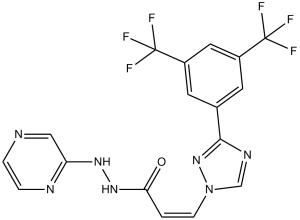
C1=CN=C(C=N1)NNC(=O)/C=C\N2C=NC(=N2)C3=CC(=CC(=C3)C(F)(F)F)C(F)(F)F
|
| InChi Key |
DEVSOMFAQLZNKR-RJRFIUFISA-N
|
| InChi Code |
InChI=1S/C17H11F6N7O/c18-16(19,20)11-5-10(6-12(7-11)17(21,22)23)15-26-9-30(29-15)4-1-14(31)28-27-13-8-24-2-3-25-13/h1-9H,(H,25,27)(H,28,31)/b4-1-
|
| 化学名 |
(Z)-3-(3-(3,5-bis(trifluoromethyl)phenyl)-1H-1,2,4-triazol-1-yl)-N'-(pyrazin-2-yl)acrylohydrazide
|
| 别名 |
KPT-330; KPT 330; 1393477-72-9; Xpovio; Selinexor (KPT-330); KPT 330; (Z)-3-(3-(3,5-Bis(trifluoromethyl)phenyl)-1H-1,2,4-triazol-1-yl)-N'-(pyrazin-2-yl)acrylohydrazide; Selinexor free base; KPT330
|
| HS Tariff Code |
2934.99.9001
|
| 存储方式 |
Powder -20°C 3 years 4°C 2 years In solvent -80°C 6 months -20°C 1 month |
| 运输条件 |
Room temperature (This product is stable at ambient temperature for a few days during ordinary shipping and time spent in Customs)
|
| 溶解度 (体外实验) |
|
|||
|---|---|---|---|---|
| 溶解度 (体内实验) |
配方 1 中的溶解度: ≥ 2.5 mg/mL (5.64 mM) (饱和度未知) in 10% DMSO + 90% Corn Oil (这些助溶剂从左到右依次添加,逐一添加), 澄清溶液。
例如,若需制备1 mL的工作液,可将100 μL 25.0 mg/mL 澄清 DMSO 储备液加入900 μL 玉米油中,混合均匀。 配方 2 中的溶解度: ≥ 2.08 mg/mL (4.69 mM) (饱和度未知) in 10% DMSO + 40% PEG300 + 5% Tween80 + 45% Saline (这些助溶剂从左到右依次添加,逐一添加), 澄清溶液。 例如,若需制备1 mL的工作液,可将 100 μL 20.8 mg/mL澄清的DMSO储备液加入到400 μL PEG300中,混匀;再向上述溶液中加入50 μL Tween-80,混匀;然后加入450 μL生理盐水定容至1 mL。 *生理盐水的制备:将 0.9 g 氯化钠溶解在 100 mL ddH₂O中,得到澄清溶液。 View More
配方 3 中的溶解度: 2.08 mg/mL (4.69 mM) in 10% DMSO + 90% (20% SBE-β-CD in Saline) (这些助溶剂从左到右依次添加,逐一添加), 悬浊液; 超声助溶。 配方 4 中的溶解度: 2% DMSO +49% PEG 300 +dd H2O: 5mg/mL 1、请先配制澄清的储备液(如:用DMSO配置50 或 100 mg/mL母液(储备液)); 2、取适量母液,按从左到右的顺序依次添加助溶剂,澄清后再加入下一助溶剂。以 下列配方为例说明 (注意此配方只用于说明,并不一定代表此产品 的实际溶解配方): 10% DMSO → 40% PEG300 → 5% Tween-80 → 45% ddH2O (或 saline); 假设最终工作液的体积为 1 mL, 浓度为5 mg/mL: 取 100 μL 50 mg/mL 的澄清 DMSO 储备液加到 400 μL PEG300 中,混合均匀/澄清;向上述体系中加入50 μL Tween-80,混合均匀/澄清;然后继续加入450 μL ddH2O (或 saline)定容至 1 mL; 3、溶剂前显示的百分比是指该溶剂在最终溶液/工作液中的体积所占比例; 4、 如产品在配制过程中出现沉淀/析出,可通过加热(≤50℃)或超声的方式助溶; 5、为保证最佳实验结果,工作液请现配现用! 6、如不确定怎么将母液配置成体内动物实验的工作液,请查看说明书或联系我们; 7、 以上所有助溶剂都可在 Invivochem.cn网站购买。 |
| 制备储备液 | 1 mg | 5 mg | 10 mg | |
| 1 mM | 2.2558 mL | 11.2788 mL | 22.5576 mL | |
| 5 mM | 0.4512 mL | 2.2558 mL | 4.5115 mL | |
| 10 mM | 0.2256 mL | 1.1279 mL | 2.2558 mL |
1、根据实验需要选择合适的溶剂配制储备液 (母液):对于大多数产品,InvivoChem推荐用DMSO配置母液 (比如:5、10、20mM或者10、20、50 mg/mL浓度),个别水溶性高的产品可直接溶于水。产品在DMSO 、水或其他溶剂中的具体溶解度详见上”溶解度 (体外)”部分;
2、如果您找不到您想要的溶解度信息,或者很难将产品溶解在溶液中,请联系我们;
3、建议使用下列计算器进行相关计算(摩尔浓度计算器、稀释计算器、分子量计算器、重组计算器等);
4、母液配好之后,将其分装到常规用量,并储存在-20°C或-80°C,尽量减少反复冻融循环。
计算结果:
工作液浓度: mg/mL;
DMSO母液配制方法: mg 药物溶于 μL DMSO溶液(母液浓度 mg/mL)。如该浓度超过该批次药物DMSO溶解度,请首先与我们联系。
体内配方配制方法:取 μL DMSO母液,加入 μL PEG300,混匀澄清后加入μL Tween 80,混匀澄清后加入 μL ddH2O,混匀澄清。
(1) 请确保溶液澄清之后,再加入下一种溶剂 (助溶剂) 。可利用涡旋、超声或水浴加热等方法助溶;
(2) 一定要按顺序加入溶剂 (助溶剂) 。
|
|---|
|
InvivoChem的所有产品仅用于作科学研究,不面向患者销售
Copyright 2020 InvivoChem LLC | All Rights Reserved 粤ICP备20063088号-1


 COA
COA
")
")
 463611831
