| 规格 | 价格 | 库存 | 数量 |
|---|---|---|---|
| 10mg |
|
||
| 25mg |
|
||
| 50mg |
|
||
| 100mg |
|
||
| Other Sizes |
|

| 靶点 |
δ-opioid receptor ( IC50 = 2.73 nM ); μ-opioid receptor ( IC50 = 5457 nM ); δ-opioid receptor) ( Ki = 1.78 nM ); μ-opioid receptor ( IC50 = 881.5 nM ); κ-opioid receptor ( IC50 = 441.8 nM )
|
|---|---|
| 体外研究 (In Vitro) |
SNC80 选择性激活 HEK293 细胞中的 μ-δ 异聚体,EC50 为 52.8 nM。 SNC80 在共表达 μ-和 δ-阿片受体的细胞中表现出比单独表达 δ-阿片受体或共表达 δ-和 κ-阿片受体的细胞更高的活性[4]。
本研究调查了SNC80的药理学,这是一种非肽配体,拟在体外和体内作为选择性δ激动剂。SNC 80能有效抑制小鼠输精管的电诱导收缩,但不能抑制豚鼠离体回肠的收缩(IC50值分别为2.73 nM和5457 nM)。δ选择性拮抗剂ICI 174864(1μM)和μ选择性拮抗剂CTAP(1μm)在小鼠输精管中的SNC 80 IC50值分别增加了236倍和1.9倍。在小鼠全脑试验中,SNC 80优先与[3H]萘屈吲哚标记的位点(δ受体)竞争,而不是与[3H]DAMGO(μ受体)或[3H]U69593κ受体标记的位点竞争。μ/delta和kappa/delta位点的SNC80的Ki值计算值之比分别为495倍和248倍,这表明该化合物在放射性配体结合分析中具有显著的δ选择性。[2] 在单独或共表达阿片受体的异源HEK293细胞中检测SNC80,这些共表达细胞系是通过共免疫沉淀建立功能性异聚体的。先前为确定阿片受体激活而建立的内部钙释放[Ca2+]i测定用于确定SNC80诱导的受体激活的选择性功效。在这项阿片类药物疗效测定中,嵌合Δ6-Gqi4-myr蛋白将阿片受体激活转变为Gq反应(细胞内钙的释放),这是通过荧光测量的。重要的是,与[35S]GTPγS测定中的阿片类药物疗效相比,[Ca2+]i测定中的类药物疗效产生了相似和一致的结果,提供了一种方便的、非放射性的全细胞疗效测量方法。SNC80在HEK293细胞中选择性激活μ-δ异聚体,EC50=52.8±27.8 nM(SEM)(图3),平均峰值ΔRFU效应为746±83(SEM),n=3(12)。表达其他阿片受体的细胞被激活的能力要低得多。在这方面,SNC80在仅表达δ-阿片受体的细胞中的效力至少比共表达μ-δ异聚体的细胞低100倍。[4] 本研究中来自完整HEK-293细胞的体外疗效数据支持了SNC80的主要靶点是异聚体μ-δ受体的概念。如图3和图S1“支持信息”所示,SNC80在共表达μ-和δ-阿片受体的细胞中的活性明显高于单独表达δ-阿片受体或共表达δ-和κ-阿片感受器的细胞。鉴于这些受体之间存在物理关联的证据,这意味着μ-δ异聚体被SNC80选择性激活,特别是因为单表达的δ受体不会产生强效激活。这些发现,再加上报告的SNC80对μ-受体的低结合亲和力,4表明它靶向μ-δ异聚体的δ-原聚体,从而导致复合物的激活[4]。 |
| 体内研究 (In Vivo) |
SNC80(10 mg/kg;腹腔注射;一次;C57BL6/J 小鼠)治疗显着减轻了过度使用舒马曲坦引起的异常疼痛[1]。动物模型:雄性和雌性C57BL6/J小鼠(20-30g)注射舒马普坦[1] 剂量:10mg/kg 给药方式:腹腔注射;一次结果:异常性疼痛显着减弱。
头痛具有高度致残性,是全球最常见的神经系统疾病之一。尽管头痛的患病率很高,但治疗选择有限。我们最近发现δ阿片受体(DOR)是偏头痛的一个新兴治疗靶点。在这项研究中,我们检查了标志性DOR激动剂SNC80在反映各种头痛疾病的疾病模型中的有效性,包括:慢性偏头痛、创伤后头痛(PTH)、曲坦类药物过度使用头痛(MOH)和阿片类药物诱导的痛觉过敏(OIH)。为了模拟慢性偏头痛,C57BL/6J小鼠接受了已知的人类偏头痛触发器硝酸甘油的慢性间歇性治疗。通过将闭式头部重量下降模型与慢性偏头痛的硝酸甘油模型相结合来模拟PTH。对于MOH和OIH,小鼠分别长期接受舒马曲坦或吗啡治疗。在所有四种模型中都观察到眶周和外周异常性疼痛的发展;SNC80在所有病例中均显著抑制了异常性疼痛。此外,我们还确定了SNC80的慢性日常治疗是否会诱导MOH/OIH,并且我们观察到相对于舒马曲坦或吗啡的有限痛觉过敏。总之,我们的结果表明,尽管病因不同,但DOR激动剂可能对多种头痛疾病有效,从而为头痛提供了一种新的治疗靶点。[1] SNC80在小鼠温水甩尾试验中,静脉注射、静脉注射和腹腔注射后产生了剂量和时间相关的镇痛作用。静脉注射、静脉注射和腹腔注射SNC 80的A50值(和95%置信区间)分别为104.9(63.7-172.7)nmol、69(51.8-92.1)nmol和57(44.5-73.1)mg/kg。在热板试验中,静脉注射SNC 80也产生了与剂量和时间相关的镇痛作用,计算出的A50值(和95%c.i.)为91.9(60.3-140.0)nmol。2. 在小鼠体内测定SNC80的镇痛活性,以研究其体内受体靶标的身份。在这方面,我们假设,由于无法形成μ-δ异聚体,SNC80在缺乏其中一种受体的敲除动物中的活性会降低。采用野生型、μ-KO和δ-KO小鼠来确定μ-和δ-阿片受体对SNC80诱导的镇痛作用的贡献。SNC80通过鞘内注射途径(i.t.)给药于小鼠,并在温水(52.2°C)尾部撤回试验中进行评估(图2),重点关注脊髓,其中体内数据支持受体共定位。选择累积给药方案部分是为了解决SNC-80的溶解度有限的问题,因为三个最高剂量总计300 nmol大大超过了单个5μL鞘内注射体积中可以溶解的化合物量。此外,与之前的非累积给药实验的多次比较显示,结果没有显著差异,验证了该方法。该图(图2)显示了两条不同的剂量-反应曲线,以说明μ-KO小鼠在低剂量下SNC80缺乏效果。在野生型(C57/129)小鼠中,SNC80的ED50=49 nmol,95%置信区间(43-56)。μ-KO小鼠的剂量-反应曲线(根据第二条较高剂量曲线计算)以平行方式右移2.7倍(ED50=131 nmol,95%CI(111-153),图2A)。在野生型(C57BL/6)小鼠中,SNC80显示ED50=53.6 nmol,95%CI(47.0-61.1),在δ-KO小鼠中右移6.1倍,ED50=327 nmol,95%CI(216-494)(约50%MPE,图2B)。结果表明,在μ或δ阿片受体敲除的小鼠中,SNC80的镇痛活性降低。[4] 我们的体内数据还表明,SNC80诱导的镇痛作用是通过脊髓中μ-δ异聚体的选择性激活产生的。μ-KO和δ-KO小鼠SNC80剂量-反应曲线的右移表明,异聚体复合物中的μ-和δ-阿片受体原聚体都有助于野生型小鼠SNC80的镇痛活性。这一结果与先前的研究一致,表明SNC80诱导的镇痛作用同时具有δ和μ阿片受体介导的成分。δ和μ敲除动物中SNC80的效力降低证明了这两种受体的体内贡献,这也是IUPHAR指南建立异聚体复合物的三个具体标准之一。[4] 在Gαo-RGS不敏感的杂合敲除小鼠中,SNC80产生抗痛觉过敏和抗抑郁样作用的效力增强,但SNC80诱导的惊厥没有变化。相反,在Gαo杂合敲除小鼠中,SNC80诱导的抗痛觉过敏被消除,而抗抑郁样作用和抽搐没有改变。在arrestin 3敲除小鼠中没有观察到SNC80诱导的行为变化。在arrestin 2基因敲除小鼠中,SNC80诱导的抽搐增强。 结论和意义:总的来说,这些发现表明,不同的信号分子可能是δ受体相对于其抗痛觉过敏和抗抑郁样作用的惊厥作用的基础[6]。 在本研究中,Gαo-RGSi和Gαo敲除小鼠的δ受体介导的抽搐没有改变。此外,我们之前观察到,在RGS4敲除小鼠中,SNC80诱导的抽搐没有改变(Dripps等人,2017)。总体而言,这些数据可能表明,介导δ受体激动剂诱导的惊厥的信号机制不同于介导抗痛觉过敏和抗抑郁样作用的信号机制。这些行为指标可能受到特定G蛋白亚基、G蛋白非依赖性信号和/或特定脑回路或区域内信号分子选择性表达的不同调节。 为了解决这个问题,我们探讨了SNC80诱导的惊厥是由G蛋白非依赖性、arrestin介导的机制产生的假设。正如Bohn等人(1999)首次表明的那样,我们观察到arrestin 3敲除小鼠吗啡诱导的镇痛作用增强。尽管A类GPCR被认为优先与arrest蛋白3相互作用(Oakley等人,2000),但在arrest蛋白-3敲除小鼠中没有观察到δ受体介导的行为(包括抽搐)的显著变化。应该指出的是,这些数据是急性服用SNC80的结果,arrestin 3可能在调节重复剂量SNC80或其他δ受体激动剂的作用中发挥作用。SNC80的这一观察结果与之前的报告一致,之前的报告发现,小鼠中arrestin 3的缺失不会改变δ受体激动剂的镇痛作用,也不会影响在慢性炎症性疼痛的完全弗氏佐剂(CFA)模型中观察到的δ受体与电压依赖性钙通道的增强偶联(Pradhan等人,2013;Pradhan等,2016)。总体而言,我们的研究结果表明,δ受体介导的抗痛觉过敏、抗抑郁药样作用或抽搐不需要arrestin 3。 在arrestin 2敲除小鼠中,我们观察到SNC80对NTG诱导的热痛觉过敏的反应没有变化。然而,之前的研究表明,SNC80对CFA诱导的机械性痛觉过敏的影响在arrestin 2敲除小鼠中得到了增强(Pradhan等人,2016)。δ受体介导的对这些不同疼痛模式(CFA与NTG;机械与热)的反应可能受到arrestin 2的不同调节。进一步的研究应调查介导不同类型δ受体介导的抗痛觉过敏的信号分子和途径的差异。SNC80的惊厥作用在arrestin 2敲除小鼠中显著增强。在arrestin 2敲除小鼠中,SNC80诱导惊厥的效力增强,表明arrestin 2中起着δ受体介导的惊厥的负调节作用。其次,arrestin 2基因敲除小鼠对单剂量SNC80有多次抽搐反应。 对δ受体介导的抽搐的耐受性通常是急性和长期的(Comer等人,1993;Hong等人,1998)。此外,癫痫发作结束后,SNC80产生的脑电图波形变化恢复到正常的基线活动(Jutkiewicz等人,2006)。据我们所知,这是首次报道啮齿动物对δ受体激动剂产生多种抽搐事件。对这一观察结果的一种可能解释是,arrestin 2的缺失通过阻止δ受体脱敏和/或上调δ受体向细胞膜的运输来产生这些行为变化,从而增强δ受体信号传导(Mittal等人,2013)。然而,在目前的研究中,δ受体介导的抗抑郁样作用和热痛觉过敏在arrestin 2敲除小鼠中没有显著改变。因此,由于区域表达、行为机制和/或信号下调和/或对SNC80惊厥作用的耐受性存在差异,SNC80的行为效应可能受到arrestin 2的不同调节。因此,arrestin 2的缺失可能会使通常会终止的信号通路持续存在并产生多个抽搐事件。未来的工作将研究arrestin 2是否也调节对δ受体激动剂其他行为影响的耐受性。 |
| 酶活实验 |
δ受体饱和结合[6]
在颈椎脱位后将小鼠斩首,立即移除前脑,并如前所述新鲜制备膜(Broom等人,2002a)。未麻醉的组织收集用于限制δ受体数量、构象和/或定位的改变,并且根据美国兽医协会动物安乐死指南,有条件地可接受。用BCA测定试剂盒测定蛋白质浓度。δ受体激动剂[3H]DPDPE的特异性结合如Broom等人(2002a)所述,使用10μM阿片类拮抗剂纳洛酮来确定非特异性结合。反应在26°C下孵育60分钟,并使用MLR‐24收割机通过浸泡在0.1%PEI中的GF/C过滤垫快速过滤停止。通过闪烁计数确定结合[3H]DPDPE,并使用GraphPad Prism 6.02版的非线性回归分析计算B max和K d值。为确保单个值的可靠性,对每只小鼠(每组n=5)的膜进行了三次检测。 |
| 细胞实验 |
细胞内钙释放测定[4]
如前所述,细胞内钙释放测定用于确定SNC80在激活阿片受体时的选择性,但需要稍作修改。简而言之,将稳定表达阿片受体的HEK293细胞在10%CO2气氛中的DMEM(10%FBS,1%P/S)中生长,使用脂质体2000用嵌合G蛋白Δ6-Gqi4-myr瞬时转染,24小时后接种到96孔半面积板中,并在转染后48小时检测细胞内钙释放。在涉及稳定表达嵌合Δ6-Gqi4-myr蛋白的HEK293细胞的实验中,除了阿片受体DNA被瞬时转染,并且共转染细胞中脂质体的量增加了一倍外,程序是相同的。检测采用Flexstation 3装置中的标准探针FLIPR钙染料试剂盒。SNC80游离碱最初溶解在DMSO中。使用DMSO,使最终DMSO浓度最大不超过0.1%v/v,该浓度不会显著改变基础反应的钙通量。为了尽量减少实验的可变性,所有实验都进行了至少三次,四次内部重复,n≥3(12),但表达κ-或μ-κ受体的细胞除外,其中n≥2(8)。对每个平板进行检测控制(空白和标准配体),以消除技术差异并确保反应均匀。 |
| 动物实验 |
雄性和雌性C57BL6/J小鼠(20-30g)腹腔注射舒马曲坦
10 mg/kg 一次 急性给予DOR激动剂[1] 末次给药后18至24小时,我们测定了SNC80的作用。在激发试验当天,测定小鼠基础后爪和头部机械阈值,之后小鼠分别接受赋形剂(VEH)或SNC80(10 mg/kg,腹腔注射)。SNC80用0.33% 1N HCl/0.9%生理盐水稀释至1 mg/mL。在基础测试后 2 小时和 SNC80 注射后 45 分钟评估 SNC80 后的阈值。 DOR 激动剂的慢性治疗[1] 为了确定慢性 DOR 激活是否会引起类似于 MOH 的过敏反应,小鼠每天接受一次载体、SUMA(0.6 mg/kg,腹腔注射)或SNC80(10 mg/kg,腹腔注射)治疗,持续 11 天。在后爪实验中,小鼠分别于第1、3、5、7、9和11天进行测试,另一组小鼠则于第1和11天进行测试。在头部实验中,根据实验的不同,小鼠分别于第1和11天以及第1、5和11天进行测试。 除SNC80外,所有药物储备液均溶于无菌水中,并用无菌生理盐水稀释。SNC80在溶于无菌水之前,先与1当量酒石酸混合。采用温水(52.5℃)尾浸试验评估SNC80的镇痛作用。本研究使用的动物包括129sv/C57BL6小鼠(μ-阿片受体野生型)、129sv/C57BL6背景的μ-KO(−/−)小鼠、C57BL/6小鼠(δ-阿片受体野生型)以及C57BL/6背景的δ-KO(−/−)小鼠,所有小鼠均可自由摄食,并保持12小时光照/12小时黑暗的昼夜节律。鞘内(it)给药采用改良的腰椎穿刺法。每个剂量组至少测试6只小鼠,每只小鼠重复测试两次,本研究共使用了48只小鼠。在所有实验中,雄性和雌性小鼠的镇痛效果均无差异。在给药前记录甩尾(TF)潜伏期,以建立药物治疗前的基线值;基线测试后不久,将最低剂量的药物溶于5 μL溶剂中,鞘内注射,并在7分钟后再次记录TF潜伏期。测试结束后,立即给予累积剂量-反应曲线的后续剂量,并在5分钟后测定TF潜伏期;由此快速连续给予三至四剂,并确定累积剂量-反应曲线。拮抗剂在首次注射激动剂前7分钟给予。若未检测到反应,则采用12秒截止时间以避免组织损伤。[4] 强迫游泳试验[6] 强迫游泳试验(FST)是一种广泛用于评估药物在啮齿动物中抗抑郁样作用的试验(Barkus,2013)。我们的实验改编自Porsolt等人(1977)的研究,并按照先前描述的方法进行(Dripps等人,2017)。简而言之,在注射SNC80(0.1、0.32、1、3.2、10 或 32 mg·kg−1)或载体后 60 分钟,将每只小鼠放入装有 15 cm 25 ± 1°C 水的 4 L 烧杯中,并使用 Sony HDR-CX220 数码摄像机记录其 6 分钟的行为。由对实验条件不知情的人员分析视频,并量化动物静止不动的时间。静止不动定义为小鼠不在水中主动移动,仅做出保持漂浮所需的动作。记录小鼠在实验开始后 30 秒的静止不动时间。 硝酸甘油诱导的痛觉过敏 [6] 硝酸甘油诱导的痛觉过敏实验参考了 Bates 等人 (2010) 的方法,并根据 Pradhan 等人的描述进行了修改。 (2014)并按照先前描述的方法进行(Dripps 等,2017)。简而言之,本研究使用雄性和雌性小鼠评估硝酸甘油(NTG)诱导的痛觉过敏。通过将小鼠尾部(距尾尖约 5 cm)浸入 46°C 水浴中,并测定小鼠缩尾潜伏期(截止时间为 60 秒)来评估痛觉过敏。在确定基线缩尾潜伏期后,每只小鼠腹腔注射 10 mg·kg⁻¹ NTG。NTG 给药 1 小时后再次评估缩尾潜伏期。NTG 给药 90 分钟后,小鼠接受 SNC80(0.32、1、3.2、10 或 32 mg·kg⁻¹)或载体注射,并在单独的笼子中连续观察小鼠 30 分钟,以观察是否有惊厥(见下文)。在给予SNC80 30分钟后再次评估尾部缩回潜伏期。 SNC80诱导的惊厥[6] 将小鼠单独饲养在笼中,持续观察其惊厥情况。除非另有说明,NTG处理对SNC80诱导的惊厥的频率或性质没有显著影响(参见补充信息)。惊厥通常由一个强直期组成,其特征是肌肉突然紧张和前爪伸展,随后是沿身体长度方向的阵挛性收缩。小鼠经常失去平衡并侧翻,但很少观察到所谓的桶滚。惊厥之后是持续2-5分钟的僵直期,之后动物活动过度,但除此之外与未处理的对照组无区别。每次抽搐的严重程度均采用改良的Racine(1972)量表进行量化,该量表改编自Jutkiewicz等人(2006):1 – 牙齿打颤或面部抽搐;2 – 头部点头或抽搐;3 – 持续时间小于3秒的强直性伸展或阵挛性抽搐;4 – 持续时间大于3秒的强直性伸展或阵挛性抽搐;5 – 持续时间大于3秒且伴有平衡丧失的强直性伸展或阵挛性抽搐。抽搐后僵直样行为的评估方法为:将一根水平杆置于小鼠前爪下方,若小鼠在30秒后仍未移开前爪,则判定为僵直阳性。两只接受 32 mg·kg−1 SNC80 治疗的阻遏蛋白 2 基因敲除小鼠在观察期结束后出现持续性惊厥,并被戊巴比妥过量注射处死。 SNC80 溶于 1 M HCl,并用无菌水稀释至 3% HCl 浓度。 |
| 参考文献 | |
| 其他信息 |
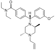
4-[(R)-[(2S,5R)-2,5-二甲基-4-丙-2-烯基-1-哌嗪基]-(3-甲氧基苯基)甲基]-N,N-二乙基苯甲酰胺是一种二芳基甲烷化合物。
已证实,在培养细胞和体内,μ-和δ-阿片受体以异源二聚体的形式共表达和共定位。然而,阿片受体异源二聚体激活的生物学意义尚不明确。为了探究其意义,本研究采用嵌合G蛋白介导的钙荧光测定法,在体外评估了SNC80选择性激活阿片受体的效力。分别在单表达和共表达阿片受体的细胞中进行了检测。结果表明,SNC80在表达μ-δ异源二聚体的细胞中产生的反应明显强于其他所有细胞系。在μ-和δ-阿片受体敲除小鼠中鞘内注射SNC80后,其镇痛活性较野生型小鼠显著降低。体内和体外实验结果共同表明,SNC80选择性激活μ-δ异源二聚体以产生最大镇痛作用。这些数据与目前认为SNC80选择性激活δ-阿片受体同源二聚体以产生镇痛作用的观点相悖。因此,数据提示,当SNC80作为体内药理学工具时,应将μ-δ异源二聚体受体作为靶点。[4]近年来,δ阿片受体作为治疗慢性疼痛和情绪障碍的靶点,引起了越来越多的关注。由于其治疗潜力,人们开发了多种工具从分子和功能角度研究δ阿片受体。本文综述了最常用的工具,并重点介绍了它们的用途和局限性。本文将介绍以下内容:(1) 用于研究δ阿片受体的细胞分析方法;(2) 几种δ阿片受体配体的特性,包括肽类和非肽类药物;(3) 检测固定组织中δ阿片受体的现有方法,以及围绕这些技术的争论;(4) 用于研究δ阿片受体激动剂体内效应的行为学分析方法,包括某些配体可诱发的运动兴奋和惊厥,而其他配体则不诱发;(5) 专门用于研究δ阿片受体的基因修饰小鼠的特性。总而言之,本综述旨在为这些工具的使用提供指导,最终目标是增进我们对δ阿片受体生理学的理解。[5] 背景和目的:G蛋白偶联受体(GPCR)存在多种构象,可以激活不同的信号通路,进而导致不同的行为输出。在啮齿动物模型中,δ阿片受体(δ受体)的激活已被证实可引起抗痛觉过敏、抗抑郁样作用和惊厥。我们最近的研究表明,这些δ受体介导的行为受到GTP酶激活蛋白G蛋白信号调节因子4(RGS4)的差异性调控,RGS4可促进G蛋白信号的终止。为了进一步评估δ受体介导的抗痛觉过敏、抗抑郁样作用和惊厥的信号机制,我们观察了体内Gαo或阻遏蛋白的变化如何影响δ受体激动剂SNC80在小鼠中诱发的行为。实验方法:本研究使用了多种信号分子表达发生改变的转基因小鼠。采用硝酸甘油诱导的热痛觉过敏实验评估抗痛觉过敏作用。采用强迫游泳实验评估抗抑郁样作用。我们还观察了小鼠在接受SNC80治疗后的惊厥活动。[6] 我们的研究目标之一是确定长期使用DOR激动剂是否会导致阿片类药物诱导的痛觉过敏/药物过度使用性痛觉过敏(OIH/MOH)。有趣的是,我们发现每日使用SNC80进行治疗和测试会导致后续的痛觉过敏,但与舒马曲坦不同,单独每日使用SNC80并不会导致疼痛敏感性增加。这些结果表明,DOR的药理学激活不会导致OIH/MOH,然而,如果长期使用SNC80并进行重复测试,则DOR激活可能促进联想学习,从而导致行为敏感化。DOR在多个脑区表达,这些脑区可以调节不同类型的学习,包括海马、杏仁核和纹状体(Le Merrer等人,2009;Pellissier等人,2016;Pradhan等人,2011)。敲除DOR会导致物体识别任务受损(Le Merrer等人,2013),以及位置条件反射任务缺陷(Le Merrer等人,2011)。此外,伏隔核壳部的DOR已被证明可以调节预测学习(Bertran-Gonzalez等人,2013;Laurent等人,2014;Laurent等人,2015a;Laurent等人,2015b)。我们此前已证实,对SNC80的耐受性显著依赖于联想学习(Pradhan等人,2010;Vicente-Sanchez等人,2018),并且与记忆和学习相关的环境线索可以调节重复接触阿片类药物后的行为结果(Gamble等人,1989;Mitchell等人,2000)。在开发用于治疗头痛的DOR激动剂时,应考虑我们的研究结果,因为慢性DOR激活可能促进偏头痛患者的相关学习行为。[1]此外,我们的体外功效数据表明,SNC80选择性激活μ-δ异源二聚体,这一结果也得到了转运和结合研究的进一步支持。这些转运研究揭示了SNC80诱导的μ-和δ-阿片受体的共内化,这与μ-δ异源二聚体作为信号单元的激活相一致。本研究表明,SNC80的强效活性需要μ-和δ-阿片受体在体外和体内均存在。综合考虑并结合先前的研究,本文提出的结果表明,SNC80在体内选择性地与μ-δ异源二聚体的δ亚基相互作用,从而激活该异源二聚体复合物。虽然这一结论并不否定先前研究中SNC80在受体同源二聚体模型中是δ选择性配体的观点,但它强烈提示μ-δ异源二聚体是该信号通路中的一个选择性靶点。 μ-δ受体异源二聚体作为SNC80的靶点,其意义在于提示该配体在体内发挥着更为复杂的作用。这是因为μ选择性配体(基于结合)也靶向μ-δ异源二聚体,但其作用靶点是μ亚基。因此,激动剂的活性似乎取决于μ-δ异源二聚体中哪个亚基被激活。与μ激动剂不同,SNC80不产生物理依赖性,这一事实表明异源二聚体系统的复杂性,并提示不同的信号通路依赖于μ-δ异源二聚体复合物中被激活的亚基。[4] |
| 分子式 |
C28H39N3O2
|
|---|---|
| 分子量 |
449.64
|
| 精确质量 |
449.304
|
| 元素分析 |
C, 74.80; H, 8.74; N, 9.35; O, 7.12
|
| CAS号 |
156727-74-1
|
| PubChem CID |
123924
|
| 外观&性状 |
Off-white to light yellow solid powder
|
| 密度 |
1.0±0.1 g/cm3
|
| 沸点 |
564.8±50.0 °C at 760 mmHg
|
| 熔点 |
122-123ºC
|
| 闪点 |
295.4±30.1 °C
|
| 蒸汽压 |
0.0±1.5 mmHg at 25°C
|
| 折射率 |
1.545
|
| LogP |
3.37
|
| tPSA |
36.02
|
| 氢键供体(HBD)数目 |
0
|
| 氢键受体(HBA)数目 |
4
|
| 可旋转键数目(RBC) |
9
|
| 重原子数目 |
33
|
| 分子复杂度/Complexity |
614
|
| 定义原子立体中心数目 |
3
|
| SMILES |
C=CCN1C[C@H](C)N(C[C@H]1C)[C@H](C2=CC=C(C=C2)C(=O)N(CC)CC)C3=CC(=CC=C3)OC
|
| InChi Key |
KQWVAUSXZDRQPZ-UMTXDNHDSA-N
|
| InChi Code |
InChI=1S/C28H39N3O2/c1-7-17-30-19-22(5)31(20-21(30)4)27(25-11-10-12-26(18-25)33-6)23-13-15-24(16-14-23)28(32)29(8-2)9-3/h7,10-16,18,21-22,27H,1,8-9,17,19-20H2,2-6H3/t21-,22+,27-/m1/s1
|
| 化学名 |
4-[(R)-[(2S,5R)-2,5-dimethyl-4-prop-2-enylpiperazin-1-yl]-(3-methoxyphenyl)methyl]-N,N-diethylbenzamide
|
| 别名 |
SNC80; SNC-80; 156727-74-1; 4-(alpha-(4-allyl-2,5-dimethyl-1-piperazinyl)-3-methoxybenzyl)-N,N-diethylbenzamide; 4-[(R)-[(2S,5R)-2,5-dimethyl-4-prop-2-enylpiperazin-1-yl]-(3-methoxyphenyl)methyl]-N,N-diethylbenzamide; Benzamide, 4-[(R)-[(2S,5R)-2,5-dimethyl-4-(2-propen-1-yl)-1-piperazinyl](3-methoxyphenyl)methyl]-N,N-diethyl-; CHEMBL13470; SNC80
|
| HS Tariff Code |
2934.99.9001
|
| 存储方式 |
Powder -20°C 3 years 4°C 2 years In solvent -80°C 6 months -20°C 1 month |
| 运输条件 |
Room temperature (This product is stable at ambient temperature for a few days during ordinary shipping and time spent in Customs)
|
| 溶解度 (体外实验) |
DMSO: ~33.33 mg/mL (~74.1 mM)
|
|---|---|
| 溶解度 (体内实验) |
配方 1 中的溶解度: ≥ 3.33 mg/mL (7.41 mM) (饱和度未知) in 10% DMSO + 40% PEG300 + 5% Tween80 + 45% Saline (这些助溶剂从左到右依次添加,逐一添加), 澄清溶液。
例如,若需制备1 mL的工作液,可将100 μL 33.3 mg/mL澄清DMSO储备液加入400 μL PEG300中,混匀;然后向上述溶液中加入50 μL Tween-80,混匀;加入450 μL生理盐水定容至1 mL。 *生理盐水的制备:将 0.9 g 氯化钠溶解在 100 mL ddH₂O中,得到澄清溶液。 配方 2 中的溶解度: ≥ 3.33 mg/mL (7.41 mM) (饱和度未知) in 10% DMSO + 90% (20% SBE-β-CD in Saline) (这些助溶剂从左到右依次添加,逐一添加), 澄清溶液。 例如,若需制备1 mL的工作液,可将 100 μL 33.3 mg/mL 澄清 DMSO 储备液加入 900 μL 20% SBE-β-CD 生理盐水溶液中,混匀。 *20% SBE-β-CD 生理盐水溶液的制备(4°C,1 周):将 2 g SBE-β-CD 溶解于 10 mL 生理盐水中,得到澄清溶液。 View More
配方 3 中的溶解度: ≥ 3.33 mg/mL (7.41 mM) (饱和度未知) in 10% DMSO + 90% Corn Oil (这些助溶剂从左到右依次添加,逐一添加), 澄清溶液。 1、请先配制澄清的储备液(如:用DMSO配置50 或 100 mg/mL母液(储备液)); 2、取适量母液,按从左到右的顺序依次添加助溶剂,澄清后再加入下一助溶剂。以 下列配方为例说明 (注意此配方只用于说明,并不一定代表此产品 的实际溶解配方): 10% DMSO → 40% PEG300 → 5% Tween-80 → 45% ddH2O (或 saline); 假设最终工作液的体积为 1 mL, 浓度为5 mg/mL: 取 100 μL 50 mg/mL 的澄清 DMSO 储备液加到 400 μL PEG300 中,混合均匀/澄清;向上述体系中加入50 μL Tween-80,混合均匀/澄清;然后继续加入450 μL ddH2O (或 saline)定容至 1 mL; 3、溶剂前显示的百分比是指该溶剂在最终溶液/工作液中的体积所占比例; 4、 如产品在配制过程中出现沉淀/析出,可通过加热(≤50℃)或超声的方式助溶; 5、为保证最佳实验结果,工作液请现配现用! 6、如不确定怎么将母液配置成体内动物实验的工作液,请查看说明书或联系我们; 7、 以上所有助溶剂都可在 Invivochem.cn网站购买。 |
| 制备储备液 | 1 mg | 5 mg | 10 mg | |
| 1 mM | 2.2240 mL | 11.1200 mL | 22.2400 mL | |
| 5 mM | 0.4448 mL | 2.2240 mL | 4.4480 mL | |
| 10 mM | 0.2224 mL | 1.1120 mL | 2.2240 mL |
1、根据实验需要选择合适的溶剂配制储备液 (母液):对于大多数产品,InvivoChem推荐用DMSO配置母液 (比如:5、10、20mM或者10、20、50 mg/mL浓度),个别水溶性高的产品可直接溶于水。产品在DMSO 、水或其他溶剂中的具体溶解度详见上”溶解度 (体外)”部分;
2、如果您找不到您想要的溶解度信息,或者很难将产品溶解在溶液中,请联系我们;
3、建议使用下列计算器进行相关计算(摩尔浓度计算器、稀释计算器、分子量计算器、重组计算器等);
4、母液配好之后,将其分装到常规用量,并储存在-20°C或-80°C,尽量减少反复冻融循环。
计算结果:
工作液浓度: mg/mL;
DMSO母液配制方法: mg 药物溶于 μL DMSO溶液(母液浓度 mg/mL)。如该浓度超过该批次药物DMSO溶解度,请首先与我们联系。
体内配方配制方法:取 μL DMSO母液,加入 μL PEG300,混匀澄清后加入μL Tween 80,混匀澄清后加入 μL ddH2O,混匀澄清。
(1) 请确保溶液澄清之后,再加入下一种溶剂 (助溶剂) 。可利用涡旋、超声或水浴加热等方法助溶;
(2) 一定要按顺序加入溶剂 (助溶剂) 。
|
|
|
|
|
InvivoChem的所有产品仅用于作科学研究,不面向患者销售
Copyright 2020 InvivoChem LLC | All Rights Reserved 粤ICP备20063088号-1


 463611831
