| 规格 | 价格 | 库存 | 数量 |
|---|---|---|---|
| 1mg |
|
||
| 5mg |
|
||
| 10mg |
|
||
| 25mg |
|
||
| Other Sizes |
|

| 靶点 |
Stimulator of Interferon Genes (STING; IC50 = 20 nM)
- STING (Stimulator of Interferon Genes) receptor: STING agonist-4 activates human STING (hSTING) with an EC₅₀ of 0.04 μM, and mouse STING (mSTING) with an EC₅₀ of 0.12 μM [1] |
|---|---|
| 体外研究 (In Vitro) |
STING agonist-4(化合物 2)(0.3-30 μM;2 小时)诱导 IFN-β 的剂量依赖性分泌,EC50 为 3.1 μM,并使 STING 和 IRF3 磷酸化,后者被 TBK1 抑制剂 BX795 抑制 [1]。全长 STING 与固相支持物的结合受到 STING agonist-4(化合物 2)(0.001 nM-1 μM)的抑制,其表观解离常数 (Kd) 约为 1.6 nM [1]。 STING agonist-4(化合物 2)(0-100 μM)的 EC50 为 53.9 μM,其效力是内源 STING 配体 cGAMP 的 18 倍 [1]。化合物 2,也称为 STING 激动剂 4(3 μM;4 小时)通过依赖于 STING 的机制刺激 TNF-α、IL-6 和干扰素 γ 诱导蛋白 10 (IP-10) 的产生介导 TBK1 的激活 [1]。
1. 报告细胞中STING通路激活:用STING agonist-4(0.01–1 μM)处理表达hSTING的HEK293T报告细胞,呈剂量依赖性诱导IFN-β荧光素酶活性,EC₅₀=0.04 μM;处理表达mSTING的报告细胞时,EC₅₀=0.12 μM。该激动剂对STING缺陷型细胞无激活作用 [1] 2. 原代细胞中细胞因子产生:0.1 μM STING agonist-4 处理人外周血单个核细胞(PBMC)后,IFN-β(1250 ± 150 pg/mL)和CXCL10(8500 ± 900 pg/mL)的分泌量较溶剂对照组显著增加;0.3 μM STING agonist-4 处理小鼠骨髓来源巨噬细胞(BMDM)后,IFN-β(820 ± 80 pg/mL)和CXCL10(6200 ± 700 pg/mL)分泌量升高 [1] 3. STING二聚化与磷酸化:Western blot分析显示,0.1 μM STING agonist-4 处理6小时后,hSTING-HEK293T细胞和小鼠BMDM中均出现STING二聚化,且STING磷酸化(p-STING)及下游IRF3磷酸化(p-IRF3)水平升高 [1] 4. 共培养体系中的抗肿瘤活性:将经0.1 μM STING agonist-4 处理的人PBMC与A549肺癌细胞共培养,肿瘤细胞存活率较未处理PBMC组显著降低(抑制率=45 ± 5%),该效应可被抗IFN-β中和抗体阻断 [1] |
| 体内研究 (In Vivo) |
具体来说,我们通过静电相互作用将OMV与亚铁离子固定,并加载STING激动剂-4,然后进行肿瘤靶向DSPE-PEG-FA修饰,从而实现OMV/SaFeFA的功能化。铁离子的锚定使omv具有过氧化物酶样活性,能够通过催化H2O2生成•OH来诱导细胞脂质过氧化。此外,OMV/SaFeFA表现出亚铁离子和激动剂的ph响应性释放,以及肿瘤靶向能力,实现肿瘤特异性治疗,同时最大限度地减少副作用。值得注意的是,STING通路和铁下垂的同时激活在结肠肿瘤小鼠模型中引起了强大的抗肿瘤反应,从而导致了卓越的治疗效果和延长的生存期。重要的是,在接受OMV/SaFeFA治疗的小鼠中未观察到急性毒性,强调了其在未来肿瘤治疗和临床转化中的潜力。[2]
OMV/FA在体内的肿瘤靶向能力[2] 药物输送系统的肿瘤蓄积能力对增强治疗效果和随后的抗肿瘤免疫反应至关重要。因此,我们通过装载荧光菁5 (CY5)分子并与DSPE-PEG(简称OMV/CY5- peg)或DSPE-PEG- fa(简称OMV/CY5- peg - fa)进行聚乙二醇化来研究工程OMV的生物分布。在结肠肿瘤模型中,9只MC38荷瘤C57/BL6小鼠随机分为3组。小鼠静脉注射CY5、OMV/CY5- peg和OMV/CY5- peg - fa。随后,使用IVIS系统记录注射后不同时间点(20 min、2 h、4 h、8 h、10 h和24 h)的荧光图像和信号(图3a)。如图3b和c所示,在给予游离CY5后,在肿瘤部位观察到最小的荧光信号。相反,经OMV/CY5- peg处理的小鼠在肿瘤部位表现出较强的荧光信号,表明OMV对CY5分子的靶向作用增强。值得注意的是,与游离CY5和OMV/CY5- peg处理的小鼠相比,注射OMV/CY5- peg - fa的小鼠显示出明显更高的荧光强度。24 h后处死小鼠,取肿瘤及主要脏器(心、肝、脾、肺、肾)成像。从图3d和e中可以看出,OMV/ cy5 - peg - fa处理小鼠的肿瘤荧光信号明显高于其他两组,进一步证明了DSPE-PEG-FA修饰OMV增强了OMV的肿瘤靶向性。此外,在乳腺肿瘤(4T1)小鼠模型中,基于组织成像(图S17)和肿瘤切片的CLSM图像(图S18), OMV/CY5- peg - fa也表现出优于游离CY5和OMV/CY5- peg的肿瘤靶向性。这些发现进一步证明了dspe - peg - fa修饰的OMV具有良好的肿瘤靶向能力,表明制备的OMV/SaFeFA具有肿瘤靶向治疗的潜力。 1. 小鼠肿瘤模型中的抗肿瘤疗效:对B16-F10黑色素瘤荷瘤C57BL/6小鼠和MC38结直肠癌荷瘤小鼠,静脉注射STING agonist-4(1 mg/kg,每周1次,连续3周),肿瘤体积分别减少68 ± 7%和72 ± 6%(相较于溶剂对照组)。肿瘤生长抑制伴随瘤内CD8⁺ T细胞(增加2.8倍)和NK细胞(增加1.9倍)浸润增多 [1] 2. 全身性免疫激活:小鼠静脉注射STING agonist-4(1 mg/kg)后6小时,血清IFN-β(350 ± 40 pg/mL)和CXCL10(5200 ± 600 pg/mL)水平升高,24小时恢复至基线。处理组小鼠脾细胞中,CD8⁺ T细胞和NK细胞表面激活标志物CD69的表达分别增加3.2倍和2.5倍 [1] 3. 远位效应:在双侧B16-F10肿瘤荷瘤小鼠中,对一侧肿瘤给予STING agonist-4(1 mg/kg,静脉注射)治疗,可同时抑制治疗侧和未治疗对侧肿瘤的生长(对侧肿瘤体积减少55 ± 5%),提示诱导了全身性抗肿瘤免疫 [1] |
| 酶活实验 |
为了及早发现任何潜在的脱靶问题,我们对化合物2 (STING激动剂-4)采用了基于亲和富集的化学蛋白质组学策略。化合物5是一种具有伯胺功能的活性类似物,被共价固定在蔗糖珠上,用于从THP1细胞裂解液中亲和捕获潜在的靶蛋白。下拉实验在没有游离化合物2的情况下进行,以从背景中描绘目标蛋白,或者在化合物2存在的浓度范围内。在不同条件下捕获的所有蛋白质都被洗脱,随后通过色氨酸同位素标记进行定量,然后进行LC-MS /MS分析,建立竞争结合曲线并确定半最大抑制(IC50)值。在这些实验中获得的IC50值代表了靶亲和力的量度,但也受到靶对头固定配体亲和力的影响。后一种效应可以通过测定靶蛋白被小珠消耗来推断,这样就可以确定表观解离常数,这在很大程度上与小珠配体无关(详见补充方法)。值得注意的是,在1000倍的窗口内,只有两种蛋白质被捕获并以剂量依赖的方式竞争,即STING和orosomucoid1 (ORM1, α -1-酸性糖蛋白1前体)。STING的平均值为1.6 nM,表明化合物2不仅在截断蛋白的人工生化检测系统中对目标蛋白具有高效能,而且对全长内源人蛋白也具有高效能。唯一鉴定出的脱靶蛋白ORM1的平均值为79 nM,选择性窗口约为40倍。ORM1是一种急性期反应物,是一种丰富的血浆蛋白,已知具有药物结合特性,已知在单核细胞中表达[1]。
释放行为试验[2] 制备OMV/SaFeFA并在不同pH值的缓冲溶液中孵育不同时间(0,1,2,4,8和12 h)。随后,样品在14,000 rpm下离心10分钟,收集上清以评估STING激动剂-4和Fe2+离子的水平。STING激动剂水平通过测量320 nm处的紫外-可见光谱吸光度来确定,而Fe2+离子水平通过菲罗啉分光光度法来评估。STING激动剂-4来自Invivochem[2] 1. STING报告基因实验:将hSTING或mSTING表达质粒与IFN-β荧光素酶报告质粒共转染HEK293T细胞,24小时后用系列浓度的STING agonist-4(0.001–10 μM)处理16小时。检测荧光素酶活性,根据剂量-反应曲线计算EC₅₀值 [1] 2. STING磷酸化实验:接种hSTING-HEK293T细胞或小鼠BMDM,用STING agonist-4(0.01–1 μM)处理6小时后裂解细胞,通过SDS-PAGE分离蛋白,采用STING、磷酸化STING(p-STING)、磷酸化IRF3(p-IRF3)及内参GAPDH抗体进行Western blot,检测通路激活情况 [1] |
| 细胞实验 |
细胞活力测定[1]
细胞类型: 人外周血单核细胞 (PBMC) 测试浓度: 0.3 μM、1 μM、3 μM、10 μM 和 30 μM 孵育时间: 2 小时 实验结果: 导致 IRF3 和 STING 磷酸化并诱导分泌干扰素-β 。 CCK-8化验[2] MC38细胞(5000个)接种于96孔板,培养过夜。接着,将PBS、OMV/FA、OMV/FeFA、OMV/SaFA和OMV/SaFeFA分别添加到细胞培养基中,OMV浓度为15µg/mL, STING激动剂-4浓度为7.5µg/mL。24 h后,使用CCK-8试剂盒评估细胞活力。[2] ROS和•OH成像[2] MC38细胞(1 × 10⁵)接种于共聚焦培养皿中,培养过夜。然后,在细胞培养基中加入PBS、OMV/FA、OMV/FeFA、OMV/SaFA、OMV/SaFeFA、H2O2(200µM)和OMV/SaFeFA + H2O2(200µM)。OMV和STING激动剂-4浓度分别为15和7.5µg/mL。24 h后,按照制造商的方案,用DCFH-DA (ROS荧光探针)和BboxiProbe O26(•OH荧光探针)对细胞进行染色,并用CLSM成像。用Image J软件测量图像的荧光强度。 Ferroptosis研究[2] MC38细胞(1 × 10⁵)接种于共聚焦培养皿中,培养过夜。随后,在细胞培养基中加入PBS、OMV/FA、OMV/FeFA、OMV/SaFA、OMV/SaFeFA和FeCl2(30µg/mL)。OMV和STING激动剂-4浓度分别为15和7.5µg/mL。24小时孵育后,根据制造商的方案,用Liperfluo探针对细胞进行染色,并使用CLSM成像。STING激动剂-4来自Invivochem 1. PBMC细胞因子产生实验:分离人PBMC并接种,用STING agonist-4(0.01–1 μM)处理24小时后收集培养上清,通过酶联免疫吸附试验(ELISA)定量IFN-β和CXCL10浓度;小鼠BMDM按类似方法制备,用0.03–3 μM STING agonist-4 处理后检测细胞因子 [1] 2. 肿瘤细胞-PBMC共培养实验:将A549肺癌细胞接种于96孔板,贴壁后按效应细胞:靶细胞=10:1的比例加入人PBMC,同时加入STING agonist-4(0.01–1 μM)。共培养72小时后,用细胞增殖检测试剂盒测定肿瘤细胞存活率;中和实验中向共培养体系加入抗IFN-β抗体 [1] |
| 动物实验 |
生物分布[2]
所有小鼠实验均按照福建师范大学动物实验伦理委员会批准的方案进行(批准号:IACUC-20220005)。实验采用4-6周龄的C57/BL6和BALB/c小鼠。将9只荷MC38肿瘤的C57/BL6小鼠随机分为3组。分别静脉注射CY5、OMV/CY5-PEG和OMV/CY5-PEG-FA,其中CY5和OMV的剂量分别为50 µg和60 µg。然后,使用IVIS系统在不同时间点(20分钟、2小时、4小时、8小时、10小时和24小时)记录小鼠的荧光信号。24小时后,处死小鼠,并记录肿瘤和主要器官的荧光信号。对于4T1乳腺肿瘤模型,将9只荷瘤BALB/c小鼠随机分为3组。这些小鼠分别接受静脉注射CY5、OMV/CY5-PEG和OMV/CY5-PEG-FA。注射后24小时,处死小鼠,并记录肿瘤和主要器官的荧光信号。使用共聚焦激光扫描显微镜(CLSM)采集肿瘤切片的荧光图像。体内治疗[2] C57/BL6小鼠右后肢腹部注射1 × 10⁶个MC38细胞。十天后,将携带MC38肿瘤的小鼠随机分为5组(n = 4),分别于第0天和第4天静脉注射PBS、OMV/FA、OMV/SaFA、OMV/FeFA和OMV/SaFeFA。在整个治疗期间,每两天测量一次肿瘤大小和体重,并使用公式V = (长 × 宽²)/2计算肿瘤体积。第16天,处死小鼠并收集肿瘤。称量肿瘤重量,切片,并进行H&E染色、TUNEL染色以及IFN-γ、SLC7A11、GPX4、NCOA4和FTH1的免疫组化染色分析。收集血清,使用商业试剂盒检测IFN-γ。此外,采用相同方法评估长期生存率,但监测期延长至58天。生物安全性[2] 通过尾静脉向C57/BL6小鼠注射PBS、OMV/FA、OMV/FeFA、OMV/SaFA和OMV/SaFeFA。治疗16天后,采集C57/BL6小鼠的血液样本进行生化分析。检测的参数包括血尿素氮(BUN)、天冬氨酸氨基转移酶(AST)、丙氨酸氨基转移酶(ALT)、碱性磷酸酶(ALP)和γ-谷氨酰转移酶(γ-GT),使用Solarbio公司的试剂盒(BC1555、BC1535、BC2145、BC1565和BC1225)。随后,处死小鼠,取出主要器官(心脏、肝脏、脾脏、肺脏和肾脏),进行苏木精-伊红(H&E)染色,用于组织学分析。 1. 皮下肿瘤模型:将B16-F10黑色素瘤细胞(1×10⁶个细胞/只)或MC38结肠癌细胞(5×10⁵个细胞/只)皮下接种于C57BL/6小鼠。当肿瘤体积达到100–150 mm³时,将小鼠随机分为治疗组和对照组。治疗组每周静脉注射一次STING激动剂-4(1 mg/kg),持续3周;对照组注射溶剂。每隔2天测量一次肿瘤体积,并在治疗结束后处死小鼠,收集肿瘤和脾脏进行免疫细胞分析[1] 2. 远隔效应模型:将B16-F10细胞皮下接种于C57BL/6小鼠左右两侧。当肿瘤体积达到100–150 mm³时,每周一次静脉注射STING激动剂-4(1 mg/kg),持续3周。每隔2天测量治疗侧和对侧肿瘤的体积[1] 3. 全身免疫激活评估:将STING激动剂-4(1 mg/kg)或载体静脉注射至C57BL/6小鼠体内。分别于给药后6、12和24小时采集血液,通过ELISA法检测血清中IFN-β和CXCL10的水平。 24小时后取出脾脏,并通过流式细胞术分析脾细胞中CD8⁺ T细胞和NK细胞上CD69的表达[1] |
| 药代性质 (ADME/PK) |
1. 血浆药代动力学:对 C57BL/6 小鼠静脉注射 STING 激动剂-4 (1 mg/kg) 后,血浆半衰期 (t₁/₂) 为 4.2 ± 0.5 小时,最大血浆浓度 (Cₘₐₓ) 为 8.5 ± 1.2 μM,曲线下面积 (AUC₀₋₂₄h) 为 32.6 ± 3.8 μM·h [1]
2. 组织分布:静脉注射 STING 激动剂-4 (1 mg/kg) 2 小时后,主要组织中均有分布,包括肝脏 (6.8 ± 0.8 μg/g)、脾脏 (5.2 ± 0.6 μg/g)、肺 (4.5 ± 0.5 μg/g) 和肿瘤 (3.9 ± 0.4 μg/g)。 μg/g)。脑组织中观察到的积累量较低(0.3 ± 0.1 μg/g)[1] 3. 代谢:体外肝微粒体孵育实验表明,STING激动剂-4代谢缓慢,在人肝微粒体中的代谢稳定性半衰期(t₁/₂)为38 ± 4分钟,在小鼠肝微粒体中为45 ± 5分钟[1] |
| 毒性/毒理 (Toxicokinetics/TK) |
1. 急性毒性:对 C57BL/6 小鼠静脉注射剂量高达 5 mg/kg(治疗剂量的 5 倍)的 STING 激动剂-4,14 天内未观察到死亡。小鼠体重无显著变化,肝肾组织病理学分析未见明显组织损伤[1]
2. 血液学和生化指标:与溶剂对照组相比,每周一次、连续 3 周每周注射一次 STING 激动剂-4 的小鼠,其白细胞计数、红细胞计数、血小板计数、肝功能指标(ALT、AST)和肾功能指标(BUN、肌酐)均无显著异常[1] |
| 参考文献 |
[1]. Design of amidobenzimidazole STING receptor agonists with systemic activity. Nature. 2018 Dec;564(7736):439-443.
[2]. Metal ions-anchored bacterial outer membrane vesicles for enhanced ferroptosis induction and immune stimulation in targeted antitumor therapy. J Nanobiotechnology . 2024 Aug 9;22(1):474. |
| 其他信息 |
干扰素基因刺激因子 (STING) 是内质网上的一种受体,它介导先天免疫系统对胞质病原体来源和自身DNA的识别。近年来,开发能够调节STING的化合物已成为癌症和传染病治疗以及作为疫苗佐剂的研究热点。据我们所知,目前的研究主要集中在开发模拟内源性STING配体cGAMP的修饰环状二核苷酸;这些化合物已进入临床试验阶段,用于治疗适合瘤内给药的实体瘤患者。本文报道了一种小分子STING激动剂的发现,该激动剂并非环状二核苷酸,且对小鼠肿瘤具有全身治疗效果。我们开发了一种连接策略,利用两种对称相关的氨基苯并咪唑 (ABZI) 类化合物的协同作用,构建了具有增强的STING结合能力和细胞功能的连接ABZI (diABZI)。对已建立同源结肠肿瘤的免疫功能正常小鼠进行静脉注射二苯并噻唑啉酮类STING激动剂,可诱导强烈的抗肿瘤活性,并使肿瘤完全且持久地消退。我们的发现是快速发展的免疫调节癌症疗法领域的一个里程碑。[1]
激活铁死亡是增强癌症治疗中抗肿瘤免疫反应的一种多功能策略。然而,开发兼具高生物相容性和治疗效率的铁死亡诱导剂仍然是一个挑战。在本研究中,我们提出了一种利用源自大肠杆菌外膜囊泡(OMV)的生物纳米颗粒进行肿瘤治疗的新方法,旨在激活铁死亡并刺激免疫反应。具体而言,我们通过静电相互作用将亚铁离子锚定在OMV上,并负载STING激动剂-4,随后进行肿瘤靶向DSPE-PEG-FA修饰,从而对OMV进行功能化修饰,以下简称OMV/SaFeFA。亚铁离子的锚定赋予OMV类过氧化物酶活性,能够通过催化H₂O₂生成•OH来诱导细胞脂质过氧化。此外,OMV/SaFeFA还表现出pH响应性亚铁离子和激动剂的释放,以及肿瘤靶向能力,从而实现肿瘤特异性治疗并最大限度地减少副作用。值得注意的是,STING通路和铁死亡的同步激活在结肠肿瘤小鼠模型中引发了强大的抗肿瘤反应,从而实现了卓越的治疗效果和延长生存期。重要的是,接受 OMV/SaFeFA 治疗的小鼠未观察到急性毒性,这凸显了其在未来肿瘤治疗和临床转化方面的潜力。[2] 1. STING 激动剂-4 是一种酰胺基苯并咪唑衍生的 STING 受体激动剂,对 STING 的选择性高于其他固有免疫受体(TLR4、TLR9、RIG-I)。[1] 2. STING 激动剂-4 的抗肿瘤机制包括激活 STING-IRF3 通路、诱导 I 型干扰素和促炎细胞因子,以及通过增加细胞毒性 T 细胞和 NK 细胞向肿瘤的浸润来增强抗肿瘤免疫反应。[1] 3. STING 激动剂-4 具有良好的药代动力学特性(半衰期长、组织分布良好)和低急性毒性,支持其作为全身抗肿瘤疗法的潜力。[1] |
| 分子式 |
C34H38N12O4
|
|---|---|
| 分子量 |
678.7435
|
| 精确质量 |
678.313
|
| 元素分析 |
C, 60.16; H, 5.64; N, 24.76; O, 9.43
|
| CAS号 |
2138300-40-8
|
| PubChem CID |
132000066
|
| 外观&性状 |
Typically exists as white to gray solids
|
| LogP |
2
|
| tPSA |
216Ų
|
| 氢键供体(HBD)数目 |
4
|
| 氢键受体(HBA)数目 |
8
|
| 可旋转键数目(RBC) |
13
|
| 重原子数目 |
50
|
| 分子复杂度/Complexity |
1150
|
| 定义原子立体中心数目 |
0
|
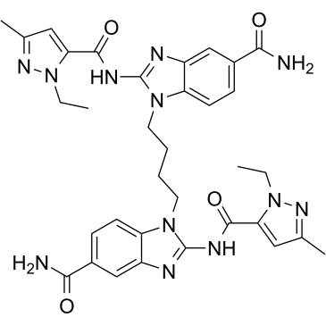
| SMILES |
CCN1C(=CC(=N1)C)C(=O)NC2=NC3=C(N2CCCCN4C5=C(C=C(C=C5)C(=O)N)N=C4NC(=O)C6=CC(=NN6CC)C)C=CC(=C3)C(=O)N
|
| InChi Key |
ICZSAXDKFXTSGL-UHFFFAOYSA-N
|
| InChi Code |
InChI=1S/C34H38N12O4/c1-5-45-27(15-19(3)41-45)31(49)39-33-37-23-17-21(29(35)47)9-11-25(23)43(33)13-7-8-14-44-26-12-10-22(30(36)48)18-24(26)38-34(44)40-32(50)28-16-20(4)42-46(28)6-2/h9-12,15-18H,5-8,13-14H2,1-4H3,(H2,35,47)(H2,36,48)(H,37,39,49)(H,38,40,50)
|
| 化学名 |
1-[4-[5-carbamoyl-2-[(2-ethyl-5-methylpyrazole-3-carbonyl)amino]benzimidazol-1-yl]butyl]-2-[(2-ethyl-5-methylpyrazole-3-carbonyl)amino]benzimidazole-5-carboxamide
|
| 别名 |
STING agonist-4; 2138300-40-8; diABZI STING agonist-2; STING agonist 2; 1,1'-(1,4-butanediyl)bis[2-[[(1-ethyl-3-methyl-1H-pyrazol-5-yl)carbonyl]amino]-1H-benzimidazole-5-carboxamide; CHEMBL4440744; STING agonist diABZI compound 2; 1-[4-[5-carbamoyl-2-[(2-ethyl-5-methylpyrazole-3-carbonyl)amino]benzimidazol-1-yl]butyl]-2-[(2-ethyl-5-methylpyrazole-3-carbonyl)amino]benzimidazole-5-carboxamide;
|
| HS Tariff Code |
2934.99.9001
|
| 存储方式 |
Powder -20°C 3 years 4°C 2 years In solvent -80°C 6 months -20°C 1 month 注意: 请将本产品存放在密封且受保护的环境中(例如氮气保护),避免吸湿/受潮。 |
| 运输条件 |
Room temperature (This product is stable at ambient temperature for a few days during ordinary shipping and time spent in Customs)
|
| 溶解度 (体外实验) |
DMSO : ~10 mg/mL (~14.73 mM)
H2O : < 0.1 mg/mL |
|---|---|
| 溶解度 (体内实验) |
配方 1 中的溶解度: 1 mg/mL (1.47 mM) in 10% DMSO + 40% PEG300 + 5% Tween80 + 45% Saline (这些助溶剂从左到右依次添加,逐一添加), 悬浮液;超声助溶。
例如,若需制备1 mL的工作液,可将100 μL 10.0 mg/mL澄清DMSO储备液加入400 μL PEG300中,混匀;然后向上述溶液中加入50 μL Tween-80,混匀;加入450 μL生理盐水定容至1 mL。 *生理盐水的制备:将 0.9 g 氯化钠溶解在 100 mL ddH₂O中,得到澄清溶液。 配方 2 中的溶解度: 1 mg/mL (1.47 mM) in 10% DMSO + 90% (20% SBE-β-CD in Saline) (这些助溶剂从左到右依次添加,逐一添加), 悬浊液; 超声助溶。 例如,若需制备1 mL的工作液,可将 100 μL 10.0 mg/mL澄清DMSO储备液加入900 μL 20% SBE-β-CD生理盐水溶液中,混匀。 *20% SBE-β-CD 生理盐水溶液的制备(4°C,1 周):将 2 g SBE-β-CD 溶解于 10 mL 生理盐水中,得到澄清溶液。 请根据您的实验动物和给药方式选择适当的溶解配方/方案: 1、请先配制澄清的储备液(如:用DMSO配置50 或 100 mg/mL母液(储备液)); 2、取适量母液,按从左到右的顺序依次添加助溶剂,澄清后再加入下一助溶剂。以 下列配方为例说明 (注意此配方只用于说明,并不一定代表此产品 的实际溶解配方): 10% DMSO → 40% PEG300 → 5% Tween-80 → 45% ddH2O (或 saline); 假设最终工作液的体积为 1 mL, 浓度为5 mg/mL: 取 100 μL 50 mg/mL 的澄清 DMSO 储备液加到 400 μL PEG300 中,混合均匀/澄清;向上述体系中加入50 μL Tween-80,混合均匀/澄清;然后继续加入450 μL ddH2O (或 saline)定容至 1 mL; 3、溶剂前显示的百分比是指该溶剂在最终溶液/工作液中的体积所占比例; 4、 如产品在配制过程中出现沉淀/析出,可通过加热(≤50℃)或超声的方式助溶; 5、为保证最佳实验结果,工作液请现配现用! 6、如不确定怎么将母液配置成体内动物实验的工作液,请查看说明书或联系我们; 7、 以上所有助溶剂都可在 Invivochem.cn网站购买。 |
| 制备储备液 | 1 mg | 5 mg | 10 mg | |
| 1 mM | 1.4733 mL | 7.3666 mL | 14.7332 mL | |
| 5 mM | 0.2947 mL | 1.4733 mL | 2.9466 mL | |
| 10 mM | 0.1473 mL | 0.7367 mL | 1.4733 mL |
1、根据实验需要选择合适的溶剂配制储备液 (母液):对于大多数产品,InvivoChem推荐用DMSO配置母液 (比如:5、10、20mM或者10、20、50 mg/mL浓度),个别水溶性高的产品可直接溶于水。产品在DMSO 、水或其他溶剂中的具体溶解度详见上”溶解度 (体外)”部分;
2、如果您找不到您想要的溶解度信息,或者很难将产品溶解在溶液中,请联系我们;
3、建议使用下列计算器进行相关计算(摩尔浓度计算器、稀释计算器、分子量计算器、重组计算器等);
4、母液配好之后,将其分装到常规用量,并储存在-20°C或-80°C,尽量减少反复冻融循环。
计算结果:
工作液浓度: mg/mL;
DMSO母液配制方法: mg 药物溶于 μL DMSO溶液(母液浓度 mg/mL)。如该浓度超过该批次药物DMSO溶解度,请首先与我们联系。
体内配方配制方法:取 μL DMSO母液,加入 μL PEG300,混匀澄清后加入μL Tween 80,混匀澄清后加入 μL ddH2O,混匀澄清。
(1) 请确保溶液澄清之后,再加入下一种溶剂 (助溶剂) 。可利用涡旋、超声或水浴加热等方法助溶;
(2) 一定要按顺序加入溶剂 (助溶剂) 。
 INI3069
 CDN prodrug-1
 STING agonist-45
 STING agonist-46
InvivoChem的所有产品仅用于作科学研究,不面向患者销售
Copyright 2020 InvivoChem LLC | All Rights Reserved 粤ICP备20063088号-1


 COA
COA
 463611831
