| 规格 | 价格 | 库存 | 数量 |
|---|---|---|---|
| 50mg |
|
||
| 100mg |
|
||
| 250mg |
|
||
| Other Sizes |
|

| 靶点 |
Endogenous Metabolite; ERK; Caspase-3/12
|
|---|---|
| 体外研究 (In Vitro) |
牛磺熊去氧胆酸 (TUDCA) 通过 PKCα 激活丝裂原激活的磷酸磷酸酶 1 (MKP-1) 并抑制 ERK 磷酸化,最终降低血管平滑肌细胞 (VSMC) 的存活和迁移。牛磺熊去氧胆酸通过 Ca2+ 诱导 PKCα 易位抑制 ERK 牛磺熊去氧胆酸 (200 μM) 可以恢复牛磺熊去氧胆酸 (200 μM) 降低的 VSMC,从而限制 VSMC 的增殖和迁移 活性,这意味着牛磺熊去氧胆酸的抗应激作用取决于 MKP-1 的表达 [1]。
TUDCA转运机制及作用通路研究 [1] 通过RT-PCR和蛋白质印迹法分析了人血管平滑肌细胞(hVSMCs)摄取TUDCA的转运蛋白。特异性siRNA敲降实验证实,TUDCA通过有机阴离子转运蛋白2(OATP2)进入hVSMCs。TUDCA通过蛋白激酶Cα(PKCα)诱导丝裂原活化蛋白激酶磷酸酶-1(MKP-1),进而抑制细胞外信号调节激酶(ERK)的活性,最终降低hVSMCs的存活率。PKC抑制剂7-羟基星形孢菌素或MKP-1敲降均可逆转TUDCA的抗增殖作用。此外,TUDCA通过ERK抑制下调基质金属蛋白酶9(MMP-9)的表达,从而抑制hVSMCs迁移能力。 结论 [1] hVSMCs对TUDCA的摄取由OATP2介导。TUDCA通过PKCα/MKP-1通路抑制ERK磷酸化,从而同时抑制VSMCs的存活和迁移能力。研究还证实TUDCA可显著抑制PDGF诱导的VSMCs增殖和迁移。[1] |
| 体内研究 (In Vivo) |
使用转移酶 dUTP 缺口切割标记 (TUNEL) 和免疫组织化学进行肿胀细胞核因子 (PCNA) 测定,研究牛磺熊去氧胆酸 (TUDCA) 对体内 VSMC 肿胀和炎症的影响。牛磺熊去氧胆酸盐(10、50 和 100 mg/kg)实验性地增强了受损组织中的 Caspase 3 活性,并且牛磺熊去氧胆酸盐引起了新内膜中 VSMC 的突变。损伤后一周,使用损伤组织测量 ERK 和 MMP-9 表达的磷酸化水平,并与正常对照进行比较。球囊损伤会增加组织中 MMP-9 的产生和 ERK 的磷酸化。在模拟调节方法中,牛磺熊去氧胆酸(10、50 和 100 mg/kg)抑制 ERK 和 MMP-9 的磷酸化 [1]。胆汁酸牛脱氧胆酸盐 (TUDCA) 是亲水性的。牛脱氧胆酸盐可减少内质网中间细胞和胰腺的数量,从而作为细胞保护剂增强肝功能,并可能预防肝细胞癌。当tauodeoxycholate给予Ang II诱导的ApoE-/-小鼠时,它显着降低Andrew分子的表达,包括eIF2α、caspase-3、caspase-12、C/EBP同源蛋白、c-Jun N-终止子蛋白(JNK) )、激活转录因子 4 (ATF4)、X-box 结合蛋白 (XBP) 和 JNK。这种影响是显着的(p<0.05)。在 ApoE-/- 小鼠中,牛磺熊去氧胆酸可减少血管紧张素 II 引起的腹主动脉收缩。将 0.5 g/kg/天的牛磺熊去氧胆酸盐给予 Ang II 诱导的 ApoE-/- 小鼠(ER 对)。 ..总胆固醇水平(663.6±88.7 mg/dL 与 655.7±65.4 mg/dL;p>0.05)或收缩压(141.3±5.6 mmHg 与 145.9±8.9 mmHg;p>0.05)之间没有差异。牛磺熊去氧胆酸盐组和AAA模型组。此外,与AAA模型组相比,牛磺熊去氧胆酸盐组的AAA损伤面积小于AAA模型组的损伤面积(0.37±0.03mm 2 比1.51±0.06mm;p<0.05)。 )[2]。
TUDCA在体内减少新生内膜增生的作用[1] 体外实验结果表明,TUDCA可能通过PKCα介导的ERK抑制作用抑制血管损伤后的新生内膜形成。采用大鼠颈动脉损伤模型分析TUDCA对新生内膜形成的影响。球囊损伤后,口服TUDCA治疗2周能显著抑制新生内膜形成,且呈剂量依赖性(内膜/中膜比值;50 mg/kg组1.48±0.11;100 mg/kg组1.17±0.16 vs 对照组1.86±0.17,n=8,P<0.05)(图4A和B)。此外,TUDCA给药显著减少了新生内膜面积(对照组100±12.9%;TUDCA 10 mg/kg组80.0±19.7%;50 mg/kg组75.5±6.8%;100 mg/kg组56.9±20%,每组n=8)和新生内膜厚度(对照组133.5±26.7 μm;10 mg/kg组92.1±23.3 μm;50 mg/kg组78.5±15.7 μm;100 mg/kg组61.8±16.3 μm,每组n=8)(图4C和D)。同时,TUDCA以剂量依赖性方式增加了管腔面积(正常组100±5.6%;对照组63.3±7.1%;10 mg/kg组73.1±11.1%;50 mg/kg组78.7±5.8%;100 mg/kg组91.2±8.2%,每组n=8),该增加与新生内膜面积的减少呈正相关(图4E)。值得注意的是,中膜面积和中膜细胞数量未受影响(在线补充材料,图S5A和B)。 |
| 酶活实验 |
FITC-TUDCA摄取实验 [1]
人血管平滑肌细胞(hVSMCs)使用平滑肌细胞生长培养基2(Smooth Muscle Cell Growth Medium 2)培养。本研究采用4名供体来源、传代4-7次的hVSMCs。将1×10⁴个hVSMCs接种于96孔板培养后,分别加入指定浓度的FITC-TUDCA,并用预冷磷酸盐缓冲液洗涤。通过检测488 nm处吸光度评估FITC信号。竞争实验中将100 μM FITC-TUDCA与不同浓度的未标记TUDCA混合,处理hVSMCs 15分钟。具体方法详见在线补充材料。 明胶酶谱分析 [1] 细胞经TUDCA处理24小时后收集上清,上样至含1 mg/mL明胶的SDS-PAGE凝胶。电泳结束后,凝胶在反应缓冲液中孵育24小时,随后用0.15%考马斯亮蓝R250染色。具体方法详见在线补充材料。 |
| 细胞实验 |
细胞活力与增殖实验 [1]
采用Ez-Cytox试剂检测细胞活力与增殖能力。将血管平滑肌细胞(VSMCs,5×10³个/孔)接种于96孔板,使用平滑肌细胞生长培养基2(Smooth Muscle Cell Growth Medium 2)培养。血清饥饿处理后,分别向hVSMCs中加入不同浓度TUDCA(0、50、100及200 μM),并设置添加/不添加1,2-双(邻氨基苯氧基)乙烷-N,N,N′,N′-四乙酸四(乙酰氧甲酯)(BAPTA,10 μM)和7-羟基星形孢菌素(H7,10 μM)的对照组,继续培养24小时。为评估TUDCA对PDGF刺激的hVSMCs增殖影响,在血清饥饿处理后同时加入TUDCA(浓度梯度同上)与PDGF-BB(50 ng/mL)。每孔加入10 μL Ez-Cytox后,通过检测450 nm波长光密度评估细胞活力。 划痕愈合实验 [1] 采用划痕法评估hVSMCs迁移能力。将1×10⁶个hVSMCs接种于60 mm培养皿,培养至细胞完全融合。分别用不同浓度TUDCA预处理1小时后,使用改良枪头在细胞单层中央制造划痕。划痕后用无血清SMCGM2培养基洗涤,继续含TUDCA培养36小时。由三位独立研究者对划痕边缘迁移细胞进行计数分析。 流式细胞术检测 [1] 通过流式细胞术评估TUDCA诱导的hVSMCs凋亡。血清饥饿处理24小时后,分别用浓度梯度(0、50、100及200 μM)TUDCA处理细胞。收集固定细胞后,通过DNA含量分析检测凋亡率。具体方法详见在线补充材料。 |
| 动物实验 |
大鼠颈动脉球囊损伤[1]
大鼠采用联合麻醉剂(氯胺酮,70 mg/kg;赛拉嗪,7 mg/kg,腹腔注射)麻醉。实验过程中,不时对肢体施加有害刺激,同时监测呼气末二氧化碳、心率、血压和心律的变化,以确定麻醉深度。暴露左侧颈外动脉后,经颈外动脉切口插入2 F Fogarty取栓导管,推进至颈总动脉,注入0.2 mL生理盐水,并旋转回抽10次。TUDCA随后每日口服一次,不同浓度(即溶剂、10、50和100 mg/kg),持续2周。颈动脉组织经4%甲醛灌注固定后,进行石蜡包埋,切片(8 μm)后进行苏木精-伊红(H&E)染色。详细方法见在线补充材料。 Ang II 诱导的小鼠腹主动脉瘤模型 [2] 将 30 只 8 周龄的 ApoE−/− C57BL/6 雄性小鼠随机分为三组(每组 n = 10):(i)假手术组,注射生理盐水(0.9%)作为载体(“正常组”);(ii)在 ApoE−/− 小鼠右侧腹部皮下植入微型渗透泵,持续 28 天释放 Ang II(1000 ng/kg/min)(“AAA 模型组”);(iii)在 AAA 模型小鼠饮用水中加入内质网应激抑制剂(TUDCA),每日一次,持续 4 周,剂量为 0.5 g/kg/天(“内质网应激抑制剂组”)。Ang II 输注 28 天后处死小鼠。如前所述22,在植入微型渗透泵前 1 周测量收缩压。在小鼠体内植入输注泵,并在输注28天后,采用无创尾套系统采集血液样本。在异氟烷麻醉下,从眼眶后静脉丛采集血液。采用酶法测定血清总胆固醇水平。 |
| 药代性质 (ADME/PK) |
吸收、分布和排泄
有证据表明,牛磺熊去氧胆酸能够穿过人体内血脑屏障。 代谢/代谢产物 牛磺熊去氧胆酸的生物转化很少。它会被肠道菌群部分去结合,形成非结合型胆汁酸。 |
| 毒性/毒理 (Toxicokinetics/TK) |
9848818 大鼠口服LD50 >5 gm/kg 日本公会东京公报专利,专利号:92-235918
9848818 大鼠静脉注射LD50 300 mg/kg 日本公会东京公报专利,专利号:92-235918 9848818 小鼠口服LD50 >6 gm/kg 日本公会东京公报专利,专利号:92-235918 9848818 小鼠静脉注射LD50 350 mg/kg 日本公会东京公报专利,专利号:92-235918 |
| 参考文献 |
|
| 其他信息 |
牛磺熊去氧胆酸是一种源自熊去氧胆酸的牛磺酸胆汁酸结合物。它是一种人体代谢产物,具有抗炎、神经保护、抑制细胞凋亡、心脏保护和维持骨密度等作用。其功能与熊去氧胆酸相关,是牛磺熊去氧胆酸盐的结合酸。
牛磺熊去氧胆酸,又称熊去氧胆酸碱,是一种高度亲水的叔胆汁酸,在人体内含量较低。它是熊去氧胆酸的牛磺酸结合物,具有与熊去氧胆酸相当的治疗效果和安全性,但亲水性更高。通常情况下,亲水性胆汁酸调节疏水性胆汁酸及其细胞毒性作用。牛磺熊去氧胆酸可减少小肠对胆固醇的吸收,从而降低人体对膳食胆固醇的摄入量和体内胆固醇含量。牛磺熊去氧胆酸目前在欧洲被用作胆汁酸衍生物,用于治疗和预防胆结石。由于其一系列分子特性——特别是其抗细胞凋亡作用——牛磺熊去氧胆酸已被用于研究炎症性代谢疾病和神经退行性疾病。 已有关于牛磺熊去氧胆酸在智人体内的研究报道,并有相关数据。 药物适应症 牛磺熊去氧胆酸用于预防和治疗胆结石的形成。牛磺熊去氧胆酸与苯丁酸联合用于治疗成人肌萎缩侧索硬化症 (ALS)。 作用机制 约90%的胆结石由胆固醇形成,而胆固醇的形成可能与高脂饮食和其他因素导致的肠道菌群失调有关。肠道菌群调节胆汁酸代谢;因此,肠道菌群组成的改变可能显著改变胆汁酸池并影响胆固醇的分泌。虽然牛磺熊去氧胆酸减少和预防胆结石形成的确切作用机制尚不清楚,但它可能通过多种途径发挥作用。最近一项小鼠研究表明,牛磺熊去氧胆酸通过上调胆汁酸从肝脏向胆囊的排泄来抑制肠道对胆固醇的吸收并降低肝脏胆固醇水平。牛磺熊去氧胆酸可降低胆囊中胆汁的胆固醇饱和度,从而提高胆汁中胆固醇的溶解度。它还能维持特定的肠道菌群组成,促进胆汁酸的合成,并减轻血液中脂多糖引起的肝脏炎症。最终,牛磺熊去氧胆酸可增强肝脏中胆汁酸的合成,并降低血清和肝脏中的胆固醇水平。牛磺熊去氧胆酸通过干扰线粒体细胞死亡途径来抑制细胞凋亡。其作用机制包括抑制氧自由基的产生、改善内质网应激以及稳定未折叠蛋白反应。牛磺熊去氧胆酸介导的其他抗凋亡过程包括细胞色素c释放、半胱天冬酶激活、DNA和核碎裂以及抑制p53转录激活。牛磺熊去氧胆酸被认为作用于多个细胞靶点,抑制细胞凋亡并上调细胞存活通路。 牛磺熊去氧胆酸是一种小分子药物,其临床试验阶段最高为IV期(涵盖所有适应症),于2022年首次获批,并有2个在研适应症。 目的:血管损伤后血管平滑肌细胞(VSMCs)增生是与新生内膜相关的主要病理生理机制之一。牛磺熊去氧胆酸(TUDCA)是一种细胞保护剂,对包括肝细胞在内的多种细胞均有效,同时也是癌细胞凋亡的诱导剂。本研究旨在探讨TUDCA是否能够通过抑制VSMCs的生长和迁移来预防新生内膜增生。方法和结果:采用RT-PCR和Western blot分析人VSMCs中TUDCA的转运蛋白。利用特异性siRNA进行的基因敲低实验表明,TUDCA通过有机阴离子转运蛋白2 (OATP2) 进入人血管平滑肌细胞(hVSMC)。TUDCA通过蛋白激酶Cα (PKCα) 诱导丝裂原活化蛋白激酶磷酸酶-1 (MKP-1) 的表达,进而抑制细胞外信号调节激酶(ERK),从而降低hVSMC的活力。使用PKC抑制剂7-羟基星形孢菌素处理或敲低MKP-1均可逆转TUDCA的抗增殖作用。此外,TUDCA通过抑制ERK并降低基质金属蛋白酶-9 (MMP-9) 的表达,从而抑制hVSMC的迁移,并降低hVSMC的活力。对颈动脉球囊损伤的大鼠口服TUDCA后,球囊损伤引起的ERK和MMP-9水平升高有所降低。 TUDCA通过减少血管平滑肌细胞(VSMC)的增殖并诱导其凋亡,显著降低了内膜与中膜的比例。结论:TUDCA通过PKCα介导的MKP-1诱导抑制ERK,从而减少平滑肌细胞的增殖并诱导其凋亡,进而抑制新生内膜增生。[1] 目的/背景:腹主动脉瘤(AAA)的特征是平滑肌细胞(SMC)浸润、凋亡、炎症细胞浸润、新生血管形成以及细胞外基质降解。既往研究表明,在小鼠模型和人类胸主动脉瘤中,内质网(ER)应激和SMC凋亡均增加。然而,ER应激是否在AAA形成过程中被激活,以及抑制ER应激是否能减轻AAA,目前尚不清楚。方法:收集人类AAA和对照主动脉样本。采用免疫组织化学染色法检测内质网应激分子伴侣葡萄糖调节蛋白(GRP)-78 和 GRP-94 的表达。探讨内质网应激抑制剂牛磺熊去氧胆酸(TUDCA)对血管紧张素II(Ang II)诱导的载脂蛋白E-/-(ApoE-/-)小鼠腹主动脉瘤(AAA)形成的影响。采用弹性蛋白染色观察弹性蛋白断裂情况。通过免疫组织化学和蛋白质印迹分析检测内质网应激分子伴侣和凋亡相关分子的蛋白表达。结果:人腹主动脉瘤和Ang II诱导的ApoE-/-小鼠的动脉瘤区GRP-78和GRP-94表达显著上调(p < 0.05)。TUDCA显著减小Ang II诱导的ApoE-/-小鼠腹主动脉的最大直径(p < 0.05)。 TUDCA显著降低了内质网应激分子伴侣的表达和凋亡细胞的数量(p < 0.05)。此外,TUDCA显著降低了血管紧张素II(Ang II)诱导的ApoE-/-小鼠中凋亡分子的表达,例如caspase-3、caspase-12、C/EBP同源蛋白、c-Jun N端激酶激活转录因子4、X-box结合蛋白和真核起始因子2α(eIF2α)(p < 0.05)。结论:结果表明,内质网应激参与了人类和Ang II诱导的ApoE-/-小鼠腹主动脉瘤(AAA)的形成。TUDCA通过抑制内质网应激介导的细胞凋亡来减轻Ang II诱导的ApoE-/-小鼠AAA的形成。[2] |
| 分子式 |
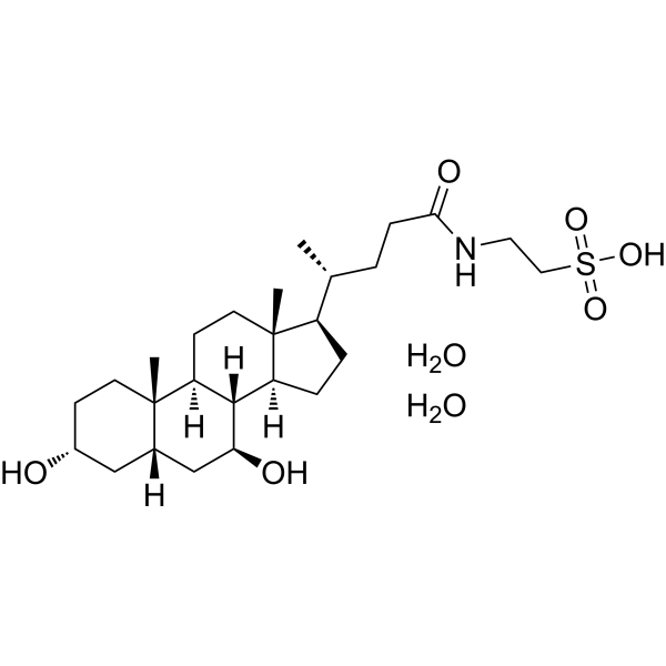
C26H49NO8S
|
|---|---|
| 分子量 |
535.734167814255
|
| 精确质量 |
535.317
|
| CAS号 |
117609-50-4
|
| 相关CAS号 |
Tauroursodeoxycholate;14605-22-2;Tauroursodeoxycholate sodium;35807-85-3;Tauroursodeoxycholate-d4 sodium;2410279-95-5
|
| PubChem CID |
71306862
|
| 外观&性状 |
White to off-white solid powder
|
| tPSA |
134
|
| 氢键供体(HBD)数目 |
6
|
| 氢键受体(HBA)数目 |
8
|
| 可旋转键数目(RBC) |
7
|
| 重原子数目 |
36
|
| 分子复杂度/Complexity |
858
|
| 定义原子立体中心数目 |
10
|
| SMILES |
S(CCNC(CC[C@@H](C)[C@H]1CC[C@H]2[C@@H]3[C@H](C[C@@H]4C[C@@H](CC[C@]4(C)[C@H]3CC[C@@]21C)O)O)=O)(=O)(=O)O.O.O
|
| InChi Key |
BNXLUNVCHFIPFY-GUBAPICVSA-N
|
| InChi Code |
InChI=1S/C26H45NO6S.2H2O/c1-16(4-7-23(30)27-12-13-34(31,32)33)19-5-6-20-24-21(9-11-26(19,20)3)25(2)10-8-18(28)14-17(25)15-22(24)29;;/h16-22,24,28-29H,4-15H2,1-3H3,(H,27,30)(H,31,32,33);2*1H2/t16-,17+,18-,19-,20+,21+,22+,24+,25+,26-;;/m1../s1
|
| 化学名 |
2-[[(4R)-4-[(3R,5S,7S,8R,9S,10S,13R,14S,17R)-3,7-dihydroxy-10,13-dimethyl-2,3,4,5,6,7,8,9,11,12,14,15,16,17-tetradecahydro-1H-cyclopenta[a]phenanthren-17-yl]pentanoyl]amino]ethanesulfonic acid;dihydrate
|
| HS Tariff Code |
2934.99.9001
|
| 存储方式 |
Powder -20°C 3 years 4°C 2 years In solvent -80°C 6 months -20°C 1 month |
| 运输条件 |
Room temperature (This product is stable at ambient temperature for a few days during ordinary shipping and time spent in Customs)
|
| 溶解度 (体外实验) |
DMSO : ~83.33 mg/mL (~155.54 mM)
|
|---|---|
| 溶解度 (体内实验) |
配方 1 中的溶解度: ≥ 2.08 mg/mL (3.88 mM) (饱和度未知) in 10% DMSO + 40% PEG300 + 5% Tween80 + 45% Saline (这些助溶剂从左到右依次添加,逐一添加), 澄清溶液。
例如,若需制备1 mL的工作液,可将100 μL 20.8 mg/mL澄清DMSO储备液加入400 μL PEG300中,混匀;然后向上述溶液中加入50 μL Tween-80,混匀;加入450 μL生理盐水定容至1 mL。 *生理盐水的制备:将 0.9 g 氯化钠溶解在 100 mL ddH₂O中,得到澄清溶液。 配方 2 中的溶解度: ≥ 2.08 mg/mL (3.88 mM) (饱和度未知) in 10% DMSO + 90% (20% SBE-β-CD in Saline) (这些助溶剂从左到右依次添加,逐一添加), 澄清溶液。 例如,若需制备1 mL的工作液,可将 100 μL 20.8 mg/mL澄清DMSO储备液加入900 μL 20% SBE-β-CD生理盐水溶液中,混匀。 *20% SBE-β-CD 生理盐水溶液的制备(4°C,1 周):将 2 g SBE-β-CD 溶解于 10 mL 生理盐水中,得到澄清溶液。 View More
配方 3 中的溶解度: ≥ 2.08 mg/mL (3.88 mM) (饱和度未知) in 10% DMSO + 90% Corn Oil (这些助溶剂从左到右依次添加,逐一添加), 澄清溶液。 1、请先配制澄清的储备液(如:用DMSO配置50 或 100 mg/mL母液(储备液)); 2、取适量母液,按从左到右的顺序依次添加助溶剂,澄清后再加入下一助溶剂。以 下列配方为例说明 (注意此配方只用于说明,并不一定代表此产品 的实际溶解配方): 10% DMSO → 40% PEG300 → 5% Tween-80 → 45% ddH2O (或 saline); 假设最终工作液的体积为 1 mL, 浓度为5 mg/mL: 取 100 μL 50 mg/mL 的澄清 DMSO 储备液加到 400 μL PEG300 中,混合均匀/澄清;向上述体系中加入50 μL Tween-80,混合均匀/澄清;然后继续加入450 μL ddH2O (或 saline)定容至 1 mL; 3、溶剂前显示的百分比是指该溶剂在最终溶液/工作液中的体积所占比例; 4、 如产品在配制过程中出现沉淀/析出,可通过加热(≤50℃)或超声的方式助溶; 5、为保证最佳实验结果,工作液请现配现用! 6、如不确定怎么将母液配置成体内动物实验的工作液,请查看说明书或联系我们; 7、 以上所有助溶剂都可在 Invivochem.cn网站购买。 |
| 制备储备液 | 1 mg | 5 mg | 10 mg | |
| 1 mM | 1.8666 mL | 9.3331 mL | 18.6661 mL | |
| 5 mM | 0.3733 mL | 1.8666 mL | 3.7332 mL | |
| 10 mM | 0.1867 mL | 0.9333 mL | 1.8666 mL |
1、根据实验需要选择合适的溶剂配制储备液 (母液):对于大多数产品,InvivoChem推荐用DMSO配置母液 (比如:5、10、20mM或者10、20、50 mg/mL浓度),个别水溶性高的产品可直接溶于水。产品在DMSO 、水或其他溶剂中的具体溶解度详见上”溶解度 (体外)”部分;
2、如果您找不到您想要的溶解度信息,或者很难将产品溶解在溶液中,请联系我们;
3、建议使用下列计算器进行相关计算(摩尔浓度计算器、稀释计算器、分子量计算器、重组计算器等);
4、母液配好之后,将其分装到常规用量,并储存在-20°C或-80°C,尽量减少反复冻融循环。
计算结果:
工作液浓度: mg/mL;
DMSO母液配制方法: mg 药物溶于 μL DMSO溶液(母液浓度 mg/mL)。如该浓度超过该批次药物DMSO溶解度,请首先与我们联系。
体内配方配制方法:取 μL DMSO母液,加入 μL PEG300,混匀澄清后加入μL Tween 80,混匀澄清后加入 μL ddH2O,混匀澄清。
(1) 请确保溶液澄清之后,再加入下一种溶剂 (助溶剂) 。可利用涡旋、超声或水浴加热等方法助溶;
(2) 一定要按顺序加入溶剂 (助溶剂) 。
 匹伐加宾
匹伐加宾
 磷酸氢化可的松
 BAY-784
 Gly-PEG3-endo-BCN
InvivoChem的所有产品仅用于作科学研究,不面向患者销售
Copyright 2020 InvivoChem LLC | All Rights Reserved 粤ICP备20063088号-1


 463611831
