| 规格 | 价格 | 库存 | 数量 |
|---|---|---|---|
| 10 mM * 1 mL in DMSO |
|
||
| 5mg |
|
||
| 10mg |
|
||
| 25mg |
|
||
| 50mg |
|
||
| 100mg |
|
||
| 250mg |
|
||
| 500mg |
|
||
| 1g |
|
||
| Other Sizes |
|

| 靶点 |
DNA-damaging chemotherapeutic agent
|
|---|---|
| 体外研究 (In Vitro) |
体外活性:Trapazamine(也称为 SR-4233;SR259075;Win59075;SR4233)是一种实验辅助药物和 DNA 损伤剂,具有治疗宫颈癌、头颈癌的潜力。当替拉扎明优先在实体瘤的缺氧区域激活其毒性形式时,可以通过减少 HIF-1α 蛋白合成来下调 HIF-1α 表达。当与阿霉素 (DOX) 和 SN-38(伊立替康的活性代谢物)等化疗药物联合使用时,曲扎明可显着抑制 HIF-1α 蛋白的积累,降低 HIF-1α 转录激活,并损害参与相关蛋白的磷酸化。同源重组修复途径,最终导致这两种药物的协同作用。激酶测定:细胞测定:与替拉扎明联合使用,拓扑异构酶 I 抑制剂在几种肝细胞癌细胞系中表现出协同细胞毒性并诱导显着的细胞凋亡。替拉扎明加 SN-38(伊立替康的活性代谢物)诱导的细胞凋亡增强伴随着线粒体去极化和 caspase 通路激活的增加。联合治疗显着抑制了 HIF-1α 蛋白的积累,降低了 HIF-1α 转录激活,并损害了同源重组修复途径中涉及的蛋白的磷酸化,最终导致了这两种药物的协同作用。
|
| 体内研究 (In Vivo) |
在人癌症Bel-7402异种移植小鼠模型中进一步验证了替拉帕扎明与伊立替康联合提高的抗癌功效。
由于替拉帕扎明加SN-38对人肝细胞癌Bel-7402细胞表现出最有效的协同作用,CI值最低(补充表S1),因此选择替拉帕扎明和伊立替康联合治疗的体内疗效,以在裸鼠体内测试Bel-7402异种移植物。如图6A和B以及补充表S2所示,与对照组相比,每2天腹腔注射25mg/kg剂量的替拉帕扎明或每天2.5mg/kg剂量的伊立替康,平均RTV没有显著差异。然而,替拉帕扎明联合伊立替康可引起明显的肿瘤生长抑制(T/C值:36.9%),明显大于替拉帕扎明(T/C值:88.1%)或单独伊立替康治疗(T/C比值:86.4%)。此外,与初始体重相比,用该组合治疗的小鼠在第26天没有明显的体重减轻(图6C)。因此,与单药治疗组相比,替拉帕扎明和伊立替康的组合具有更有效的肿瘤生长抑制作用,但不会导致动物体重减轻。[1]
大鼠每周一次腹腔注射6次替拉帕扎明,分为5(5TP)和10mg/kg(10TP)两种剂量,而阿霉素的剂量为1.8mg/kg(DOX)。随后两组同时接受两种药物(5TP+DOX和10TP+DOX)。替拉帕扎明降低了心脏脂质过氧化,使阿霉素改变的RyR2蛋白水平恢复正常。DOX组和TP+DOX组的GSH/GSSG比值、总谷胱甘肽、cTnI、AST和SERCA2水平没有显著变化。在10TP和10TP+DOX组中观察到心肌细胞坏死。[2] 通过尾静脉注射到携带肿瘤的小鼠体内后,TPZ(替拉帕扎明)-Pba-NPs在注射后3小时和24小时的血液浓度分别高出3.17倍,在肿瘤组织中的积聚分别高出4.12倍。在激光照射肿瘤组织后,TPZ-Pba-NPs通过有效的药物递送和体内协同作用成功抑制了肿瘤生长。[3] |
| 酶活实验 |
拓扑异构酶I抑制剂是一类具有广泛临床活性的抗癌药物。然而,它们对癌症的疗效有限。在这里,我们提出了体外和体内证据,表明肝细胞癌中缺氧诱导因子-1α(HIF-1α)的极高水平与拓扑异构酶I抑制剂的耐药性密切相关。在我们小组之前进行的一项研究中,我们发现替拉帕扎明可以通过减少HIF-1α蛋白合成来下调HIF-1α的表达。因此,我们假设替拉帕扎明与拓扑异构酶I抑制剂联合使用可能会克服化疗耐药性。在这项研究中,我们研究了拓扑异构酶I抑制剂与替拉帕扎明联合使用时,在几种肝细胞癌细胞系中表现出协同细胞毒性并诱导显著凋亡。替拉帕扎明加SN-38(伊立替康的活性代谢产物)诱导的细胞凋亡增强伴随着线粒体去极化和胱天蛋白酶途径激活的增加。联合治疗显著抑制了HIF-1α蛋白的积累,降低了HIF-1β的转录激活,并损害了参与同源重组修复途径的蛋白质的磷酸化,最终导致这两种药物的协同作用[1]。
|
| 细胞实验 |
替拉帕明/Tirapazamine 和拓扑异构酶 I 抑制剂一起表现出协同细胞毒性,并显着减少几种肝细胞癌细胞系中的细胞数量。线粒体去极化和 caspase 通路激活的增加与替拉扎明加 SN-38(伊立替康的活性代谢物)诱导的细胞凋亡增强相关。这两种药物通过联合治疗协同作用,显着减少 HIF-1α 蛋白积累,减少 HIF-1α 转录激活,并阻碍同源重组修复途径中涉及的蛋白磷酸化。
凋亡和线粒体膜电位(ΔΨm)的流式细胞术分析[1] 细胞在常氧或缺氧条件下用SN-38、Tirapazamine 或其组合处理12小时。在收获并用冷PBS缓冲液洗涤两次后,使用Annexin V-FITC/PI凋亡检测试剂盒分析凋亡细胞。PI染色后亚G1期的分析也用于评估细胞凋亡。对于PI染色,收获处理过的细胞,在-20°C下用70%乙醇固定,然后用RNaseA孵育,然后在黑暗中进行PI染色30分钟。为了测定线粒体电位,将细胞重新悬浮在含有0.1μmol/L JC-1的PBS中,并在37°C的黑暗中孵育15分钟。所有样本均使用FACS Calibur细胞仪进行分析。 克隆形成试验[1] 用替拉帕扎明或SN-38/TPT/HCPT/MONCPT或其组合处理的细胞在软琼脂上以三份的形式铺在60毫米的培养皿中。一旦设置好,将培养皿覆盖2.5 mL培养基,在37°C的缺氧条件下孵育10天,然后对菌落进行评分和拍照。 免疫荧光[1] 将细胞铺在玻璃培养玻片上,并与SN-38/替拉帕扎明或SN-38+Tirapazamine /替拉帕扎明或载体[0.1%DMSO(v/v)]一起孵育不同时间。然后用4%多聚甲醛固定细胞,并用含有0.1%Triton X-100的PBS渗透。用5%牛血清白蛋白阻断30分钟后,将细胞与HIF-1α或γ-H2AX原代抗体(1:100稀释)孵育1小时,用PBS洗涤三次,然后分别在黑暗中与Alexa Fluor 488偶联抗体和罗丹明二抗孵育。通过DAPI(4′6-二脒基-2-苯基吲哚)染色观察细胞核。使用Olympus Fluorview 1000共聚焦显微镜分析荧光信号。 |
| 动物实验 |
每周六次,大鼠接受腹腔注射替拉帕明(5 mg/kg (5TP) 和 10 mg/kg (10TP))和阿霉素(1.8 mg/kg,DOX)。接下来的两组(5TP+DOX 和 10TP+DOX)同时接受两种药物。替拉帕明使受阿霉素影响的 RyR2 蛋白水平恢复正常,并降低了心脏脂质过氧化水平。
体内活性测定[1] 将 Bel-7402 细胞(每只动物 5 × 10⁶ 个细胞,皮下注射到腋窝)注射到 5 至 6 周龄的 BALB/c 雄性无胸腺小鼠体内,建立肿瘤模型。当肿瘤平均体积达到约 100 mm³ 时开始治疗。使用游标卡尺测量肿瘤体积(mm³),并计算公式为 (W²×L)/2,其中 W 为宽度,L 为长度。无胸腺小鼠每日腹腔注射一次溶于生理盐水的 CPT-11(2.5 mg/kg),每 2 天腹腔注射一次溶于 Cremophor:乙醇:0.9% 无菌氯化钠溶液(体积比 1:1:8)的替拉帕明(25 mg/kg)。每 2 天记录一次小鼠体重和肿瘤体积,直至处死动物。动物饲养符合机构指南。本研究采用性成熟的雄性 Wistar CRL: (WI)WUBR 品系白化大鼠,购自商业养殖场。初始体重为 160–195 g 的动物饲养于 22°C、12 小时光照/12 小时黑暗循环的稳定条件下,并喂以标准化的颗粒状饲料 LSM。大鼠经腹腔注射(ip)多柔比星和/或替拉帕明[2]。 动物随机分为六组(n = 7):DOX组:多柔比星1.8 mg/kg;5TP组:替拉帕明5 mg/kg;10TP组:替拉帕明10 mg/kg;5TP+DOX组:多柔比星1.8 mg/kg和替拉帕明5 mg/kg;10TP+DOX组:多柔比星1.8 mg/kg和替拉帕明10 mg/kg;对照组腹腔注射0.9%氯化钠溶液。所有研究组均每周腹腔注射一次多柔比星(1.8 mg/kg)和两种剂量的替拉帕明,持续六周。给药一周后终止研究。这些动物被处死,并在尸检期间采集了血液和心脏样本[2]。 |
| 毒性/毒理 (Toxicokinetics/TK) |
135413511 大鼠腹腔注射 LD50 59390 ug/kg 毒理学档案,66(100),1992 [PMID:1605723]
135413511 大鼠静脉注射 LD >36 mg/kg 肾脏、输尿管和膀胱:肾小管变化(包括急性肾衰竭、急性肾小管坏死);内分泌:其他变化;血液:骨髓变化未包含在上述内容中。《毒理学家》,12(154),1992 135413511 小鼠腹腔注射LD50 89 mg/kg。《国际放射肿瘤学、生物学、物理学杂志》,16(977),1989 [PMID:2703405] 135413511 小鼠静脉注射LD50 101 mg/kg 行为:嗜睡(总体活动减少);皮肤及附属器官(皮肤);毛发;其他。《英国癌症杂志》增刊,20(84),1993 |
| 参考文献 |
|
| 其他信息 |
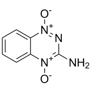
替拉帕明属于苯并三嗪类化合物,其结构为1,2,4-苯并三嗪,在3位带有氨基取代基,在1位和4位带有两个氧基取代基。它具有抗肿瘤、诱导细胞凋亡和抗菌的活性。替拉帕明是一种N-氧化物,属于苯并三嗪类化合物,也是一种芳香胺。它在功能上与1,2,4-苯并三嗪相关。
替拉帕明,也称为SR-4233,是一种在缺氧条件下激活的实验性抗癌药物。这种激活机制非常有用,因为缺氧状态常见于人类实体瘤中,这种现象被称为肿瘤缺氧。因此,替拉帕明仅在实体瘤的缺氧区域被激活。值得注意的是,通常情况下,这些缺氧区域的细胞对放射疗法和大多数抗癌药物具有耐药性。鉴于以上所有原因,强烈建议将替拉帕明与其他抗癌疗法联合使用。替拉帕明于2006年进入III期临床试验,用于治疗头颈癌、妇科肿瘤以及其他类型的实体瘤。 替拉帕明是一种具有潜在抗肿瘤活性的苯并三嗪二氧化物。替拉帕明可被多种还原酶选择性激活,在缺氧细胞中形成自由基,从而诱导DNA单链和双链断裂、碱基损伤和细胞死亡。该药物还能增强缺氧细胞对电离辐射的敏感性,并通过抑制拓扑异构酶II来抑制辐射诱导的DNA链断裂的修复。 (NCI04) 一种三嗪衍生物,可导致缺氧细胞DNA链断裂,从而使肿瘤细胞对其他药物和放射线的细胞毒活性更加敏感。 药物适应症 用于治疗头颈癌。 作用机制 大量的临床前试验已证实,其对缺氧细胞的选择性毒性机制是母体分子发生单电子还原反应生成自由基,该自由基与DNA相互作用,产生单链和双链断裂以及致命的染色体畸变。该药物与分次放射治疗以及某些化疗药物(特别是顺铂和卡铂)联合使用时也显示出活性。 药效学 替拉帕明是一种抗癌药物,在氧合良好的正常组织中无活性,但在实体瘤的低氧环境中则具有活性。因此,该药物能够杀死这些缺氧或低氧细胞,同时限制对正常组织的毒性。由于这些低氧细胞通常对放射线和常用抗癌药物具有耐药性,因此,当替拉帕明与标准抗癌疗法联合使用时,可能非常有效。拓扑异构酶I抑制剂是一类具有广泛临床活性的抗癌药物。然而,它们对肝细胞癌的疗效有限。本文中,我们提供了体外和体内证据,表明肝细胞癌中极高水平的缺氧诱导因子-1α (HIF-1α) 与对拓扑异构酶I抑制剂的耐药性密切相关。在我们课题组之前的研究中,我们发现替拉帕明可以通过降低HIF-1α蛋白的合成来下调HIF-1α的表达。因此,我们假设替拉帕明与拓扑异构酶I抑制剂联合使用可能克服化疗耐药性。本研究发现,拓扑异构酶I抑制剂与替拉帕明联用可产生协同细胞毒性,并显著诱导多种肝癌细胞系发生凋亡。替拉帕明联合SN-38(伊立替康的活性代谢物)增强的凋亡伴随着线粒体去极化和caspase通路激活。联合治疗显著抑制了HIF-1α蛋白的积累,降低了HIF-1α的转录激活,并削弱了同源重组修复通路相关蛋白的磷酸化,最终导致两种药物的协同作用。此外,在人肝癌Bel-7402异种移植小鼠模型中进一步验证了替拉帕明联合伊立替康的抗癌疗效增强。综合来看,我们的数据首次表明,HIF-1α与肝细胞癌中拓扑异构酶I抑制剂的耐药性密切相关。这些结果提示HIF-1α是一个有前景的靶点,并为开展临床试验以研究拓扑异构酶I抑制剂联合替拉帕明治疗肝细胞癌的疗效提供了理论依据。[1] 阿霉素(DOX)可引起长期心肌病,其病因与氧化应激和收缩功能障碍有关。替拉帕明(TP)是一种实验性佐剂药物,其氧化还原转化过程与DOX相同。本研究旨在评估替拉帕明对接受阿霉素治疗的大鼠的氧化应激、收缩蛋白水平和心肌细胞坏死的影响。大鼠每周腹腔注射一次替拉帕明,共注射六次,剂量分别为5 mg/kg (5TP) 和10 mg/kg (10TP),同时给予阿霉素,剂量为1.8 mg/kg (DOX)。随后两组大鼠同时接受两种药物治疗 (5TP+DOX 和 10TP+DOX)。替拉帕明降低了心脏脂质过氧化水平,并使阿霉素引起的RyR2蛋白水平改变恢复正常。DOX组和TP+DOX组之间GSH/GSSG比值、总谷胱甘肽、cTnI、AST和SERCA2水平无显著变化。10TP组和10TP+DOX组均观察到心肌细胞坏死。[2] 联合治疗中,药物的协同作用及其有效递送至关重要。本文筛选了12种抗癌药物与脱镁叶绿酸a (Pba) 光动力疗法 (PDT) 联合应用。基于细胞活力测试中的组合指数(CI)值,我们筛选出替拉帕明(TPZ),并制备了含有Pba和TPZ的自组装明胶纳米颗粒(NPs)。所得TPZ-Pba-NPs表现出协同杀伤肿瘤细胞的作用,这是因为在Pba光动力疗法(PDT)和激光照射产生的缺氧条件下,TPZ被激活。将TPZ-Pba-NPs经尾静脉注射到荷瘤小鼠体内后,注射后3小时和24小时,其血药浓度和肿瘤组织蓄积量分别提高了3.17倍和4.12倍。激光照射肿瘤组织后,TPZ-Pba-NPs通过高效的药物递送和体内协同作用,成功抑制了肿瘤的生长。这些总体结果表明,基于CI值的体外药物筛选、缺氧条件下的机制研究以及实时体内成像,是开发用于优化联合疗法的纳米颗粒的有效策略。[3] 替拉帕明(TP)已被证明能增强缺氧细胞中电离辐射的细胞毒性作用,因此可作为放射增敏剂的候选药物。这种选择性行为通常与氧气(O2)的丰度直接相关。本文研究了TP在真空、微水合(含1至3个水分子)以及嵌入连续水体中的电子性质。我们讨论了TP的电子亲和力、电荷分布和键解离能,发现这些性质在水合后并未发生显著变化。与其较大的电子亲和力相符,电子附着引发的键断裂需要高于2.5 eV的能量,这排除了生物活性TP自由基的直接形成。因此,我们的结果表明,TP的选择性行为不能用邻近O2分子的单电子还原来解释。我们提出,TP的低氧选择性可能是由于O2清除氢自由基所致。[4] |
| 分子式 |
C7H6N4O2
|
|
|---|---|---|
| 分子量 |
178.05
|
|
| 精确质量 |
178.049
|
|
| 元素分析 |
C, 47.19; H, 3.39; N, 31.45; O, 17.96
|
|
| CAS号 |
27314-97-2
|
|
| 相关CAS号 |
|
|
| PubChem CID |
135413511
|
|
| 外观&性状 |
Orange to dark orange-red solid powder
|
|
| 密度 |
1.7±0.1 g/cm3
|
|
| 沸点 |
493.6±28.0 °C at 760 mmHg
|
|
| 熔点 |
220ºC
|
|
| 闪点 |
252.3±24.0 °C
|
|
| 蒸汽压 |
0.0±1.3 mmHg at 25°C
|
|
| 折射率 |
1.777
|
|
| LogP |
-0.31
|
|
| tPSA |
89.83
|
|
| 氢键供体(HBD)数目 |
1
|
|
| 氢键受体(HBA)数目 |
4
|
|
| 可旋转键数目(RBC) |
0
|
|
| 重原子数目 |
13
|
|
| 分子复杂度/Complexity |
191
|
|
| 定义原子立体中心数目 |
0
|
|
| SMILES |
[O-][N+]1=C(N([H])[H])N=[N+](C2=C([H])C([H])=C([H])C([H])=C12)[O-]
|
|
| InChi Key |
ORYDPOVDJJZGHQ-UHFFFAOYSA-N
|
|
| InChi Code |
InChI=1S/C7H6N4O2/c8-7-9-11(13)6-4-2-1-3-5(6)10(7)12/h1-4H,(H2,8,9)
|
|
| 化学名 |
1,4-dioxido-1,2,4-benzotriazine-1,4-diium-3-amine
|
|
| 别名 |
SR 4233; SR-4233; TIRAPAZAMINE; 27314-97-2; 3-Aminobenzo[e][1,2,4]triazine 1,4-dioxide; 1,2,4-Benzotriazin-3-amine, 1,4-dioxide; 3-Amino-1,2,4-benzotriazine 1,4-dioxide; Tirazone; Win-59075; WIN 59075; SR4233; SR259075; SR-259075; SR 259075; WIN 59075; WIN-59075; WIN59075; NSC130181; NSC-130181; NSC 130181; Tirazone; TP; Tirapazamine; 3-Aminobenzo[e][1,2,4]triazine 1,4-dioxide; 3-Amino-1,2,4-benzotriazine 1,4-dioxide; 1,2,4-Benzotriazin-3-amine, 1,4-dioxide; Tirazone; Win-59,075; SR-4233;
|
|
| HS Tariff Code |
2934.99.9001
|
|
| 存储方式 |
Powder -20°C 3 years 4°C 2 years In solvent -80°C 6 months -20°C 1 month |
|
| 运输条件 |
Room temperature (This product is stable at ambient temperature for a few days during ordinary shipping and time spent in Customs)
|
| 溶解度 (体外实验) |
|
|||
|---|---|---|---|---|
| 溶解度 (体内实验) |
配方 1 中的溶解度: 2.5 mg/mL (14.03 mM) in 10% DMSO + 90% Corn Oil (这些助溶剂从左到右依次添加,逐一添加), 悬浮液;超声助溶。
例如,若需制备1 mL的工作液,可将100 μL 25.0 mg/mL 澄清 DMSO 储备液加入900 μL 玉米油中,混合均匀。 配方 2 中的溶解度: ≥ 2.08 mg/mL (11.68 mM) (饱和度未知) in 10% DMSO + 40% PEG300 + 5% Tween80 + 45% Saline (这些助溶剂从左到右依次添加,逐一添加), 澄清溶液。 例如,若需制备1 mL的工作液,可将 100 μL 20.8 mg/mL澄清的DMSO储备液加入到400 μL PEG300中,混匀;再向上述溶液中加入50 μL Tween-80,混匀;然后加入450 μL生理盐水定容至1 mL。 *生理盐水的制备:将 0.9 g 氯化钠溶解在 100 mL ddH₂O中,得到澄清溶液。 View More
配方 3 中的溶解度: ≥ 2.08 mg/mL (11.68 mM) (饱和度未知) in 10% DMSO + 90% (20% SBE-β-CD in Saline) (这些助溶剂从左到右依次添加,逐一添加), 澄清溶液。 配方 4 中的溶解度: 10 mg/mL (56.13 mM) in 50% PEG300 50% Saline (这些助溶剂从左到右依次添加,逐一添加), 悬浊液; 超声助溶。 *生理盐水的制备:将 0.9 g 氯化钠溶解在 100 mL ddH₂O中,得到澄清溶液。 1、请先配制澄清的储备液(如:用DMSO配置50 或 100 mg/mL母液(储备液)); 2、取适量母液,按从左到右的顺序依次添加助溶剂,澄清后再加入下一助溶剂。以 下列配方为例说明 (注意此配方只用于说明,并不一定代表此产品 的实际溶解配方): 10% DMSO → 40% PEG300 → 5% Tween-80 → 45% ddH2O (或 saline); 假设最终工作液的体积为 1 mL, 浓度为5 mg/mL: 取 100 μL 50 mg/mL 的澄清 DMSO 储备液加到 400 μL PEG300 中,混合均匀/澄清;向上述体系中加入50 μL Tween-80,混合均匀/澄清;然后继续加入450 μL ddH2O (或 saline)定容至 1 mL; 3、溶剂前显示的百分比是指该溶剂在最终溶液/工作液中的体积所占比例; 4、 如产品在配制过程中出现沉淀/析出,可通过加热(≤50℃)或超声的方式助溶; 5、为保证最佳实验结果,工作液请现配现用! 6、如不确定怎么将母液配置成体内动物实验的工作液,请查看说明书或联系我们; 7、 以上所有助溶剂都可在 Invivochem.cn网站购买。 |
| 制备储备液 | 1 mg | 5 mg | 10 mg | |
| 1 mM | 5.6164 mL | 28.0820 mL | 56.1640 mL | |
| 5 mM | 1.1233 mL | 5.6164 mL | 11.2328 mL | |
| 10 mM | 0.5616 mL | 2.8082 mL | 5.6164 mL |
1、根据实验需要选择合适的溶剂配制储备液 (母液):对于大多数产品,InvivoChem推荐用DMSO配置母液 (比如:5、10、20mM或者10、20、50 mg/mL浓度),个别水溶性高的产品可直接溶于水。产品在DMSO 、水或其他溶剂中的具体溶解度详见上”溶解度 (体外)”部分;
2、如果您找不到您想要的溶解度信息,或者很难将产品溶解在溶液中,请联系我们;
3、建议使用下列计算器进行相关计算(摩尔浓度计算器、稀释计算器、分子量计算器、重组计算器等);
4、母液配好之后,将其分装到常规用量,并储存在-20°C或-80°C,尽量减少反复冻融循环。
计算结果:
工作液浓度: mg/mL;
DMSO母液配制方法: mg 药物溶于 μL DMSO溶液(母液浓度 mg/mL)。如该浓度超过该批次药物DMSO溶解度,请首先与我们联系。
体内配方配制方法:取 μL DMSO母液,加入 μL PEG300,混匀澄清后加入μL Tween 80,混匀澄清后加入 μL ddH2O,混匀澄清。
(1) 请确保溶液澄清之后,再加入下一种溶剂 (助溶剂) 。可利用涡旋、超声或水浴加热等方法助溶;
(2) 一定要按顺序加入溶剂 (助溶剂) 。
| NCT Number | Recruitment | interventions | Conditions | Sponsor/Collaborators | Start Date | Phases |
| NCT02174549 | Active Recruiting |
Procedure: Conventional Transarterial Embolization (TAE) Drug: Tirapazamine |
Hepatocellular Carcinoma Neuroendocrine Tumors |
Teclison Ltd. | September 2014 | Phase 1 Phase 2 |
| NCT00003369 | Completed | Drug: tirapazamine Drug: cisplatin |
Cervical Cancer | SWOG Cancer Research Network | August 1998 | Phase 2 |
| NCT00098995 | Completed | Drug: tirapazamine Drug: cisplatin |
Cervical Cancer | Peter MacCallum Cancer Centre, Australia |
December 2004 | Phase 1 |
| NCT00094081 | Completed | Drug: tirapazamine (SR259075) Drug: cisplatin |
Head and Neck Neoplasms | Sanofi | October 2002 | Phase 3 |
| NCT00020696 | Completed | Drug: cisplatin Drug: tirapazamine |
Primary Peritoneal Cavity Cancer Ovarian Cancer |
Gynecologic Oncology Group | June 2001 | Phase 21 |
 DNA oxidative damage (AP/100 kbp) in heart homogenates.
Schematic presentation of one-electron reduction of DOX and TP in hypoxic and normoxic conditions.Oxid Med Cell Longev. 2012; 2012: 890826. |
|---|
(a) Increased eosinophilia of scattered cardiomyocytes (10TP+DOX group; H&E, objective magnification 10x). (b) Positive color reaction detecting necrosis (group 10TP+DOX; Selyes method, objective magnification 20x).Oxid Med Cell Longev. 2012; 2012: 890826. |
Representative Western blot analysis for RyR2 protein in cardiac muscle homogenates (beta-actin is shown as a loading control) and densitometric analysis (mean ± SD) of total RyR2 content expressed as percent changes with respect to the control group, which was established at 100%.Oxid Med Cell Longev. 2012; 2012: 890826. |
 (S,S)-D211
 Di(MMY-SJG)-Ph-prop-2-yne-1-sulfonic acid
 MOMA-341
 Morpholino G phosphoramidite
InvivoChem的所有产品仅用于作科学研究,不面向患者销售
Copyright 2020 InvivoChem LLC | All Rights Reserved 粤ICP备20063088号-1


 COA
COA


 463611831
