| 规格 | 价格 | 库存 | 数量 |
|---|---|---|---|
| 5mg |
|
||
| 10mg |
|
||
| 25mg |
|
||
| 50mg |
|
||
| 100mg |
|
||
| 250mg |
|
||
| 500mg |
|
||
| Other Sizes |
|

| 靶点 |
Plasminogen activator inhibitor-1 (PAI-1) (IC50 = 6.95 μM)
|
|---|---|
| 体外研究 (In Vitro) |
根据对接实验,TM5275 附着在 PAI-1 的 A β-折叠 (s4A) 的第 4 链上。 TM5275 是一种选择性 PAI-1,浓度高达 100 μM 时不会干扰其他丝氨酸蛋白酶抑制剂/丝氨酸蛋白酶系统[1]。 TM5275 抑制 tPA-GFP-PAI-1 高分子量复合物的形成,因此在 20 和 100 μM 剂量下可显着延长 tPA-GFP 在 VEC 上的保留时间。 TM5275 加速纤溶酶原的时间依赖性积聚以及纤维蛋白凝块在表达 tPA-GFP 的细胞上及其周围的分解[2]。用 70-100 μM TM5275 处理的 ES-2 和 JHOC-9 细胞 72 小时后细胞活力降低。使用 100 μM TM5275,细胞生长被抑制 48-96 小时。当用 100 μM TM5275 处理时,与对照组相比,细胞培养基中的活性 PAI-1 显着减少。有人提出,TM5275 可能对 PAI-1 表达升高的卵巢癌具有抗增殖作用 [3]。
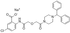
TM5275,5-氯-2-[({2-[4-(二苯甲基)哌嗪-1-基]-2-氧代乙氧基}乙酰基)氨基]苯甲酸酯(图1B),是通过对基于TM5007结构设计和合成的90多种化合物进行广泛的构效关系研究而发现的(图1A)。在考虑了体外PAI-1抑制活性和药代动力学研究(Tmax、Cmax、T½)后,TM5275最终被选为试验化合物(见下文)。 通过肽底物的tPA依赖性水解测量的TM5275的PAI-1抑制活性与TM5007和PAI-749相当:TM5275、TM5007、PAI-749的半最大抑制(IC50)值分别为6.95、5.60和8.37 μmol/L。 对PAI-1部分和TM5275进行对接模拟,以了解TM5275的作用机制。TM5275与PAI-1的Aβ-折叠(s4A)位置的链4结合。尽管TM5275和TM5007都在缝隙内与PAI-1的s4A片段结合,但仔细检查后发现它们的结合位点不同,如图2所示:TM5275的大体积二苯甲基不能容纳在TM5007的结合位点上,因此TM5275在s4A缝隙中明显移位。 在体外,TM5275(高达100 μmol/L)不会干扰其他丝氨酸蛋白酶系统(即α1抗胰蛋白酶/胰蛋白酶和α2抗纤溶酶/纤溶酶)。因此,其PAI-1抑制活性具有特异性。在十二烷基硫酸钠聚丙烯酰胺凝胶电泳上,当PAI-1与TM5275预孵育时,没有观察到与tPA形成的PAI-1共价复合物(数据未显示)。[1] TM5275在20和100μM的浓度下,通过抑制tPA-GFP-PAI-1高分子量复合物的形成,显著延长了tPA-GFP在VEC上的保留时间。TM5275增强了纤溶酶原的时间依赖性积聚,以及表达tPA-GFP的细胞上和周围纤维蛋白凝块的溶解。 TM5275的促纤维蛋白溶解作用通过PAI-1抑制导致的VEC表面tPA保留时间延长和纤溶酶生成增强得到了清楚的证明。[2] TM5275抑制纯化系统中rtPA和rPAI-1之间的高分子量复合物形成。 TM5275延长了tPA-GFP在VEC表面的保留时间。 TM5275抑制了EA.hy926细胞表面tPA和PAI-1之间的高分子量复合物形成,并减少了上清液中tPA-PAI-1复合物的量。 TM5275对VEC及其周围纤溶酶原积聚的影响。 TM5275对VEC纤维蛋白凝块溶解的疗效。 [2] TM5275对PAI-1的药理抑制降低卵巢癌症细胞的细胞增殖[3] TM5275是一种对PAI-1特异性的小分子抑制剂(图4A),已被开发为PAI-1相关疾病的治疗试剂。17,18我们研究了其作为靶向卵巢癌症细胞增殖的治疗试剂的潜力。用不同浓度的TM5275处理卵巢癌症细胞。70–100μM TM5275在ES-2和JHOC-9细胞中处理72小时后,细胞存活率降低,而其他细胞系对TM5275处理相对不敏感(图4B)。TM5275在卵巢癌症细胞系中的IC50值如表1所示。此外,在TM5275处理后的指定时间点测量细胞生长。从48小时到96小时,100μM TM5275抑制了细胞生长(图4C)。此外,与对照组相比,用100μM TM5275处理的细胞在细胞培养基中的活性PAI-1显著降低,证实了TM5275对PAI-1抑制的有效性(图4D)。这些结果表明,具有高表达PAI-1的卵巢癌症细胞,如ES-2和JHOC-9,易于受到TM5275的生长抑制。总之,TM5275对PAI-1的药理抑制作用可能在高表达PAI-1卵巢癌症中发挥抗增殖作用。 为了确定TM5275对ES-2细胞周期进程的影响,用或不用TM5275处理细胞24小时,并分析细胞周期分布。与载体处理相比,TM5275处理显著降低了G0/G1期细胞的百分比(52.0±1.5%至33.0±4.9%),增加了G2/M期细胞的比例(18.3±0.8%至27.7±2.0%)(图4E)。此外,用TM5275处理的细胞显示凋亡细胞百分比显著增加(图4F)。这些结果表明,PAI-1的药理学抑制诱导了卵巢癌症细胞中的G2/M细胞周期阻滞,并根据siRNA敲低PAI-1促进了细胞凋亡。 |
| 体内研究 (In Vivo) |
对于小鼠和大鼠,TM5275具有非常低的毒性和良好的药代动力学特征。在大鼠血栓形成模型中。与给予媒介物治疗的大鼠(72.5±2.0 mg)相比,以10和50 mg/kg剂量给予TM5275的大鼠具有显着更小的血凝块重量(分别为60.9±3.0和56.8±2.8 mg)。 TM5275(50 mg/kg)的抗血栓功效与参考抗血栓药物噻氯匹定(500 mg/kg)相当。剂量为10 mg/kg后,TM5275的血浆浓度达到17.5±5.2 μM。当 TM5275 (5 mg/kg) 与 tPA (0.3 mg/kg) 一起服用时,tPA (0.3 mg/kg) 的抗血栓作用大大增加,产生的益处与高剂量 tPA (3 mg/kg) 相当。 /千克)[1]。
大鼠血栓形成模型[1] 表1描述了TM5275在大鼠动静脉分流模型中的抗血栓作用。给药10和50的大鼠的血栓重量明显较低 TM5275(60.9±3.0和56.8±2.8)mg/kg mg)比赋形剂处理的大鼠(72.5±2.0 mg).高达300 需要mg/kg的TM5007才能达到50 同一模型中TM5275的浓度为mg/kg。TM5275(50mg/kg)的抗血栓作用与参考抗血栓化合物噻氯匹啶(500mg/kg)相当。TM5275血浆浓度达到17.5±5.2 μmol/L,剂量为10 mg/kg。 TM5275在大鼠FeCl3颈动脉血栓形成模型中的抗血栓作用如图3所示。TM5275和另一种标准抗血栓药物氯吡格雷以剂量依赖的方式证明了抗血栓作用(图3)。TM5275和氯吡格雷的最小有效剂量分别为1和3 mg/kg。它对应于TM5275的血浆浓度为4.9±3.6 μmol/L。正如预期的那样,氯吡格雷以剂量依赖的方式延长出血时间(图3)。相比之下,TM5275不影响出血时间,这是作为抗血栓药物的潜在益处。口服两小时后,TM5275(10mg/kg)不影响ADP和胶原蛋白诱导的血小板聚集(数据未显示)。因此,其抗血栓作用与对血小板的任何影响无关。 如图4所示,TM5275在同一型号中进一步与tPA结合。单独使用tPA(0.3mg/kg)不能提供显著的抗血栓作用。然而,TM5275(5 mg/kg)与tPA(0.3 mg/kg)联合使用显著增强了单独使用tPA(0.3mg/kg)的抗血栓作用,并提供了与高tPA剂量(3 mg/kg)相似的益处。联合治疗的出血时间与单独使用低剂量tPA(0.3mg/kg)的出血时间相似。 猴血栓模型[1] 在光化学诱导血栓形成的食蟹猴模型中评估了TM5275的抗血栓作用(表2)。TM5275的总闭塞时间显著缩短(53.9±19.9 分钟)和氯吡格雷(39.4±25.8 与赋形剂组(119.0±17.4)相比 分钟)。TM5275血浆浓度达到18.9±3.7 TM5275组为μmol/L。 TM5275作为一种对出血时间没有影响的抗血栓药物,其益处已在非人灵长类动物中得到证实(表3)。给药50小时后 mg/kg(比有效剂量高5倍)的TM5275,出血时间仅稍长(146.7±3.3 与给药前相比(83.3±6.7秒) secs),而在10 mg/kg氯吡格雷组(>600秒)高于给药前观察值(113.3±8.8秒) 秒)。 |
| 酶活实验 |
体外PAI-1活性测定[1]
PAI-1抑制活性通过之前描述的显色测定法进行评估(Izuhara等人,2008)。调整培养基的成分以提高测定的灵敏度:0.15 摩尔/升氯化钠,50 mmol/L Tris-HCl pH8,0.2 mmol/L CHAPS、0.1%PEG-6000、1%二甲亚砜、5 nmol/L人活性PAI-1,2 nmol/L人2链tPA和0.2 终浓度为mmol/L的Spectrozyme tPA。以不同浓度加入受试化合物,并通过logit-log分析计算IC50。 PAI-1/tPA复合物对十二烷基硫酸钠聚丙烯酰胺凝胶电泳的影响[1] 在混合PAI-1、tPA和化合物的培养基中估计受试化合物对PAI-1/tPA复合物形成的影响。最终的组成包括100 mmol/L HEPES,pH 7.4,150 mmol/L氯化钠、0.05%吐温20、0.8%二甲亚砜、0.875 μmol/L PAI-1,0.7 μmol/L tPA和化合物(160μmol/L)。蛋白质通过十二烷基硫酸钠聚丙烯酰胺凝胶电泳分离,并通过考马斯染色进行可视化。 SDS聚丙烯酰胺凝胶电泳(SDS-PAGE)[2] 在纯化系统中使用SDS-PAGE评估了TM5275对tPA-PAI-1复合物形成的影响。在37°C下与浓度为0(单独溶剂)、20和100μM的TM5275在HBS中孵育10分钟后,rPAI-1(终浓度为250 nM)与rtPA(终浓度270 nM)在37°C下孵育30分钟。与样品缓冲液(非还原性)混合后,对混合物进行10%SDS-PAGE,并用考马斯亮蓝对蛋白质条带进行染色。 纤维蛋白自动造影[2] 为了评估TM5275对PAI-1在培养的EA.hy926细胞或上清液中与tPA形成高分子量复合物的能力的影响,通过纤维蛋白自体移植对tPA-PAI-1复合物和游离tPA的量进行了半定量。在37°C下,在0(单独溶剂)、20和100μM TM5275存在下孵育3小时后,收集表达tPA-GFP或不表达EA.hy926细胞的培养基,在3000×g下离心10分钟以去除细胞碎片,与SDS样品缓冲液混合,并进行10%SDS-PAGE。如前所述,在分离蛋白质条带后,通过富含纤溶酶原的纤维蛋白指示剂凝胶检测tPA依赖性活性[17]。 纤溶酶原积累分析[2] 在37°C下用100μMTM5275或HBS/3%BSA溶剂处理30分钟后,将表达tPA-GFP的细胞与含有plg-568(20 nM)的人纤溶酶原(0.5μM)一起孵育。然后每10分钟用配备有60X油浸物镜的共聚焦激光扫描显微镜分析细胞表面或周围plg-568的积聚,该物镜捕获了波长为570nm至670nm的plg-568荧光。在每个实验结束前,我们添加了2.5μg/mL的Cell Mask Deep Red质膜染色,以鉴定表达tPA-GFP的细胞的定位。我们在单个细胞周围创建了一个感兴趣区域(ROI),包括最基底焦平面处的细胞周区域,并使用FV10-ASW软件(Olympus)测量了荧光强度。由于ROI内的平均荧光强度在10分钟内呈线性增加,我们计算了荧光随时间增加的斜率,表示plg-568的时间依赖性累积,并将其称为dF-plg。 纤维蛋白凝块溶解成像[2] tPA-GFP转染的细胞与100μM TM5275或HBS/3%BSA溶剂在37°C下预孵育30分钟。然后通过在HBS/3%BSA中混合0.5μM的人纤溶酶原(含有20 nM plg-568、2 U/mL凝血酶和1 mg/mL的人纤维蛋白原(含有10μg/mL fbg-647),在细胞上形成纤维蛋白凝块。在VEC上形成纤维蛋白凝块后,我们开始使用FV1000通过自动选择的二向色镜和每种荧光染料的适当波长范围每10分钟收集一次图像。然后,我们使用FV10-ASW软件计算了来自单个随机选择的tPA-GFP表达细胞的裂解面积,该细胞位于培养皿底部上方约3μm的焦平面处。 |
| 细胞实验 |
细胞活力测定[3]
用CellTiter Glo发光细胞活力测定法评估细胞活力。细胞以1-2×10³/孔的密度接种在96孔板上。用siRNA TM5275处理后,向每个孔中加入80μl CellTiter-Glo试剂,然后在轨道振荡器上混合平板内容物。在标准光度计上对发光进行定量。 细胞周期分析[3] 对ES-2细胞(1×10⁶/100mm培养皿)进行适当的处理。将细胞胰蛋白酶化,并用冰冷的70%乙醇固定过夜。用PBS洗涤固定细胞,并在100μg/ml RNase存在下用50μg/ml碘化丙啶(PI)染色。使用流式细胞仪的FL3通道定量细胞荧光。使用FlowJo软件确定细胞周期分布。 膜联蛋白V染色[3] 对ES-2细胞(1×10⁶/100mm培养皿)进行适当的处理。将细胞胰蛋白酶消化,用PBS洗涤,并用来自死细胞凋亡试剂盒的AlexaFluor-488偶联的膜联蛋白V和PI染色。使用流式细胞仪分析染色细胞。 半胱天冬酶测定[3] 通过Caspase-Glo 3/7或Caspase Glo 8检测试剂盒测定Caspase活化。42细胞以2×10³/孔的密度接种在96孔板上,用siRNA转染。72小时后,加入80μl单独的Caspase-Glo试剂。在室温下孵育0.5小时后,在微孔板光度计上读取样品。 |
| 动物实验 |
药代动力学研究[1]
将TM5275悬浮于0.5%羧甲基纤维素钠(CMC)溶液中,通过灌胃法分别给予雄性ICR小鼠(50 mg/kg)、雄性Wistar大鼠(50 mg/kg)和雄性食蟹猴(1 mg/kg)。分别于给药前(0 h)和给药后1、2、6和24 h采集肝素化血样。采用反相高效液相色谱法测定血浆药物浓度。计算最大药物浓度时间(Tmax)、最大药物浓度(Cmax)和药物半衰期(T½)。在猴子的生物利用度 (BA) 研究中,分别在口服给药前 (0 小时) 和给药后 0.5、1、2、4、6、8、24、48、72、120 和 168 小时,以及静脉注射给药前 (0 小时) 和给药后 0.08、0.25、0.5、1、2、4、6、8、24、48、72、120 和 168 小时,从静脉采集肝素化血样。采用WinNonlin Professional软件5.01版(Pharsight公司,美国北卡罗来纳州)进行非房室模型分析计算生物利用度(BA)。 毒性[1] 为评估急性毒性,将TM5275(小鼠1000 mg/kg,大鼠和猴子2000 mg/kg)悬浮于0.5% CMC溶液中,通过灌胃法给予雄性(n=5)和雌性(n=5)ICR小鼠(CLEA Japan公司)、雄性(n=5)和雌性(n=5)Sprague-Dawley大鼠(Charles River Japan公司,日本神奈川县)以及雄性食蟹猴(n=2)(Japan SLC公司)。每周监测动物体重一次。给药后2周(小鼠)和1周(大鼠)对各器官进行组织学研究。 为评估亚急性毒性,分别以三种不同剂量(200、600和2000 mg/kg/天)的TM5275灌胃法,连续2周给予雄性(n=5)和雌性(n=5)Sprague-Dawley大鼠以及雄性食蟹猴(n=2)(日本SLC公司)。研究结束时,检测血糖、总胆固醇、甘油三酯、天冬氨酸氨基转移酶、丙氨酸氨基转移酶、肌酐、尿素氮、总蛋白、白蛋白、血红蛋白、红细胞计数和血细胞比容水平,以及活化部分凝血活酶时间和凝血酶原时间。测量体重并进行尿液分析。 采用以下安全药理学核心测试:对Sprague-Dawley大鼠进行改良的Irwin中枢神经系统测试,口服剂量高达2 g/kg的TM5275;以及三项心血管测试。(1) 比格犬口服2 g/kg TM5275后进行遥测心电图记录,测定QT间期;(2) 豚鼠右心室乳头肌在5 μmol/L TM5275剂量下的动作电位; (3) 在稳定转染的人胚肾 (HEK) 293 细胞中,以 5 μmol/L 的 TM5275 浓度测量 hERGIkr 电流。 动静脉分流血栓形成大鼠模型 [1] 采用先前描述的方法(Morishima 等,1997)在雄性 CD 大鼠中建立动静脉分流血栓形成模型。将 TM5275(10 和 50 mg/kg,n=9)或噻氯匹定(500 mg/kg,n=6)悬浮于 0.5% CMC 溶液中,于实验前 90 分钟灌胃给药。对照组大鼠仅灌胃 0.5% CMC 溶液(n=10)。血液在分流道内循环 30 分钟。最终测量了覆盖丝线的血栓的湿重。 氯化铁治疗的颈动脉血栓形成大鼠模型[1] 体重280至310克的雄性Sprague-Dawley大鼠用戊巴比妥钠(50 mg/kg,腹腔注射)麻醉,并固定在加热垫上。实验过程中,直肠温度维持在38℃。暴露左侧颈总动脉,并将一张滤纸(2.5 × 4.2 mm)折叠包裹在动脉周围。将脉冲多普勒血流仪的探头置于滤纸上测量动脉血流量。获得稳定的基线血流后,向滤纸上滴加2 μL氯化铁(FeCl3)生理盐水(35% (w/w))。5分钟后,取出滤纸,并用生理盐水冲洗动脉。在 FeCl3 生理盐水暴露后,连续监测颈总动脉血流 30 分钟。计算首次闭塞时间。 在 FeCl3 暴露前 2 小时,通过灌胃法给予不同浓度的 TM5275(0.3、1、5 mg/kg)和氯吡格雷(1、3、10 mg/kg),这些药物均悬浮于 0.5% CMC 溶液中。(每组 n=8)监测血流 30 分钟后,将血压计袖带置于尾部并充气至 40 mmHg。用动物采血针(Goldenrod,Medipoint Inc.,Mineola,NY,USA)切开伤口,并每隔 30 秒在伤口上插入一小段滤纸,直至不再出现染色。出血时间精确到30秒。如果出血时间超过10分钟,则终止实验。 进一步确定了TM5275与tPA联合应用的益处。分别口服(TM5275)或静脉注射(tPA)TM5275(5 mg/kg,n=10)、tPA(0.3或3 mg/kg,各n=10)或TM5275(5 mg/kg)加tPA(0.3 mg/kg)(n=10)。实验按照上述条件进行。 光化学诱导动脉血栓猴模型[1] 3~4岁、体重2.8~3.5 kg的雄性食蟹猴,先经肌内注射10 mg/kg盐酸氯胺酮进行麻醉,再经静脉注射25 mg/kg戊巴比妥钠。将动物固定在加热垫上,直肠温度维持在36.5~37.5℃。通过2 cm的切口暴露大隐动脉,并根据Umemura等人(1993)改进的方法,通过光化学反应诱导血栓形成。简而言之,使用氙灯(配备吸热滤光片和绿色滤光片)产生绿光(波长 540 nm,强度 900,000 lx)照射大隐动脉。照射光束由安装在微操纵器上的直径 3 mm 的光纤引导。将脉冲多普勒血流仪的探头置于大隐动脉上以测量动脉血流量。待基线血流量稳定后,进行 20 分钟的光照射,并开始静脉注射玫瑰红(20 mg/kg),持续 6 分钟。在光化学血栓形成前 2 小时,通过灌胃给予悬浮于 0.5% CMC 溶液中的 TM5275(10 mg/kg)或氯吡格雷(10 mg/kg)(每组 n=6)。在光照射开始后,监测大隐动脉血流量 3 小时。与啮齿动物不同,在猴子中,光化学诱导的动脉血栓形成会逐渐降低脑血流量,随后发生再通,最终再次发生血栓形成,这一过程在卒中患者中也有观察到,被称为循环性血流减少(Maeda 等,2005a,2005b)。因此,在实验过程中计算了总闭塞时间。 在实验前,对已适应椅子约束的猴子进行了多次重复训练,并测量了出血时间。将血压计袖带置于固定在猴椅上的清醒猴子的上肢,并充气至 40 mmHg。在实验开始前 2 小时,通过灌胃法给予悬浮于 0.5% CMC 溶液中的 TM5275(50 mg/kg)或氯吡格雷(10 mg/kg)(每组 n=3)。使用21号针头的微型采血针在小腿内侧进行采血。每隔10秒,在伤口处放置一片滤纸,直至不再出现血迹。出血时间精确到10秒。最长观察时间为10分钟。 |
| 药代性质 (ADME/PK) |
药代动力学[1]
与TM5007相比,TM5275的药代动力学显著改善。大鼠口服50 mg/kg TM5275后,计算得到的血浆Tmax、Cmax和T½分别为2 h、34 μmol/L和2.5 h,而相同剂量TM5007的相应值分别为18 h、8.8 μmol/L和124 h。TM5275因此使Cmax提高了四倍,并显著缩短了Tmax和T½。小鼠口服50 mg/kg TM5275后,这些参数的值分别为:1 h、6.9 μmol/L和6.5 h。在猴子中,口服 1 mg/kg 的 TM5275 后,Tmax、Cmax 和 T½ 值分别为 6 小时、10.5 μmol/L 和 114.7 小时。TM5275 在猴子体内的生物利用度达到 96%。 |
| 毒性/毒理 (Toxicokinetics/TK) |
毒性[1]
已在体内评估了急性毒性。小鼠单次给予1000 mg/kg的TM5275,大鼠和猴子单次给予2000 mg/kg的TM5275,小鼠在2周后未出现任何症状,大鼠和猴子在1周后未出现任何症状。体重和各器官的组织学检查均未见异常。 已在大鼠和猴子中评估了亚急性毒性,每日分别给予三种不同剂量的TM5275(200、600和2000 mg/kg/天),持续2周。体重和各器官的组织学检查均未见异常。血浆和尿液的生化指标,包括活化部分凝血酶原时间、凝血酶原时间和红细胞计数,均未见异常。 在安全性药理学研究中,TM5275不会改变中枢神经系统(大鼠改良的Irwin试验)或心血管系统的测试结果:(1) 犬心电图记录中的QT间期;(2) 豚鼠右心室乳头肌的动作电位;以及 (3) HEK293细胞中的hERGIkr电流。 |
| 参考文献 |
|
| 其他信息 |
纤溶酶原激活物抑制剂 (PAI)-1 抑制剂可用于治疗多种疾病,包括血栓形成。一项基于先导化合物 (TM5007) 的构效关系研究,通过对接模拟虚拟筛选获得了一种新的 PAI-1 抑制剂 (TM5275)。该抑制剂的抗血栓疗效和不良反应已在大鼠和非人灵长类动物(食蟹猴)体内进行了测试。在大鼠中,口服 TM5275(1~10 mg/kg)的抗血栓作用在动静脉分流血栓模型中与噻氯匹定 (500 mg/kg) 相当,在氯化铁处理的颈动脉血栓模型中与氯吡格雷 (3 mg/kg) 相当。 TM5275 不影响活化部分凝血酶原时间、凝血酶原时间或血小板活性,也不会延长出血时间。与组织型纤溶酶原激活剂 (tPA) 联用时,TM5275 可提高 tPA 的疗效并降低其不良反应。在光化学诱导的猴动脉血栓模型中,TM5275 (10 mg/kg) 的抗血栓作用与氯吡格雷 (10 mg/kg) 相同,且不会增加出血风险。本研究证实了一种新型、更强效的 PAI-1 抑制剂在大鼠以及首次在非人灵长类动物中具有抗血栓作用。这些作用的实现并未对出血时间产生不良影响。[1]
引言:血浆中纤溶酶原激活剂抑制剂-1 (PAI-1) 水平升高会降低纤溶活性,从而导致血栓性疾病。因此,抑制 PAI-1 活性具有临床意义。我们最近证明,组织型纤溶酶原激活剂 (tPA) 在分泌后以重链依赖的方式保留在血管内皮细胞 (VEC) 表面,这对于 VEC 表面的高纤溶活性至关重要,并且 PAI-1 会因高分子量复合物的形成而将保留的 tPA 从细胞表面解离。基于tPA含量及其与PAI-1平衡在胞吐后动态变化的模型,我们利用显微镜技术研究了新合成的小分子PAI-1抑制剂TM5275如何调节tPA的滞留和VEC表面来源的纤溶活性。 材料与方法:采用全内反射荧光显微镜分析了TM5275对绿色荧光蛋白(GFP)标记的tPA(tPA-GFP)在VEC上的分泌和滞留动力学的影响。本研究采用共聚焦激光扫描显微镜,通过检测表达tPA-GFP的VEC细胞上的纤溶酶原积累和纤维蛋白凝块溶解情况,评估了TM5275对纤溶酶活性生成的影响。 结果:浓度为20和100 μM的TM5275通过抑制tPA-GFP-PAI-1高分子量复合物的形成,显著延长了tPA-GFP在VEC细胞上的滞留时间。 TM5275 增强了纤溶酶原在表达 tPA-GFP 的细胞表面及周围随时间推移的积累,并促进了纤维蛋白凝块的溶解。 结论:TM5275 的促纤溶作用通过延长 tPA 的滞留时间和增强血管内皮细胞 (VEC) 表面纤溶酶的生成(由于 PAI-1 抑制)得到了清晰的证实。 关键词:共聚焦激光扫描显微镜 (CLSM);HEPES 缓冲液;PAI-1;全内反射荧光 (TIRF);TM5275;血管内皮细胞 (VEC);共聚焦激光扫描显微镜;纤溶;纤溶酶原激活剂抑制剂-1;纤溶酶原激活剂抑制剂-1 (PAI-1);纤溶酶原激活剂抑制剂-1 (PAI-1) 抑制剂;tPA;组织型纤溶酶原激活剂;全内反射荧光全内反射荧光显微镜 (TIRF);血管内皮细胞。[2] 纤溶酶原激活物抑制剂 (PAI)-1 可预测多种癌症的不良预后。本研究探讨了 PAI-1 在卵巢癌中的生物学作用及其靶向药物治疗的潜力。在卵巢癌患者中,肿瘤组织中 PAI-1 mRNA 的表达与不良预后呈正相关。为了确定 PAI-1 在卵巢癌细胞增殖中的作用,我们检测了 PAI-1 抑制剂对表达 PAI-1 的卵巢癌细胞的影响。利用小干扰 RNA 敲低 PAI-1 可显著抑制细胞生长,并伴有 G2/M 期细胞周期阻滞和内源性凋亡。同样,使用小分子 PAI-1 抑制剂 TM5275 治疗可有效抑制高表达 PAI-1 的卵巢癌细胞的增殖。这些结果共同表明,PAI-1促进卵巢癌细胞的生长。有趣的是,与浆液性肿瘤相比,卵巢透明细胞癌中PAI-1的表达增加。我们的结果表明,PAI-1抑制剂可促进卵巢癌细胞的周期阻滞和凋亡,PAI-1抑制剂可能代表一类新型抗肿瘤药物。[3] |
| 分子式 |
C28H27CLN3NAO5
|
|
|---|---|---|
| 分子量 |
543.98
|
|
| 精确质量 |
543.153
|
|
| 元素分析 |
C, 61.82; H, 5.00; Cl, 6.52; N, 7.72; Na, 4.23; O, 14.71
|
|
| CAS号 |
1103926-82-4
|
|
| 相关CAS号 |
1103928-13-7 (free acid);1103926-82-4 (sodium);
|
|
| PubChem CID |
53240409
|
|
| 外观&性状 |
White to off-white solid powder
|
|
| LogP |
2.541
|
|
| tPSA |
102.01
|
|
| 氢键供体(HBD)数目 |
1
|
|
| 氢键受体(HBA)数目 |
6
|
|
| 可旋转键数目(RBC) |
9
|
|
| 重原子数目 |
38
|
|
| 分子复杂度/Complexity |
752
|
|
| 定义原子立体中心数目 |
0
|
|
| SMILES |
C1CN(CCN1C(C2=CC=CC=C2)C3=CC=CC=C3)C(=O)COCC(=O)NC4=C(C=C(C=C4)Cl)C(=O)[O-].[Na+]
|
|
| InChi Key |
JSHSGBIWNPQCQZ-UHFFFAOYSA-M
|
|
| InChi Code |
InChI=1S/C28H28ClN3O5.Na/c29-22-11-12-24(23(17-22)28(35)36)30-25(33)18-37-19-26(34)31-13-15-32(16-14-31)27(20-7-3-1-4-8-20)21-9-5-2-6-10-21;/h1-12,17,27H,13-16,18-19H2,(H,30,33)(H,35,36);/q;+1/p-1
|
|
| 化学名 |
sodium;2-[[2-[2-(4-benzhydrylpiperazin-1-yl)-2-oxoethoxy]acetyl]amino]-5-chlorobenzoate
|
|
| 别名 |
|
|
| HS Tariff Code |
2934.99.9001
|
|
| 存储方式 |
Powder -20°C 3 years 4°C 2 years In solvent -80°C 6 months -20°C 1 month 注意: 请将本产品存放在密封且受保护的环境中,避免吸湿/受潮。 |
|
| 运输条件 |
Room temperature (This product is stable at ambient temperature for a few days during ordinary shipping and time spent in Customs)
|
| 溶解度 (体外实验) |
|
|||
|---|---|---|---|---|
| 溶解度 (体内实验) |
配方 1 中的溶解度: ≥ 2.5 mg/mL (4.60 mM) (饱和度未知) in 10% DMSO + 40% PEG300 + 5% Tween80 + 45% Saline (这些助溶剂从左到右依次添加,逐一添加), 澄清溶液。
例如,若需制备1 mL的工作液,可将100 μL 25.0 mg/mL澄清DMSO储备液加入到400 μL PEG300中,混匀;然后向上述溶液中加入50 μL Tween-80,混匀;加入450 μL生理盐水定容至1 mL。 *生理盐水的制备:将 0.9 g 氯化钠溶解在 100 mL ddH₂O中,得到澄清溶液。 配方 2 中的溶解度: ≥ 2.5 mg/mL (4.60 mM) (饱和度未知) in 10% DMSO + 90% (20% SBE-β-CD in Saline) (这些助溶剂从左到右依次添加,逐一添加), 澄清溶液。 例如,若需制备1 mL的工作液,可将 100 μL 25.0 mg/mL澄清DMSO储备液加入900 μL 20% SBE-β-CD生理盐水溶液中,混匀。 *20% SBE-β-CD 生理盐水溶液的制备(4°C,1 周):将 2 g SBE-β-CD 溶解于 10 mL 生理盐水中,得到澄清溶液。 View More
配方 3 中的溶解度: ≥ 2.5 mg/mL (4.60 mM) (饱和度未知) in 10% DMSO + 90% Corn Oil (这些助溶剂从左到右依次添加,逐一添加), 澄清溶液。 1、请先配制澄清的储备液(如:用DMSO配置50 或 100 mg/mL母液(储备液)); 2、取适量母液,按从左到右的顺序依次添加助溶剂,澄清后再加入下一助溶剂。以 下列配方为例说明 (注意此配方只用于说明,并不一定代表此产品 的实际溶解配方): 10% DMSO → 40% PEG300 → 5% Tween-80 → 45% ddH2O (或 saline); 假设最终工作液的体积为 1 mL, 浓度为5 mg/mL: 取 100 μL 50 mg/mL 的澄清 DMSO 储备液加到 400 μL PEG300 中,混合均匀/澄清;向上述体系中加入50 μL Tween-80,混合均匀/澄清;然后继续加入450 μL ddH2O (或 saline)定容至 1 mL; 3、溶剂前显示的百分比是指该溶剂在最终溶液/工作液中的体积所占比例; 4、 如产品在配制过程中出现沉淀/析出,可通过加热(≤50℃)或超声的方式助溶; 5、为保证最佳实验结果,工作液请现配现用! 6、如不确定怎么将母液配置成体内动物实验的工作液,请查看说明书或联系我们; 7、 以上所有助溶剂都可在 Invivochem.cn网站购买。 |
| 制备储备液 | 1 mg | 5 mg | 10 mg | |
| 1 mM | 1.8383 mL | 9.1915 mL | 18.3830 mL | |
| 5 mM | 0.3677 mL | 1.8383 mL | 3.6766 mL | |
| 10 mM | 0.1838 mL | 0.9192 mL | 1.8383 mL |
1、根据实验需要选择合适的溶剂配制储备液 (母液):对于大多数产品,InvivoChem推荐用DMSO配置母液 (比如:5、10、20mM或者10、20、50 mg/mL浓度),个别水溶性高的产品可直接溶于水。产品在DMSO 、水或其他溶剂中的具体溶解度详见上”溶解度 (体外)”部分;
2、如果您找不到您想要的溶解度信息,或者很难将产品溶解在溶液中,请联系我们;
3、建议使用下列计算器进行相关计算(摩尔浓度计算器、稀释计算器、分子量计算器、重组计算器等);
4、母液配好之后,将其分装到常规用量,并储存在-20°C或-80°C,尽量减少反复冻融循环。
计算结果:
工作液浓度: mg/mL;
DMSO母液配制方法: mg 药物溶于 μL DMSO溶液(母液浓度 mg/mL)。如该浓度超过该批次药物DMSO溶解度,请首先与我们联系。
体内配方配制方法:取 μL DMSO母液,加入 μL PEG300,混匀澄清后加入μL Tween 80,混匀澄清后加入 μL ddH2O,混匀澄清。
(1) 请确保溶液澄清之后,再加入下一种溶剂 (助溶剂) 。可利用涡旋、超声或水浴加热等方法助溶;
(2) 一定要按顺序加入溶剂 (助溶剂) 。
 Decreased cell viability in cancer cells treated with TM5275 and TM5441.PLoS One.2015 Jul 24;10(7):e0133786. |
|---|
 Treatment with TM5275 or TM5441 increases intrinsic apoptosis.PLoS One.2015 Jul 24;10(7):e0133786. |
 Increased apoptosis in cancer cells treated with TM5275 and TM5441.PLoS One.2015 Jul 24;10(7):e0133786. |
 Decreased proliferation in cancer cells treated with TM5275 and TM5441.PLoS One.2015 Jul 24;10(7):e0133786. |
|---|
 Pre-clinical activity of TM5441 in vivo.PLoS One.2015 Jul 24;10(7):e0133786. |
 TM5441 inhibits EC branching morphogenesis.PLoS One.2015 Jul 24;10(7):e0133786. |
 TM compounds improve kidney function and morphology in STZ-induced diabetic mice.PLoS One.2016 Jun 3;11(6):e0157012. |
|---|
 TM compounds inhibit kidney fibrosis in STZ-induced diabetic mice.PLoS One.2016 Jun 3;11(6):e0157012. |
 TM compounds inhibit kidney inflammation in STZ-induced diabetic mice.
TM compounds inhibit PAI-1-induced fibrotic and inflammatory responsesin vitro.PLoS One.2016 Jun 3;11(6):e0157012. |
InvivoChem的所有产品仅用于作科学研究,不面向患者销售
Copyright 2020 InvivoChem LLC | All Rights Reserved 粤ICP备20063088号-1


 COA
COA

 463611831
