| 规格 | 价格 | 库存 | 数量 |
|---|---|---|---|
| 10 mM * 1 mL in DMSO |
|
||
| 2mg |
|
||
| 5mg |
|
||
| 10mg |
|
||
| 50mg |
|
||
| 100mg |
|
||
| 250mg |
|
||
| 500mg |
|
||
| 1g |
|
||
| 2g |
|
||
| 5g |
|
||
| Other Sizes |
|

| 靶点 |
FAK
Y15 targets FAK (PTK2) with an IC50 of 1.5 μM (kinase activity inhibition)[1] Y15 targets FAK (PTK2) with an IC50 of 1.2 μM (kinase activity inhibition) [2] |
|---|---|
| 体外研究 (In Vitro) |
Y15 体外处理会导致结肠癌细胞、乳腺癌细胞和黑色素瘤细胞活力降低、脱离增加和细胞凋亡增加。 Y15 剂量依赖性地抑制 TPC1、BCPAP、K1 和 TT 细胞系中总 FAK 和 pY397 的表达。甲状腺癌的所有细胞系都会经历有效的剂量依赖性分离。所有髓样和乳头状甲状腺癌细胞系都表现出坏死增加,并且当暴露于 Y15 时,所有这些细胞系都表现出集落形成的剂量依赖性减少[1]。 Y15 不靶向同源 Pyk-2、c-Src、c-RAF、EGFR、IGFR、PDGFR、PI3K、VEGFR-3 和 c-Met [3]。
1. Y15对多种肿瘤细胞系具有增殖抑制作用:48h MTT实验中,对MDA-MB-231细胞的IC50为5 μM,MCF-7细胞为7 μM,HepG2细胞为6 μM,A549细胞为8 μM[1] 2. Y15(5 μM)处理MDA-MB-231细胞48h后,凋亡率从对照组的3.2%升至28.5%(Annexin V/PI染色,流式细胞术检测)[1] 3. Y15以浓度依赖性方式降低MDA-MB-231细胞中p-FAK(Tyr397)和p-STAT3(Tyr705)的蛋白表达,5 μM浓度下p-FAK表达降低约70%,p-STAT3表达降低约65%(Western blot检测)[1] 4. Y15(2.5 μM、5 μM)处理MDA-MB-231细胞24h后,细胞迁移能力分别降低40%和75%,侵袭能力分别降低35%和68%(Transwell实验)[1] 1. Y15对结肠癌细胞系具有增殖抑制作用:48h CCK-8实验中,对HCT116细胞的IC50为4 μM,SW480细胞为5 μM[2] 2. Y15(2 μM、4 μM)处理HCT116细胞后,克隆形成数从对照组的120个/孔降至45个/孔和15个/孔,抑制率分别为62.5%和87.5%[2] 3. Y15(4 μM)处理HCT116细胞24h后,将细胞周期阻滞于G1期,G1期细胞比例从对照组的55%升至72%,S期从30%降至18%,G2/M期从15%降至10%(PI染色,流式细胞术检测)[2] 4. Y15以浓度依赖性方式降低HCT116细胞中p-FAK(Tyr397)和p-Src(Tyr416)的蛋白表达,4 μM浓度下p-FAK降低约60%,p-Src降低约55%(Western blot检测)[2] 1. Y15对人正常肝细胞系L02具有毒性:24h MTT实验的IC50为25 μM,48h为18 μM;10 μM及以下浓度处理48h后,细胞存活率大于85%[3] 2. Y15对人肾小管上皮细胞系HK-2具有毒性:24h MTT实验的IC50为30 μM,48h为22 μM;10 μM及以下浓度处理后,细胞存活率大于90%[3] 3. Y15(15 μM、30 μM)处理L02细胞48h后诱导氧化应激,30 μM浓度下ROS水平升高约40%,MDA含量升高约35%,SOD活性降低约25%(试剂盒检测)[3] |
| 体内研究 (In Vivo) |
Y15 在体内抑制神经母细胞瘤、胰腺肿瘤和乳腺肿瘤的生长[2]。根据对小鼠进行的药代动力学研究,Y15在这些动物体内吸收非常快,腹腔注射30mg/kg剂量后4.8分钟内达到最大血浆浓度。 Y15 的半衰期分别为 6.9 分钟和 11.6 分钟,在小鼠和人肝微粒体中代谢迅速。在为期 7 天的研究中,Y15 单次口服给药的最大耐受剂量为 200 mg/kg,多次口服给药的最大耐受剂量为 100 mg/kg。在 28 天的研究中,腹腔注射 30 毫克/公斤,在 7 天的研究中,口服 100 毫克/公斤,Y15 不会导致任何死亡或体重的统计学显着变化。在小鼠的各个器官中,在28天内腹腔注射30mg/kg剂量和在7天内口服100mg/kg剂量时,没有出现临床、化学、血液学或组织病理学变化[3]。
1. 裸鼠荷MDA-MB-231肿瘤模型中,Y15以10 mg/kg剂量腹腔注射(每周5次,连续3周),肿瘤体积从对照组的1200 mm³降至450 mm³,肿瘤重量从对照组的1.1 g降至0.4 g,抑瘤率约63.6%[1] 2. 免疫组化检测显示,Y15处理组肿瘤组织中p-FAK和p-STAT3的阳性表达率从对照组的85%、80%分别降至25%、20%,Ki-67阳性率从75%降至30%[1] 1. 裸鼠荷HCT116肿瘤模型中,Y15以8 mg/kg剂量口服给药(每日1次,连续21天),肿瘤体积从对照组的1000 mm³降至380 mm³,肿瘤重量从对照组的0.95 g降至0.32 g,抑瘤率约66.3%[2] 2. 实时荧光定量PCR检测显示,Y15处理组肿瘤组织中Cyclin D1和CDK4的mRNA表达分别降低约50%和45%[2] 1. SD大鼠急性毒性实验:单次灌胃Y1550、100 mg/kg剂量组大鼠无死亡;200 mg/kg剂量组出现轻微腹泻,3天后恢复;400 mg/kg剂量组死亡率为20%(2/10),死亡大鼠可见胃黏膜充血、肝小叶轻度脂肪变性[3] 2. SD大鼠亚慢性毒性实验:Y1510 mg/kg剂量每日灌胃90天,大鼠无明显异常;20 mg/kg剂量组出现轻微肝酶升高(ALT升高约20%),肝组织可见少量炎性细胞浸润;40 mg/kg剂量组ALT、AST升高约50%,BUN升高约30%,肝组织出现中度脂肪变性,肾近曲小管上皮细胞轻度水肿[3] |
| 酶活实验 |
含有 10 μCi [γ-32P]-ATP 的激酶缓冲液 将含有 10 μCi [γ-32P]-ATP 的激酶缓冲液与 0.1 μg纯化的 FAK 蛋白和 20 mM HEPES,pH 7.4、5 mM MgCl2、5 mM MnCl2、0.1 mM Na3VO 4..激酶反应在室温下运行五分钟后,添加 2× Laemmli 缓冲液以停止反应。使用 Ready SDS-10% PAGE 凝胶分离蛋白质,并使用放射自显影术显示磷酸化烯醇酶。
1. FAK激酶活性检测实验:将重组人FAK蛋白与不同浓度的Y15(0.1-10 μM)在反应缓冲液中预孵育15分钟,加入ATP和含FAK磷酸化位点的底物多肽,30℃孵育30分钟;终止反应后,用酶标仪检测磷酸化底物的吸光度值,计算激酶活性抑制率,并通过非线性回归分析得出IC50值[1] 1. FAK-Src复合物激酶活性检测实验:将重组FAK和Src蛋白在反应缓冲液中孵育形成复合物,加入不同浓度的Y15(0.05-5 μM)预孵育20分钟,加入ATP和特异性底物,37℃孵育40分钟;采用放射性同位素标记的ATP检测磷酸化底物的放射性强度,计算激酶活性抑制率并确定IC50值[2] |
| 细胞实验 |
将每孔一万个细胞接种到 96 孔培养皿中,加入 100 μL 含有 10% FBS 和 1% 青霉素/链霉素的培养基。抑制剂处理 24 小时后,每孔加入 20 μL Cell Titer 96 Aqueous One Solution Cell Proliferation Assay。在两小时的试剂孵育期后,在 490 nm 处读取板的读数。
1. 细胞增殖实验(MTT法):将对数生长期的肿瘤细胞(MDA-MB-231、MCF-7等)以5×10³个/孔接种于96孔板,培养24h后加入不同浓度的Y15(0-20 μM)继续培养48h;加入MTT溶液孵育4h,弃上清后加有机溶剂溶解甲臜结晶,酶标仪检测490 nm处吸光度,计算细胞存活率和IC50[1] 2. 细胞凋亡实验(Annexin V/PI双染法):将MDA-MB-231细胞接种于6孔板,培养24h后用5 μM Y15处理48h;收集细胞并预冷PBS洗涤2次,加入Annexin V-FITC和PI染液室温避光孵育15分钟,流式细胞仪检测凋亡率[1] 3. Western blot实验:收集Y15处理后的MDA-MB-231细胞,提取总蛋白并测定浓度,进行SDS-PAGE电泳后转印至膜上,封闭后加入一抗(抗p-FAK、抗FAK、抗p-STAT3、抗STAT3、抗β-actin)孵育过夜,次日加入二抗孵育,化学发光试剂显影后分析蛋白条带灰度值[1] 4. 细胞迁移和侵袭实验(Transwell法):迁移实验中,将含不同浓度Y15的无血清培养基悬浮的MDA-MB-231细胞接种于Transwell上室,下室加含10%胎牛血清的培养基,培养24h后结晶紫染色并计数穿膜细胞;侵袭实验则在Transwell上室铺基质胶,其余步骤同迁移实验[1] 1. 细胞增殖实验(CCK-8法):将HCT116、SW480细胞以4×10³个/孔接种于96孔板,培养24h后加入不同浓度的Y15(0-15 μM)继续培养48h;加入CCK-8溶液孵育2h,酶标仪检测450 nm处吸光度,计算细胞存活率和IC50[2] 2. 克隆形成实验:将HCT116细胞以500个/孔接种于6孔板,培养24h后加入不同浓度的Y15培养14天;甲醇固定细胞后结晶紫染色,计数大于50个细胞的克隆数并计算克隆形成率[2] 3. 细胞周期实验(PI染色法):将HCT116细胞接种于6孔板,Y15处理24h后收集细胞,70%冷乙醇固定过夜,PBS洗涤后加PI染液和RNase A室温避光孵育30分钟,流式细胞仪检测细胞周期分布[2] 4. 实时荧光定量PCR实验:提取Y15处理后的HCT116细胞或肿瘤组织总RNA,反转录为cDNA,以cDNA为模板用特异性引物进行PCR扩增,以GAPDH为内参计算目标基因(Cyclin D1、CDK4)的相对表达量[2] 1. 肝细胞和肾小管上皮细胞毒性实验(MTT法):将L02和HK-2细胞以6×10³个/孔接种于96孔板,培养24h后加入不同浓度的Y15(0-50 μM),分别培养24h和48h;加入MTT溶液孵育4h,溶解甲臜结晶后检测吸光度,计算细胞存活率和IC50[3] 2. 细胞氧化应激检测实验:Y15(15 μM、30 μM)处理L02细胞48h后,采用试剂盒检测细胞内ROS水平、MDA含量和SOD活性[3] |
| 动物实验 |
小鼠:采用6周龄雌性裸鼠。在第0天,将5×10⁶个与基质胶混合的胰腺癌si5-IGF-1R细胞皮下注射到裸鼠侧腹部。到第7天,将动物随机分为两组。一组(n=5)注射30 mg/kg的Y15,另一组(n=5)注射PBS。将5×10⁶个与基质胶混合的胰腺癌si-ctrl细胞皮下注射到裸鼠侧腹部,以构建胰腺癌si-ctrl异种移植瘤。在第7天,将这些动物也随机分为两组;每组5只动物注射TAE226(30 mg/kg),另5只动物注射PBS作为对照。腹腔注射总量为 0.1 mL 的药物和 PBS。从第 10 天开始,每隔三到四天测量肿瘤的大小,测量长度(mm)和宽度(mm)。肿瘤体积的计算公式为:体积 (cm3) = 1/2 × 长度 (cm) × 宽度 (cm)²。1. 裸鼠 MDA-MB-231 异种移植模型:将 5×10⁶ 个 MDA-MB-231 细胞接种到 4-6 周龄雌性裸鼠的右侧腋窝;当肿瘤体积达到约 100 mm³ 时,将小鼠随机分为对照组和治疗组(n=6)。将Y15溶于DMSO,并用生理盐水稀释(最终DMSO浓度<0.1%),以10 mg/kg的剂量腹腔注射,每周5次,持续3周;对照组注射等体积的溶剂;每3天测量肿瘤的长和宽,计算肿瘤体积(体积=长×宽²/2);实验结束时处死小鼠,剥离肿瘤组织并称重[1]
1.裸鼠HCT116异种移植模型:将5×10⁶个HCT116细胞接种到4-6周龄裸鼠的右背部;当肿瘤体积达到约80 mm³时,将小鼠随机分为对照组和治疗组(n=8);将Y15悬浮于0.5% CMC-Na中,以8 mg/kg的剂量灌胃给药,每日一次,持续21天;对照组给予等体积的0.5% CMC-Na;每2天测量一次肿瘤体积;实验结束时处死小鼠,剥离肿瘤组织提取RNA进行PCR检测[2] 1. SD大鼠急性毒性试验:将健康的SD大鼠(雌雄各半,体重180-220 g)随机分为5组(n=10),分别单次口服给予0(生理盐水)、50、100、200、400 mg/kg的Y15;Y15溶于Tween-80和生理盐水的混合物中(最终Tween-80浓度为5%);观察大鼠死亡情况及临床症状(活动、饮食、粪便)14天,解剖死亡大鼠以观察器官病理变化[3] 2. SD大鼠亚慢性毒性试验:将健康SD大鼠(雌雄各半,体重180-220 g)随机分为4组(n=12),分别每日灌胃给予0、10、20、40 mg/kg剂量的Y15,连续90天;Y15的配制方法与急性毒性试验相同;每周称量大鼠体重;试验结束时采集血液检测生化指标,并取各器官(心、肝、脾、肺、肾)进行HE染色观察组织病理变化[3] |
| 药代性质 (ADME/PK) |
1. 血浆蛋白结合率:Y15在大鼠血浆中的血浆蛋白结合率为85.2%±2.3%(超滤法检测)[3]
2. 代谢:大鼠口服Y15(20 mg/kg)后,在肝脏中检测到两种主要代谢物(羟基化产物和去甲基化产物),并通过LC-MS进行鉴定[3] |
| 毒性/毒理 (Toxicokinetics/TK) |
1. 腹腔注射Y15(10 mg/kg)未引起裸鼠体重明显下降或肝肾功能异常(血清ALT、AST、BUN、Cr检测)[1]
1. 口服Y15(8 mg/kg)未引起裸鼠饮食、饮水、活动或体重异常,且主要器官(心、肝、脾、肺、肾)未见明显病理损伤(HE染色)[2] 1. 急性毒性:SD大鼠口服Y15的LD50约为350 mg/kg(Bliss法计算)[3] 2. 亚慢性毒性:10 mg/kg剂量的Y15未引起明显的亚慢性毒性;20 mg/kg剂量引起轻度肝毒性; 40 mg/kg 剂量下,Y15 表现出中度肝毒性和轻度肾毒性 [3] 3. 血浆蛋白结合率:Y15 在大鼠血浆中的血浆蛋白结合率为 85.2%±2.3% [3] 4. 氧化应激毒性:高浓度 Y15 (30 μM) 可诱导人肝 L02 细胞发生氧化应激,其特征为 ROS 水平升高、MDA 含量增加和 SOD 活性降低 [3] |
| 参考文献 | |
| 其他信息 |
黏着斑激酶 (FAK) 在甲状腺癌中表达上调,小分子 FAK 支架抑制剂 Y15 已被证实可在体外和体内抑制癌细胞生长。我们旨在检测 Y15 在甲状腺癌细胞系中的疗效,比较 Y15 与临床试验药物 FAK 抑制剂 PF-04554878 的基因表达谱,并探索 Y15 在新型药物联合方案中的应用。在四种甲状腺癌细胞系中,Y15 和更高剂量的 PF-04554878 均能以剂量依赖的方式降低细胞活力。Y15 可降低 Y397 细胞的 FAK 和总 FAK 水平,而 PF-04554878 的降低作用较弱。在所有细胞系中,Y15 均能以剂量依赖的方式增加细胞脱落和坏死。Y15 和 PF-04554878 均能以剂量依赖的方式降低细胞克隆形成能力。我们比较了乳头状甲状腺细胞系TPC1、BCPAP和K1的基因表达谱,结果显示,Y15处理后分别有380个、109个和74个基因的表达变化超过2倍。常见的上调基因参与细胞凋亡、细胞周期、转录和热休克;下调基因参与细胞周期、细胞间相互作用和癌症干细胞标志物。我们还比较了Y15和PF-04554878处理的TT细胞的基因表达谱。Y15处理导致144个基因的表达变化超过4倍,而PF-04554878处理导致208个基因的表达变化超过4倍(p<0.05)。在表达变化超过4倍的基因中,有11个基因在两种处理中均有表达变化,这些基因参与代谢、细胞周期、迁移和转录。 Y15 在 TT 细胞中与 PF-04554878 表现出协同作用,在耐药的 K1 细胞中与卡博替尼、索拉非尼、帕唑帕尼也表现出协同作用,与舒尼替尼表现出强烈的协同作用。本报告揭示了Y15抑制剂的生物学效应,检测了4种不同甲状腺癌细胞系对Y15的独特和共同基因特征谱,证实了Y15和PF-04554878联合治疗的差异性反应变化,并显示了Y15与PF-04554878、卡博替尼、索拉非尼、帕唑帕尼和舒尼替尼的协同作用。[1]
1. Y15是一种小分子FAK抑制剂,它通过抑制FAK磷酸化(Tyr397)阻断FAK-STAT3信号通路,从而抑制肿瘤细胞增殖、迁移和侵袭,并诱导肿瘤细胞凋亡。[1] 2. 本研究首次报道了Y15对三阴性乳腺癌细胞的抑制作用,为三阴性乳腺癌的治疗提供了一个新的潜在靶点和候选药物。[1] 1. Y15作为一种FAK抑制剂,可通过抑制FAK-Src信号通路来调节细胞周期相关基因(Cyclin D1、CDK4)的表达,使结肠癌细胞停滞在G1期,从而抑制细胞增殖和克隆形成[2]。2. 本研究证实了Y15在结直肠癌治疗中的潜力,为结直肠癌的靶向治疗提供了实验证据[2]。1. 在治疗剂量范围内(<10 mg/kg),Y15对正常细胞和实验动物未表现出明显的毒性,但高剂量(≥20 mg/kg)可诱发肝肾毒性,其毒性机制可能与氧化应激的诱导有关[3]。2. 临床应用期间应监测肝肾功能,并避免高剂量使用[3]。 |
| 分子式 |
C6H14CL4N4
|
|---|---|
| 分子量 |
284.0142
|
| 精确质量 |
281.997
|
| 元素分析 |
C, 25.37; H, 4.97; Cl, 49.93; N, 19.73
|
| CAS号 |
4506-66-5
|
| 相关CAS号 |
4506-66-5 (HCl)
|
| PubChem CID |
78260
|
| 外观&性状 |
Light green to green solid powder
|
| 沸点 |
400.9ºC at 760mmHg
|
| 熔点 |
≥300ºC(lit.)
|
| 闪点 |
233.6ºC
|
| 折射率 |
1.827
|
| LogP |
5.548
|
| tPSA |
104.08
|
| 氢键供体(HBD)数目 |
8
|
| 氢键受体(HBA)数目 |
4
|
| 可旋转键数目(RBC) |
0
|
| 重原子数目 |
14
|
| 分子复杂度/Complexity |
90.3
|
| 定义原子立体中心数目 |
0
|
| SMILES |
Cl[H].Cl[H].Cl[H].Cl[H].N([H])([H])C1C([H])=C(C(=C([H])C=1N([H])[H])N([H])[H])N([H])[H]
|
| InChi Key |
BZDGCIJWPWHAOF-UHFFFAOYSA-N
|
| InChi Code |
InChI=1S/C6H10N4.4ClH/c7-3-1-4(8)6(10)2-5(3)9;;;;/h1-2H,7-10H2;4*1H
|
| 化学名 |
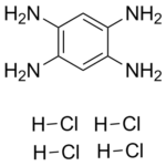
benzene-1,2,4,5-tetramine;tetrahydrochloride
|
| 别名 |
FAK Inhibitor 14; FAK Inhibitor Y15; Y15 hydrochloride; Y15 tetrahydrochloride; 1,2,4,5-Benzenetetramine tetrahydrochloride; Benzene-1,2,4,5-tetraamine tetrahydrochloride; FAK Inhibitor 14; Y15; Benzene-1,2,4,5-tetramine 4HCl; 1,2,4,5-Tetraaminobenzene tetrahydrochloride; MFCD00012970; Y 15; Y-15.
|
| HS Tariff Code |
292159
|
| 存储方式 |
Powder -20°C 3 years 4°C 2 years In solvent -80°C 6 months -20°C 1 month 注意: 请将本产品存放在密封且受保护的环境中,避免吸湿/受潮。 |
| 运输条件 |
Room temperature (This product is stable at ambient temperature for a few days during ordinary shipping and time spent in Customs)
|
| 溶解度 (体外实验) |
DMSO: 25~56 mg/mL (197.2~88.0 mM)
Water: ~56 mg/mL (~197.2 mM) |
|---|---|
| 溶解度 (体内实验) |
配方 1 中的溶解度: 10 mg/mL (35.21 mM) in PBS (这些助溶剂从左到右依次添加,逐一添加), 澄清溶液; 超声助溶。 (<60°C).
请根据您的实验动物和给药方式选择适当的溶解配方/方案: 1、请先配制澄清的储备液(如:用DMSO配置50 或 100 mg/mL母液(储备液)); 2、取适量母液,按从左到右的顺序依次添加助溶剂,澄清后再加入下一助溶剂。以 下列配方为例说明 (注意此配方只用于说明,并不一定代表此产品 的实际溶解配方): 10% DMSO → 40% PEG300 → 5% Tween-80 → 45% ddH2O (或 saline); 假设最终工作液的体积为 1 mL, 浓度为5 mg/mL: 取 100 μL 50 mg/mL 的澄清 DMSO 储备液加到 400 μL PEG300 中,混合均匀/澄清;向上述体系中加入50 μL Tween-80,混合均匀/澄清;然后继续加入450 μL ddH2O (或 saline)定容至 1 mL; 3、溶剂前显示的百分比是指该溶剂在最终溶液/工作液中的体积所占比例; 4、 如产品在配制过程中出现沉淀/析出,可通过加热(≤50℃)或超声的方式助溶; 5、为保证最佳实验结果,工作液请现配现用! 6、如不确定怎么将母液配置成体内动物实验的工作液,请查看说明书或联系我们; 7、 以上所有助溶剂都可在 Invivochem.cn网站购买。 |
| 制备储备液 | 1 mg | 5 mg | 10 mg | |
| 1 mM | 3.5210 mL | 17.6050 mL | 35.2100 mL | |
| 5 mM | 0.7042 mL | 3.5210 mL | 7.0420 mL | |
| 10 mM | 0.3521 mL | 1.7605 mL | 3.5210 mL |
1、根据实验需要选择合适的溶剂配制储备液 (母液):对于大多数产品,InvivoChem推荐用DMSO配置母液 (比如:5、10、20mM或者10、20、50 mg/mL浓度),个别水溶性高的产品可直接溶于水。产品在DMSO 、水或其他溶剂中的具体溶解度详见上”溶解度 (体外)”部分;
2、如果您找不到您想要的溶解度信息,或者很难将产品溶解在溶液中,请联系我们;
3、建议使用下列计算器进行相关计算(摩尔浓度计算器、稀释计算器、分子量计算器、重组计算器等);
4、母液配好之后,将其分装到常规用量,并储存在-20°C或-80°C,尽量减少反复冻融循环。
计算结果:
工作液浓度: mg/mL;
DMSO母液配制方法: mg 药物溶于 μL DMSO溶液(母液浓度 mg/mL)。如该浓度超过该批次药物DMSO溶解度,请首先与我们联系。
体内配方配制方法:取 μL DMSO母液,加入 μL PEG300,混匀澄清后加入μL Tween 80,混匀澄清后加入 μL ddH2O,混匀澄清。
(1) 请确保溶液澄清之后,再加入下一种溶剂 (助溶剂) 。可利用涡旋、超声或水浴加热等方法助溶;
(2) 一定要按顺序加入溶剂 (助溶剂) 。
|
|
|
|
InvivoChem的所有产品仅用于作科学研究,不面向患者销售
Copyright 2020 InvivoChem LLC | All Rights Reserved 粤ICP备20063088号-1


 COA
COA


 463611831
