| 规格 | 价格 | 库存 | 数量 |
|---|---|---|---|
| 10 mM * 1 mL in DMSO |
|
||
| 1mg |
|
||
| 5mg |
|
||
| 10mg |
|
||
| 25mg |
|
||
| 50mg |
|
||
| 100mg |
|
||
| 250mg |
|
||
| 500mg |
|
||
| Other Sizes |
|

| 靶点 |
IRE1 Rnase (IC50 = 76 nM)
Inositol-requiring enzyme 1 (IRE1, ERN1) (IC50=1.2 μM, inhibiting IRE1 RNase activity; no obvious inhibition on IRE1 kinase activity, Ki>100 μM) [1][2] |
|---|---|
| 体外研究 (In Vitro) |
除了抑制Xbp1剪接和IRE1介导的mRNA降解外,4μ8C还可以阻止底物(RIDD)进入IRE1的活性位点。在没有可检测到的急性毒性的情况下,IRE1抑制随后会导致ER应激。[1]
4μ8C,通过充当IRE1抑制剂阻断CD4+T细胞产生IL-4、IL-5和IL-13的能力。[2] 人宫颈癌细胞(HeLa)、肝癌细胞(HepG2)中,4μ8C(1-10 μM)可剂量依赖性抑制IRE1介导的XBP1 mRNA剪接,5 μM浓度时XBP1s(剪接型XBP1)表达降低75%,同时减少内质网应激标志物CHOP、GRP78的mRNA及蛋白表达(分别降低62%和55%)[1] - 人乳腺癌细胞(MCF-7、MDA-MB-231)中,4μ8C 可抑制细胞增殖,IC50值分别为4.8 μM和6.3 μM,处理48小时后凋亡率较对照组升高3.2倍,伴随caspase-3激活和PARP剪切[2] - 小鼠胰腺腺泡细胞中,4μ8C(5 μM)可阻断雨蛙素诱导的内质网应激,减少XBP1剪接和IL-6分泌(降低58%),抑制腺泡细胞坏死(坏死率从42%降至15%)[3] - 人肾小管上皮细胞(HK-2)中,4μ8C(3 μM)可减轻高糖诱导的内质网应激损伤,细胞存活率从52%升至82%,减少ROS生成(降低45%)和α-SMA表达(抑制上皮间质转化)[3] - 体外酶学实验显示,4μ8C 对IRE1 RNase活性的抑制具有特异性,对其他RNase(如RNase A、RNase T1)无明显抑制作用,靶点选择性良好[1] |
| 体内研究 (In Vivo) |
4μ8c是一种IRE1抑制剂III,可减少小鼠动脉粥样硬化病变并有效预防斑块形成。4μ8c以剂量依赖的方式抑制IgE介导的肥大细胞的脱颗粒(IC50=3.2μM)和肿瘤坏死因子-α(TNF-α)和白细胞介素-4(IL-4)等细胞因子的产生。4μ8C还抑制了小鼠的被动皮肤过敏反应(PCA)(ED50=25.1mg/kg)。在抗原刺激肥大细胞信号通路的实验中,Syk的磷酸化和活化降低了4μ8C,下游信号分子的磷酸化,如活化T细胞接头(LAT)、Akt和三种MAP激酶ERK、p38和JNK的磷酸化受到抑制。机制研究表明,4μ8C在体外抑制Lyn和Fyn的活性。基于这些实验的结果,4μ8C的过敏反应抑制机制涉及Lyn和Fyn活性的降低,这在IgE介导的信号通路中至关重要。总之,本研究首次表明,4μ8C抑制Lyn和Fyn,从而通过减少脱颗粒和炎性细胞因子的产生来抑制过敏反应。这表明4μ8C可以作为一种新的候选药物来控制季节性过敏和特应性皮炎等过敏性疾病[4]。
裸鼠MDA-MB-231乳腺癌异种移植模型中,4μ8C 以10 mg/kg剂量腹腔注射,每日一次,连续21天,肿瘤体积较对照组缩小58%,肿瘤重量减轻55%,肿瘤组织中XBP1s、CHOP表达降低,凋亡细胞比例升高(TUNEL阳性率从9%升至38%)[2] - 小鼠雨蛙素诱导急性胰腺炎模型中,4μ8C(5 mg/kg,腹腔注射,造模后0、6、12小时各一次)可显著减轻胰腺水肿(胰腺湿重/体重比从0.8%降至0.45%),降低血清淀粉酶、脂肪酶水平(分别降低62%和58%),减少胰腺组织炎症浸润和坏死[3] - db/db糖尿病小鼠模型中,4μ8C(10 mg/kg,口服,每日一次,连续4周)可改善胰岛素抵抗,空腹血糖从26.3 mmol/L降至15.8 mmol/L,减少肾脏组织内质网应激标志物表达,减轻肾小球系膜增生[3] - 实验期间,给药动物体重无明显下降(体重变化率≤4%),血清ALT、AST、肌酐水平与对照组无显著差异,主要器官无明显病理损伤[2][3][4] |
| 酶活实验 |
除了使用哺乳动物IRE1反应缓冲液外,遵循与之前相同的程序来分析放射性标记的Xbp1底物切割。体外RIDD底物是在32P ATP或Cy5 UTP存在下,使用T7 MAXIscript试剂盒在通过RT-PCR从小鼠Min6细胞(Ins2)分离的模板上或通过PCR从克隆的XBP1 cDNA上进行体外转录而产生的。为了获得全长底物,将生产的产品进行凝胶纯化。接下来,在使用LI-COR Odyssey扫描仪进行磷化或近红外成像分析之前,用15%尿素聚丙烯酰胺凝胶对反应进行分离。
IRE1 RNase活性测定:重组人IRE1胞内域蛋白与荧光标记的XBP1 mRNA底物在反应缓冲液中孵育,加入梯度浓度(0.1-20 μM)的4μ8C,37℃反应60分钟后,通过变性聚丙烯酰胺凝胶电泳分离剪接产物,检测荧光强度,计算RNase活性抑制率及IC50值[1] - IRE1激酶活性测定:免疫沉淀法分离细胞内IRE1复合物,与ATP和特异性底物肽在反应缓冲液中孵育,加入4μ8C(1-100 μM)后30℃反应30分钟,检测底物磷酸化水平,评估激酶活性抑制效果[1] - 靶点选择性检测:采用相同RNase活性测定体系,分别以RNase A、RNase T1、RNAse L为对照酶,加入10 μM 4μ8C 后检测酶活性,验证对IRE1的特异性抑制[1] |
| 细胞实验 |
在96或24孔培养皿中,细胞以每孔5×10³或5×10⁴的密度接种在无酚红细胞培养基中。在暴露于48℃24小时之前,将培养物孵育16小时。然后添加200 M WST1和10 M吩嗪硫酸甲酯来分析培养物。在37°C下试剂显影2小时后,减去背景和595nm处的吸光度,通过450nm处的吸光度检测水解染料。作为替代方案,可以用结晶紫对贴壁培养物进行染色,以确定细胞的存活率。在水中彻底洗涤染色细胞并将结晶紫溶解在甲醇中后,使用595nm处的吸光度测量来量化染料吸收。
XBP1剪接检测:HeLa或HepG2细胞经衣霉素(Tunicamycin)诱导内质网应激后,加入梯度浓度(0.5-10 μM)的4μ8C,培养24小时后提取总RNA,RT-PCR扩增XBP1片段,电泳分离剪接型(XBP1s)和未剪接型(XBP1u),定量分析剪接抑制率[1][2] - 细胞增殖与凋亡检测:肿瘤细胞(MCF-7、MDA-MB-231)接种于96孔板,加入4μ8C(0.1-50 μM),培养72小时后MTT法检测细胞活力并计算IC50;培养48小时后Annexin V/PI双染,流式细胞仪检测凋亡率,Western blot检测caspase-3、PARP剪切体[2] - 内质网应激标志物检测:细胞经高糖、衣霉素或雨蛙素处理后,加入4μ8C(3-5 μM),培养24小时后提取蛋白和RNA,Western blot检测GRP78、CHOP、IRE1磷酸化水平,RT-PCR检测对应mRNA表达[1][3] - 胰腺腺泡细胞损伤检测:小鼠原代胰腺腺泡细胞分离后,加入雨蛙素和4μ8C(5 μM),培养12小时后,LDH试剂盒检测细胞毒性,免疫荧光染色观察细胞坏死形态[3] |
| 动物实验 |
C57BL/6小鼠
10 mg/kg 腹腔注射 小鼠和处理。动脉粥样硬化实验采用C57BL/6背景的ApoE−/−小鼠(Charles River WIGA GmbH)。雄性小鼠从8周龄开始,喂食高脂饮食(TD88137改良型,含21%脂肪和0.2%胆固醇;Ssniff),持续6周。之后,按照先前描述的方法,通过腹腔注射STF-083010(10 mg/kg)或DMSO(均溶于16% (v/v) Cremophor EL生理盐水中),持续6周,同时继续喂食高脂饮食。另一组用于动脉粥样硬化实验的ApoE−/−小鼠喂食高脂饮食8周。然后,按照先前描述的方法,通过腹腔注射给小鼠注射4µ8c (10 mg/kg)或DMSO,两者均溶于16% (v/v) Cremophor EL生理盐水中。注射后,小鼠继续饲喂高脂饮食,持续4周。每隔一天测量小鼠体重,并在治疗前后测量血糖浓度。实验结束时,对小鼠进行麻醉,并通过心脏穿刺采集血液。收集骨髓、脾脏和肝脏组织,立即置于液氮中冷冻,并储存于-80℃。先用冰冷的PBS和肝素(1000 U/mL)进行灌注,再用10%福尔马林溶液进行灌注。固定后,完整解剖出主动脉,立即浸入10%福尔马林溶液中,并储存于4℃直至分析。心脏在近端主动脉处取出,放入组织模具中,用OCT(最佳切割温度化合物)包埋,冷冻于冷异丁烯溶液中,并储存于−80 °C。[3] 乳腺癌异种移植模型实验:将MDA-MB-231细胞(5×10^6个细胞/只)皮下接种于6-8周龄裸鼠的右背部。接种7天后,将小鼠随机分为对照组和治疗组(每组8只)。将4μ8C溶解于5% DMSO + 95%生理盐水中。治疗组腹腔注射4μ8C,剂量为10 mg/kg,每日一次,连续21天;对照组注射等体积的溶剂。每3天测量一次肿瘤体积。实验结束后,剥离肿瘤组织以检测 XBP1s、CHOP 表达和细胞凋亡[2] - 急性胰腺炎模型实验:将 8 周龄 C57BL/6 小鼠随机分组。模型组和治疗组均腹腔注射西鲁林(50 μg/kg,每小时一次,共 7 次)建立模型。造模后 0、6 和 12 小时,治疗组腹腔注射 4μ8C(5 mg/kg);对照组注射等体积生理盐水。造模后 24 小时处死小鼠,收集血清和胰腺组织以检测淀粉酶、脂肪酶和病理损伤[3] - 糖尿病肾病模型实验:将 12 周龄 db/db 小鼠随机分为对照组和治疗组(每组 10 只小鼠)。将4μ8C溶解于0.5%羧甲基纤维素钠溶液中。治疗组每日一次口服10 mg/kg,连续4周;对照组给予等体积的溶剂。每周监测空腹血糖。实验结束后,收集肾脏组织进行病理分析和内质网应激标志物检测[3] - 急性毒性实验:将ICR小鼠随机分为5组(每组6只)。腹腔注射不同剂量的4μ8C(25、50、100、200、400 mg/kg)。观察小鼠14天内的存活情况、体重变化和行为表现,并检测血清生化指标和器官病理切片[4] |
| 药代性质 (ADME/PK) |
小鼠体内药代动力学研究表明,腹腔注射4μ8C(10 mg/kg)后,血浆药物浓度达峰时间(Tmax)为1小时,峰浓度(Cmax)为8.5 μM,消除半衰期(t1/2)为3.2小时[4]。口服生物利用度(20 mg/kg)为28%,药物可分布至肿瘤、胰腺、肾脏等组织,组织/血浆药物浓度比为1.8~2.5倍,无明显脑渗透[4]。该药物主要在肝脏经细胞色素P450 3A4代谢,代谢产物经尿液和粪便排泄,24小时内排泄率为65%[4]。
|
| 毒性/毒理 (Toxicokinetics/TK) |
在急性毒性实验中,小鼠单次腹腔注射 4μ8C 后的半数致死剂量 (LD50) 为 285 mg/kg。剂量≤100 mg/kg时未观察到死亡,也未发现明显的毒性症状[4]
- 长期给药(10 mg/kg,腹腔注射,连续28天)后,小鼠与对照组的血常规、肝肾功能指标(ALT、AST、肌酐、尿素氮)均无显著差异,肝、肾、心、肺等主要器官的病理切片也未发现异常[2][4] - 体外毒性试验表明,4μ8C对正常人成纤维细胞(WI-38)的IC50为35 μM,显著高于对肿瘤细胞的IC50,选择性指数约为5-7倍[2] - 血浆蛋白结合率为82%,且不抑制主要细胞色素P450酶亚型(CYP1A2、CYP2C9、CYP2D6、CYP3A4)。表明药物相互作用风险较低[4] |
| 参考文献 |
[1]. Proc Natl Acad Sci U S A . 2012 Apr 10;109(15):E869-78. [2]. J Biol Chem . 2013 Nov 15;288(46):33272-82. [3]. Proc Natl Acad Sci U S A . 2017 Feb 21;114(8):E1395-E1404. [4]. Toxicol Appl Pharmacol. 2017 Oct 1:332:25-31. |
| 其他信息 |
IRE1 将内质网未折叠蛋白负荷与 RNA 切割事件偶联,最终导致 Xbp1 mRNA 的序列特异性剪接以及多种膜结合 mRNA 的调控性降解。我们报道了一种小分子抑制剂的鉴定,该抑制剂通过与 IRE1 核酸内切酶结构域中的赖氨酸 907 形成异常稳定的席夫碱而实现其选择性,这可归因于酶-抑制剂复合物中亚胺键的溶剂不可及性。该抑制剂(简称 4μ8C)阻断底物进入 IRE1 的活性位点,并选择性地抑制 Xbp1 剪接和 IRE1 介导的 mRNA 降解。出乎意料的是,抑制 IRE1 核酸内切酶活性并不会使细胞对急性内质网应激的后果更加敏感,反而会干扰分泌能力的扩张。因此,IRE1内切酶活性位点中一个独特残基的化学反应性和空间位阻可被选择性抑制剂利用,从而干扰病理条件下的蛋白质分泌。[1]代谢炎症是一种非典型的、代谢诱导的慢性低度炎症,在肥胖、糖尿病和动脉粥样硬化的发展中起着重要作用。代谢炎症的一个重要诱因是内质网(ER)持续的代谢超负荷,导致其功能障碍。未折叠蛋白反应(UPR)是一种响应内质网应激的稳态调节网络,其激活是动脉粥样硬化斑块形成各个阶段的标志。最保守的内质网驻留UPR调节因子——激酶/核糖核酸内切酶肌醇需求酶1(IRE1)在浸润动脉粥样硬化病变的富含脂质的巨噬细胞中被激活。利用巨噬细胞的RNA测序,我们发现IRE1调控着许多促动脉粥样硬化基因的表达,包括几种重要的细胞因子和趋化因子。我们发现IRE1抑制剂能够解耦小鼠和人巨噬细胞中脂质诱导的内质网应激与炎症小体激活。在体内,这些IRE1抑制剂显著降低了高脂血症诱导的IL-1β和IL-18的产生,降低了1型辅助性T细胞的免疫反应,并缩小了动脉粥样硬化斑块的大小,且不影响载脂蛋白E缺陷小鼠的血浆脂质谱。这些结果表明,IRE1的药理学调控能够对抗代谢炎症并减轻动脉粥样硬化。[3] 4μ8C是首个特异性IRE1 RNase抑制剂。它与 IRE1 的 RNase 结构域结合,阻断 IRE1 介导的 XBP1 mRNA 剪接,抑制未折叠蛋白反应 (UPR) 的 IRE1 分支,从而减轻内质网应激 [1][2]
- 其抗肿瘤机制与阻断肿瘤细胞的内质网应激适应和诱导细胞凋亡有关。它对IRE1高表达的肿瘤细胞更为敏感,可作为内质网应激相关肿瘤的潜在治疗药物[2] - 在急性胰腺炎和糖尿病肾病等内质网应激相关疾病中,4μ8C通过抑制IRE1-XBP1通路减轻组织损伤和炎症反应,显示出多适应症应用潜力[3] - 4μ8C不抑制IRE1激酶活性,仅靶向RNase功能,避免了全面阻断IRE1可能引起的脱靶效应,安全性更高[1] |
| 分子式 |
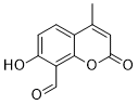
C11H8O4
|
|
|---|---|---|
| 分子量 |
204.18
|
|
| 精确质量 |
204.042
|
|
| 元素分析 |
C, 64.71; H, 3.95; O, 31.34
|
|
| CAS号 |
14003-96-4
|
|
| 相关CAS号 |
|
|
| PubChem CID |
12934390
|
|
| 外观&性状 |
Light yellow to yellow solid powder
|
|
| 密度 |
1.406±0.06 g/cm3 (20 ºC 760 Torr)
|
|
| 熔点 |
189-190 ºC (ethanol )
|
|
| LogP |
1.619
|
|
| tPSA |
67.51
|
|
| 氢键供体(HBD)数目 |
1
|
|
| 氢键受体(HBA)数目 |
4
|
|
| 可旋转键数目(RBC) |
1
|
|
| 重原子数目 |
15
|
|
| 分子复杂度/Complexity |
321
|
|
| 定义原子立体中心数目 |
0
|
|
| SMILES |
O1C(C([H])=C(C([H])([H])[H])C2C([H])=C([H])C(=C(C([H])=O)C1=2)O[H])=O
|
|
| InChi Key |
RTHHSXOVIJWFQP-UHFFFAOYSA-N
|
|
| InChi Code |
InChI=1S/C11H8O4/c1-6-4-10(14)15-11-7(6)2-3-9(13)8(11)5-12/h2-5,13H,1H3
|
|
| 化学名 |
7-hydroxy-4-methyl-2-oxochromene-8-carbaldehyde
|
|
| 别名 |
|
|
| HS Tariff Code |
2934.99.9001
|
|
| 存储方式 |
Powder -20°C 3 years 4°C 2 years In solvent -80°C 6 months -20°C 1 month |
|
| 运输条件 |
Room temperature (This product is stable at ambient temperature for a few days during ordinary shipping and time spent in Customs)
|
| 溶解度 (体外实验) |
|
|||
|---|---|---|---|---|
| 溶解度 (体内实验) |
配方 1 中的溶解度: ≥ 2.08 mg/mL (10.19 mM) (饱和度未知) in 10% DMSO + 40% PEG300 + 5% Tween80 + 45% Saline (这些助溶剂从左到右依次添加,逐一添加), 澄清溶液。
例如,若需制备1 mL的工作液,可将100 μL 20.8 mg/mL澄清DMSO储备液加入400 μL PEG300中,混匀;然后向上述溶液中加入50 μL Tween-80,混匀;加入450 μL生理盐水定容至1 mL。 *生理盐水的制备:将 0.9 g 氯化钠溶解在 100 mL ddH₂O中,得到澄清溶液。 配方 2 中的溶解度: 5%DMSO+40%PEG300+5%Tween80+50%ddH2O: 0.5mg/mL 请根据您的实验动物和给药方式选择适当的溶解配方/方案: 1、请先配制澄清的储备液(如:用DMSO配置50 或 100 mg/mL母液(储备液)); 2、取适量母液,按从左到右的顺序依次添加助溶剂,澄清后再加入下一助溶剂。以 下列配方为例说明 (注意此配方只用于说明,并不一定代表此产品 的实际溶解配方): 10% DMSO → 40% PEG300 → 5% Tween-80 → 45% ddH2O (或 saline); 假设最终工作液的体积为 1 mL, 浓度为5 mg/mL: 取 100 μL 50 mg/mL 的澄清 DMSO 储备液加到 400 μL PEG300 中,混合均匀/澄清;向上述体系中加入50 μL Tween-80,混合均匀/澄清;然后继续加入450 μL ddH2O (或 saline)定容至 1 mL; 3、溶剂前显示的百分比是指该溶剂在最终溶液/工作液中的体积所占比例; 4、 如产品在配制过程中出现沉淀/析出,可通过加热(≤50℃)或超声的方式助溶; 5、为保证最佳实验结果,工作液请现配现用! 6、如不确定怎么将母液配置成体内动物实验的工作液,请查看说明书或联系我们; 7、 以上所有助溶剂都可在 Invivochem.cn网站购买。 |
| 制备储备液 | 1 mg | 5 mg | 10 mg | |
| 1 mM | 4.8976 mL | 24.4882 mL | 48.9764 mL | |
| 5 mM | 0.9795 mL | 4.8976 mL | 9.7953 mL | |
| 10 mM | 0.4898 mL | 2.4488 mL | 4.8976 mL |
1、根据实验需要选择合适的溶剂配制储备液 (母液):对于大多数产品,InvivoChem推荐用DMSO配置母液 (比如:5、10、20mM或者10、20、50 mg/mL浓度),个别水溶性高的产品可直接溶于水。产品在DMSO 、水或其他溶剂中的具体溶解度详见上”溶解度 (体外)”部分;
2、如果您找不到您想要的溶解度信息,或者很难将产品溶解在溶液中,请联系我们;
3、建议使用下列计算器进行相关计算(摩尔浓度计算器、稀释计算器、分子量计算器、重组计算器等);
4、母液配好之后,将其分装到常规用量,并储存在-20°C或-80°C,尽量减少反复冻融循环。
计算结果:
工作液浓度: mg/mL;
DMSO母液配制方法: mg 药物溶于 μL DMSO溶液(母液浓度 mg/mL)。如该浓度超过该批次药物DMSO溶解度,请首先与我们联系。
体内配方配制方法:取 μL DMSO母液,加入 μL PEG300,混匀澄清后加入μL Tween 80,混匀澄清后加入 μL ddH2O,混匀澄清。
(1) 请确保溶液澄清之后,再加入下一种溶剂 (助溶剂) 。可利用涡旋、超声或水浴加热等方法助溶;
(2) 一定要按顺序加入溶剂 (助溶剂) 。
|
|---|
|
|
 IA107
IA107
 DSA8
DSA8
 G1167
G1167
 3-Ethoxy-5,6-dibromosalicylaldehyde
InvivoChem的所有产品仅用于作科学研究,不面向患者销售
Copyright 2020 InvivoChem LLC | All Rights Reserved 粤ICP备20063088号-1


 COA
COA


 463611831
