| 规格 | 价格 | 库存 | 数量 |
|---|---|---|---|
| 5mg |
|
||
| 10mg |
|
||
| 25mg |
|
||
| 50mg |
|
||
| 100mg |
|
||
| 250mg |
|
||
| Other Sizes |
|

| 靶点 |
Bcl-xL (Ki = 0.01 nM); Bcl-2 (Ki = 80 nM)
B-cell lymphoma 2 like 1 (BCL-XL) (binding Ki = 0.3 nM; recombinant protein binding IC50 = 0.4 nM) [2] - No significant binding activity to BCL-2 (Ki > 1000 nM), MCL-1 (Ki > 1000 nM), or BCL-W (Ki = 48 nM), with over 2500-fold selectivity for BCL-XL [2] - BCL-XL-dependent survival signaling pathway in colorectal cancer cells (cellular inhibition IC50 = 1–5 nM) [3] |
|---|---|
| 体外研究 (In Vitro) |
A-1155463 会干扰细胞中的 BCL-XL-BIM 复合物,但不会干扰 BCL-2-BIM 复合物。 A-1155463 对 BCL-2 依赖性 RS4;11 细胞没有可检测到的细胞毒性 (EC50>5 mM),但它会杀死 BCL-XL 依赖性 Molt-4 细胞 (EC50=70 nM)。在 BCL-XL 依赖性 H146 细胞中,A-1155463 会导致细胞凋亡的典型迹象,如线粒体释放细胞色素 c、激活 caspase 以及 caspase 依赖性亚 G0-G1 DNA 含量积累所示。
在BCL-XL依赖的肿瘤细胞系(H146小细胞肺癌、RS4;11急性淋巴细胞白血病、SW620结直肠癌)中,A-1155463 以剂量依赖性抑制细胞增殖,IC50范围为1–5 nM[2][3] - 处理SW620结直肠癌细胞48小时后,A-1155463 诱导凋亡率达60%,伴随caspase-3/7激活(活性升高3.5倍)和PARP切割(Western blot验证)[3] - 在BCL-2依赖的SU-DHL-6细胞或MCL-1依赖的KMS-11细胞中,A-1155463 浓度高达1000 nM时仍无抗增殖活性,验证靶点特异性[1][2] - 对BCL-XL高表达的结直肠癌患者来源肿瘤细胞(PDCs),A-1155463 抑制增殖IC50=2.8 nM,而BCL-XL低表达PDCs无明显响应[3] |
| 体内研究 (In Vivo) |
A-1155463 多次给药后可抑制体内 H146 小细胞肺癌异种移植肿瘤的生长,并导致小鼠出现基于机制的可逆性血小板减少症。单次腹腔注射给药后,SCID-Beige 小鼠的血小板迅速且可逆地减少,这证明了 A-1155463 能够发挥体内靶向活性。此外,A-1155463 治疗对携带肿瘤的 SCID-Beige 小鼠提供了适度但具有统计显着性的肿瘤生长抑制作用。随附的手稿将对 A-1155463 的活性与不同肿瘤类型的适当化疗相结合进行彻底的机制分析。在非肿瘤 SCID-Beige 小鼠中,单次腹膜内注射 5 mg/kg A-1155463 后,血小板计数显着下降,然后在 72 小时内恢复到正常水平。 SCID-Beige 小鼠接种 BCL-XL 依赖性 H146 肿瘤细胞后,每日腹膜内注射 5 mg/kg 剂量,持续 14 天,可显着抑制肿瘤生长(最大肿瘤生长抑制 = 44%),即停药后缓解。
在H146细胞异种移植裸鼠模型中,A-1155463 以15 mg/kg每日一次口服给药,连续14天,肿瘤体积较对照组减少82%,无明显体重下降[2] - 在SW620结直肠癌异种移植模型中,A-1155463 20 mg/kg每日一次口服给药,连续21天,肿瘤生长抑制率达78%,荷瘤小鼠中位生存期延长52%[3] - 单次口服15 mg/kg A-1155463 后,小鼠肿瘤组织药物浓度达峰时间(Tmax)为2小时,峰浓度(Cmax)=8.6 μM,有效浓度(>1 nM)维持16小时[2] - 在RS4;11细胞异种移植模型中,A-1155463 10 mg/kg每日一次口服给药,连续10天,可诱导肿瘤组织中caspase-3激活和凋亡细胞积累[1] |
| 酶活实验 |
和选择性 BCL-XL 抑制剂;它以皮摩尔亲和力 (Ki 0.01 nM) 与 BCL-XL 结合,同时与 BCL-2 (Ki = 80 nM) 和相关蛋白 BCL-W (Ki = 19 nM) 和 MCL-1 (Ki > 440 nM) 结合>弱1000倍。
使用帝肯Gemini机器人从500μM开始在DMSO中连续稀释化合物。使用Tecan Temo在测定缓冲液中进行1:10的中间稀释,并将10微升转移到白色384孔低容量Corning#3673测定板(2倍起始浓度;10%DMSO)上。然后,将10l蛋白质/探针/抗体混合物以上表所列的最终浓度添加到每个孔中。然后将样品在室温下孵育1小时。对于每种测定,探针/抗体和蛋白质/探针/抗体分别作为阴性和阳性对照包含在每个测定板上。使用340nm激发滤光片和520nm(f-Bak)和495nm(Tb标记的抗His抗体)发射滤光片在Envision上测量时间分辨荧光。解离常数(Ki)使用王方程确定(王Z。描述两个不同配体与蛋白质分子竞争结合的精确数学表达式。FEBS Lett.199536011-114)[2] 荧光偏振竞争结合实验:将荧光标记的BIM BH3肽与重组人BCL-XL蛋白孵育,加入梯度浓度的A-1155463 ,检测荧光偏振信号变化,计算药物与BCL-XL的结合Ki值和IC50值[2] - 表面等离子体共振(SPR)实验:将BCL-XL蛋白共价固定于传感器芯片表面,通入不同浓度的A-1155463 溶液,实时监测结合速率(ka)、解离速率(kd)及平衡解离常数(KD),验证结合特异性[2] - 等温滴定量热(ITC)实验:将A-1155463 溶液逐滴加入含BCL-XL蛋白的缓冲液中,记录热效应变化,计算结合焓(ΔH)和结合熵(ΔS),明确结合模式[2] |
| 细胞实验 |
A-1155463 以逐渐增加的浓度给予细胞。按照制造商的说明,72 小时后使用 CellTiter-Glo 发光细胞活力测定法测试细胞的活力。调整结果以反映未经处理的细胞。借助 GraphPad Prism 程序,可以计算 EC50。
细胞增殖和存活率测定[1] 将癌症乳腺细胞系以每孔5000个细胞接种在96细胞板中,并用9×3剂量基质中的化合物组合处理,其中以三倍步骤(20-0.001μM)稀释的navitoclax、venetoclax和a-1155463,以及50、5.0或0.5 nM的多烯紫杉醇。在评估存活率之前,将细胞孵育72小时。NSCLC细胞系在5×5剂量基质中用化合物组合治疗72小时,并如前所述进行评估[1]。 将卵巢癌症细胞系以每孔10000个细胞接种在96细胞板中,并在9×3剂量的基质中用化合物组合处理48小时。以三倍的步骤稀释多西他赛(10-1.1 nM)。Navitoclax、venetoclax和A-1155463分两步稀释(20-0.08μM)。[1] 菌落形成试验[1] 来源于正常人骨髓(BM)的造血前体细胞与不同浓度的navitoclax、venetoclax或A-1155463加上或减去5nM多烯紫杉醇在MethoCult 4230甲基纤维素基培养基中孵育,所述培养基补充有30ng mL-1重组人粒细胞集落刺激因子(rhGCSF)。DMSO用于制备所有测试化合物的储备溶液,并在所有孔中以<0.002%的终浓度存在。将来自三个不同批次(BM07B21195、BM0080512A和BM5H09)的冷冻BM光密度细胞在37°C下快速解冻,在补充了2%胎牛血清(IMDM+2%FBS)的10 mL Iscove改良Dulbecco's培养基(IMDM)中洗涤一次,然后重新悬浮在IMDM+2%FBS中。将2.4-4.3×10⁴个活细胞接种在6孔板的每个孔中,并在37°C(5%CO2)下在试验化合物的存在下孵育14-16天。由训练有素的技术人员使用光学显微镜计数包含至少30个粒细胞的集落形成单元。每种条件都进行了三次测试,以确定平均菌落数+/-一个标准差。 细胞增殖实验:H146、RS4;11、SW620细胞及结直肠癌PDCs分别接种于96孔板(每孔3×10³–5×10³个细胞),加入0.01–1000 nM梯度浓度的A-1155463 ,培养72小时后,采用MTT法检测细胞活力,计算增殖抑制率和IC50值[1][2][3] - 凋亡检测实验:SW620细胞经A-1155463 (5 nM)处理48小时后,收集细胞,用Annexin V-PE/7-AAD双染,通过流式细胞仪区分早期凋亡和晚期凋亡细胞比例[3] - Western blot实验:细胞经不同浓度A-1155463 处理后,提取总蛋白,经电泳、转膜、封闭,加入抗BCL-XL、caspase-3、cleaved-PARP及GAPDH一抗和荧光二抗,化学发光法检测蛋白表达及剪切水平[1][2][3] - 克隆形成实验:RS4;11细胞接种于6孔板(每孔1×10³个细胞),加入1 nM A-1155463 持续培养14天,甲醇固定后结晶紫染色,计数克隆形成数,计算克隆形成抑制率[1] |
| 动物实验 |
配制于 5% DMSO、10% EtOH、20% Cremaphor ELP 和 65% D5W 的溶液中;5 mg/kg;腹腔注射。
SCID-Beige 小鼠 小鼠及饲养 - SCID 和 SCID-bg 小鼠购自 Charles River 公司。每笼饲养 5、8 或 10 只小鼠。小鼠到达时的体重为 18-20 g。食物和水自由摄取。在实验开始前,小鼠在动物房内适应至少一周。动物在12小时光照:12小时黑暗周期(早上6:00开灯)的光照期进行测试。[2] 异种移植模型建立(小细胞肺癌/白血病):将处于对数生长期的H146或RS4;11细胞悬浮于PBS和Matrigel(体积比1:1)的混合物中,皮下接种到裸鼠右背部,每只小鼠接种2×10^6个细胞。[1][2] -异种移植模型建立(结直肠癌):将SW620细胞或结直肠癌浆细胞样树突状细胞(PDC)悬浮于PBS和Matrigel(体积比1:1)的混合物中,皮下接种到NOD-SCID小鼠右背部,每只小鼠接种3×10^6个细胞。[3] -给药方案1:当肿瘤体积达到120–150 mm³时,将小鼠随机分组。 (每组6-8只小鼠)。实验组每日口服A-1155463(10-15 mg/kg),而载体对照组连续10-14天给予含有5%二甲基亚砜、10%聚乙二醇400和85%生理盐水的混合液[1][2] - 给药方案2:在结直肠癌模型中,当肿瘤体积达到150 mm³时,实验组每日口服A-1155463(20 mg/kg),而对照组连续21天给予等体积的载体[3] - 检测指标:每2-3天测量一次肿瘤体积(公式:体积=长×宽²/2)和小鼠体重。给药期结束后,处死小鼠,解剖并称重肿瘤组织,部分组织用于凋亡标志物的Western blot检测或Ki-67(增殖标志物)的免疫组织化学检测[1][2][3] |
| 药代性质 (ADME/PK) |
小鼠口服给药后,A-1155463吸收迅速,达峰时间(Tmax)为1.5小时,口服生物利用度约为52%[2]
- 血浆半衰期(t1/2)为7.8小时,稳态分布容积(Vdss)为1.5 L/kg,血浆清除率(CL)为0.12 L/h/kg[2] - 肿瘤组织与血浆药物浓度比为4.2:1,给药16小时后仍可在肿瘤组织中检测到有效治疗浓度(>1 nM)[2] - 体外人肝微粒体代谢实验表明,A-1155463主要由CYP3A4代谢,具有良好的代谢稳定性(体外半衰期>2小时)[2] |
| 毒性/毒理 (Toxicokinetics/TK) |
在为期21天的小鼠毒性实验中,每日一次口服剂量高达30 mg/kg的A-1155463,小鼠体重增长正常(生长率>90%),肝肾功能(ALT、AST、肌酐、尿素氮)和血常规指标均未见明显异常[2][3]。
- 血浆蛋白结合率约为99%,主要与白蛋白和α1-酸性糖蛋白结合,无明显的血浆蛋白结合置换风险[2]。 - 单次高剂量(50 mg/kg)给药后,部分小鼠出现短暂的血小板计数下降(降低<25%),停药4天后恢复正常,未见其他血液学毒性[2]。 - 在结直肠癌模型中,长期(21天)给药后未观察到肠道毒性或组织病理学损伤[3]。 |
| 参考文献 |
|
| 其他信息 |
BCL-2/BCL-XL/BCL-W抑制剂ABT-263(navitoclax)在慢性淋巴细胞白血病等淋巴系统恶性肿瘤中展现出良好的临床活性。然而,由于BCL-XL抑制引起的血小板减少症限制了其在这些疾病中的疗效。这促使人们研发出BCL-2选择性抑制剂venetoclax(ABT-199/GDC-0199),该抑制剂在这些癌症中表现出强大的活性,且不影响血小板。navitoclax还被证明可以增强多西他赛在实体瘤临床前模型中的疗效,但由于中性粒细胞减少症,这种联合疗法的临床应用受到限制。我们使用维奈托克(venetoclax)以及BCL-XL选择性抑制剂A-1155463和A-1331852来评估抑制BCL-2或BCL-XL对维奈托克联合多西他赛疗效和毒性的相对贡献。体外和体内实验均表明,选择性BCL-2抑制剂可抑制粒细胞生成,这可能是维奈托克与多西他赛联合用药时临床观察到的中性粒细胞减少症加重的原因。相反,选择性BCL-XL抑制剂虽然不抑制粒细胞生成,但在与多西他赛联合治疗多种实体瘤时疗效显著。因此,BCL-XL选择性抑制剂有望增强多西他赛在实体瘤中的疗效,并避免维奈托克联合用药时观察到的中性粒细胞减少症加重。这些研究证明了这套选择性BCL-2家族抑制剂工具包的转化应用价值,并突显了它们作为改良癌症疗法的潜力。[1]
A-1155463是一种高效且选择性的BCL-XL抑制剂,是通过核磁共振(NMR)片段筛选和基于结构的药物设计发现的。与我们近期报道的抑制剂WEHI-539相比,该化合物对BCL-XL依赖性细胞系的抑制活性显著增强,且不具备WEHI-539固有的药理学缺陷。A-1155463在小鼠体内可引起机制性且可逆的血小板减少症,并在多次给药后抑制H146小细胞肺癌异种移植瘤的生长。因此,A-1155463不仅是研究BCL-XL生物学的优秀工具分子,也是进一步优化的有效先导化合物。[2] 程序性细胞死亡(或凋亡)缺陷是癌症的标志之一。包括BCL-2、BCL-X(L)和MCL-1在内的抗凋亡B细胞淋巴瘤2 (BCL-2)家族蛋白已被证实是多种癌症类型中的关键生存因子。由于BCL2和MCL1扩增的癌症类型更容易受到其各自编码蛋白的抑制,我们假设BCL2L1扩增频率较高的癌症对BCL-X(L)的依赖性更强。方法:为了识别BCL2L1扩增频率较高的肿瘤亚型,我们利用癌症基因组图谱(TCGA)数据库进行了数据挖掘。然后,我们使用选择性强效的BCL-X(L)抑制剂A-1155463和BCL2L1 siRNA,在一系列细胞系中评估了其对BCL-X(L)的依赖性。我们通过多种基因操作方法进一步研究了BCL-X(L)的作用机制。结果:我们发现,在所有检测的肿瘤类型中,结直肠癌中BCL2L1扩增的频率最高。BCL2L1拷贝数>3的结直肠癌细胞系对A-1155463更为敏感。与此一致的是,高表达BCL-XL和NOXA(一种拮抗MCL-1活性的促凋亡蛋白)的细胞系对A-1155463也敏感。通过siRNA沉默BCL-X(L)的表达可以杀死对A-1155463敏感的细胞系,而对耐药细胞系几乎没有影响。此外,在耐药细胞系中沉默MCL-1的表达可使其对A-1155463敏感,而沉默NOXA则消除这种敏感性。结论:这项工作证明了表征常见基因组改变对于识别癌症生存基因的实用性。此外,这些研究证明了高效选择性化合物 A-1155463 在研究 BCL-X(L) 在介导特定类型肿瘤细胞存活中的作用方面的实用性,并表明 BCL-X(L) 抑制剂可能是治疗 BCL-X(L) 和 NOXA 高表达的结直肠肿瘤的有效方法。[3] A-1155463 是一种高选择性、口服有效的 BCL-XL 小分子抑制剂,其作用机制包括与 BCL-XL 的 BH3 结合口袋竞争性结合,阻断其与促凋亡蛋白(BIM、BAX)的相互作用,并释放凋亡信号。[1][2][3] - 体外实验表明,A-1155463 与氟尿嘧啶(一种结直肠癌化疗药物)联合使用时,对 SW620 细胞的抗增殖活性具有协同作用。 (联合指数 CI = 0.65)[3] - 该药物对 BCL-XL 高表达且 BIM 阳性的结直肠癌亚型具有显著的治疗潜力,可作为此类患者的靶向治疗候选药物[3] - 与非选择性 BCL-2 家族抑制剂(例如 ABT-737)相比,A-1155463 对正常造血细胞的毒性更低,安全性更高[1][2] |
| 分子式 |
C35H32FN5O4S2
|
|
|---|---|---|
| 分子量 |
669.79
|
|
| 精确质量 |
669.187
|
|
| 元素分析 |
C, 62.76; H, 4.82; F, 2.84; N, 10.46; O, 9.55; S, 9.57
|
|
| CAS号 |
1235034-55-5
|
|
| 相关CAS号 |
|
|
| PubChem CID |
59447577
|
|
| 外观&性状 |
Off-white to yellow solid
|
|
| 密度 |
1.5±0.1 g/cm3
|
|
| 折射率 |
1.716
|
|
| LogP |
6.61
|
|
| tPSA |
164Ų
|
|
| 氢键供体(HBD)数目 |
2
|
|
| 氢键受体(HBA)数目 |
11
|
|
| 可旋转键数目(RBC) |
11
|
|
| 重原子数目 |
47
|
|
| 分子复杂度/Complexity |
1150
|
|
| 定义原子立体中心数目 |
0
|
|
| SMILES |
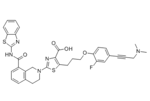
S1C(=C(C(=O)O)N=C1N1CC2C(C(NC3=NC4=CC=CC=C4S3)=O)=CC=CC=2CC1)CCCOC1C=CC(C#CCN(C)C)=CC=1F
|
|
| InChi Key |
SOYCFODXNRVBTI-UHFFFAOYSA-N
|
|
| InChi Code |
InChI=1S/C35H32FN5O4S2/c1-40(2)17-6-8-22-14-15-28(26(36)20-22)45-19-7-13-30-31(33(43)44)38-35(47-30)41-18-16-23-9-5-10-24(25(23)21-41)32(42)39-34-37-27-11-3-4-12-29(27)46-34/h3-5,9-12,14-15,20H,7,13,16-19,21H2,1-2H3,(H,43,44)(H,37,39,42)
|
|
| 化学名 |
2-[8-(1,3-benzothiazol-2-ylcarbamoyl)-3,4-dihydro-1H-isoquinolin-2-yl]-5-[3-[4-[3-(dimethylamino)prop-1-ynyl]-2-fluorophenoxy]propyl]-1,3-thiazole-4-carboxylic acid
|
|
| 别名 |
|
|
| HS Tariff Code |
2934.99.9001
|
|
| 存储方式 |
Powder -20°C 3 years 4°C 2 years In solvent -80°C 6 months -20°C 1 month |
|
| 运输条件 |
Room temperature (This product is stable at ambient temperature for a few days during ordinary shipping and time spent in Customs)
|
| 溶解度 (体外实验) |
|
|||
|---|---|---|---|---|
| 溶解度 (体内实验) |
配方 1 中的溶解度: ≥ 2.5 mg/mL (3.73 mM) (饱和度未知) in 10% DMSO + 40% PEG300 + 5% Tween80 + 45% Saline (这些助溶剂从左到右依次添加,逐一添加), 澄清溶液。
例如,若需制备1 mL的工作液,可将100 μL 25.0 mg/mL澄清DMSO储备液加入到400 μL PEG300中,混匀;然后向上述溶液中加入50 μL Tween-80,混匀;加入450 μL生理盐水定容至1 mL。 *生理盐水的制备:将 0.9 g 氯化钠溶解在 100 mL ddH₂O中,得到澄清溶液。 配方 2 中的溶解度: ≥ 2.5 mg/mL (3.73 mM) (饱和度未知) in 10% DMSO + 90% (20% SBE-β-CD in Saline) (这些助溶剂从左到右依次添加,逐一添加), 澄清溶液。 例如,若需制备1 mL的工作液,可将 100 μL 25.0 mg/mL澄清DMSO储备液加入900 μL 20% SBE-β-CD生理盐水溶液中,混匀。 *20% SBE-β-CD 生理盐水溶液的制备(4°C,1 周):将 2 g SBE-β-CD 溶解于 10 mL 生理盐水中,得到澄清溶液。 View More
配方 3 中的溶解度: ≥ 2.5 mg/mL (3.73 mM) (饱和度未知) in 10% DMSO + 90% Corn Oil (这些助溶剂从左到右依次添加,逐一添加), 澄清溶液。 1、请先配制澄清的储备液(如:用DMSO配置50 或 100 mg/mL母液(储备液)); 2、取适量母液,按从左到右的顺序依次添加助溶剂,澄清后再加入下一助溶剂。以 下列配方为例说明 (注意此配方只用于说明,并不一定代表此产品 的实际溶解配方): 10% DMSO → 40% PEG300 → 5% Tween-80 → 45% ddH2O (或 saline); 假设最终工作液的体积为 1 mL, 浓度为5 mg/mL: 取 100 μL 50 mg/mL 的澄清 DMSO 储备液加到 400 μL PEG300 中,混合均匀/澄清;向上述体系中加入50 μL Tween-80,混合均匀/澄清;然后继续加入450 μL ddH2O (或 saline)定容至 1 mL; 3、溶剂前显示的百分比是指该溶剂在最终溶液/工作液中的体积所占比例; 4、 如产品在配制过程中出现沉淀/析出,可通过加热(≤50℃)或超声的方式助溶; 5、为保证最佳实验结果,工作液请现配现用! 6、如不确定怎么将母液配置成体内动物实验的工作液,请查看说明书或联系我们; 7、 以上所有助溶剂都可在 Invivochem.cn网站购买。 |
| 制备储备液 | 1 mg | 5 mg | 10 mg | |
| 1 mM | 1.4930 mL | 7.4650 mL | 14.9301 mL | |
| 5 mM | 0.2986 mL | 1.4930 mL | 2.9860 mL | |
| 10 mM | 0.1493 mL | 0.7465 mL | 1.4930 mL |
1、根据实验需要选择合适的溶剂配制储备液 (母液):对于大多数产品,InvivoChem推荐用DMSO配置母液 (比如:5、10、20mM或者10、20、50 mg/mL浓度),个别水溶性高的产品可直接溶于水。产品在DMSO 、水或其他溶剂中的具体溶解度详见上”溶解度 (体外)”部分;
2、如果您找不到您想要的溶解度信息,或者很难将产品溶解在溶液中,请联系我们;
3、建议使用下列计算器进行相关计算(摩尔浓度计算器、稀释计算器、分子量计算器、重组计算器等);
4、母液配好之后,将其分装到常规用量,并储存在-20°C或-80°C,尽量减少反复冻融循环。
计算结果:
工作液浓度: mg/mL;
DMSO母液配制方法: mg 药物溶于 μL DMSO溶液(母液浓度 mg/mL)。如该浓度超过该批次药物DMSO溶解度,请首先与我们联系。
体内配方配制方法:取 μL DMSO母液,加入 μL PEG300,混匀澄清后加入μL Tween 80,混匀澄清后加入 μL ddH2O,混匀澄清。
(1) 请确保溶液澄清之后,再加入下一种溶剂 (助溶剂) 。可利用涡旋、超声或水浴加热等方法助溶;
(2) 一定要按顺序加入溶剂 (助溶剂) 。
 (a) Kinetics of platelet reduction and rebound following a single IP dose of A-1155463 in SCID-Beige mice.ACS Med Chem Lett.2014Aug 26;5(10):1088-93. |
|---|
X-ray crystal structure of A-1155463 (green) bound to BCL-XL.ACS Med Chem Lett.2014Aug 26;5(10):1088-93. |
 X-ray crystal structure of compound10(green) bound to BCL-XL.ACS Med Chem Lett.2014Aug 26;5(10):1088-93. |
 ReACp53 scrambled peptide TFA
 p53-hDM2 cyclic peptide inhibitor 16e
 MMRi6
MMRi6
 dsP53-285 saRNA
InvivoChem的所有产品仅用于作科学研究,不面向患者销售
Copyright 2020 InvivoChem LLC | All Rights Reserved 粤ICP备20063088号-1


 COA
COA

 463611831
