| 规格 | 价格 | 库存 | 数量 |
|---|---|---|---|
| 10 mM * 1 mL in DMSO |
|
||
| 1mg |
|
||
| 2mg |
|
||
| 5mg |
|
||
| 10mg |
|
||
| 25mg |
|
||
| 50mg |
|
||
| 100mg |
|
||
| Other Sizes |
|
| 靶点 |
Akt1 (Ki = 11 nM); PKA (Ki = 16 nM); CDK2 (Ki = 46 nM); GSK3β (Ki = 110 nM); ERK2 (Ki = 260 nM); PKCδ (Ki = 360 nM); RSK2 (Ki = 580 nM); MAPK-AP2 (Ki = 1.1 μM nM); PKCγ (Ki = 1.2 μM); Chk1 (Ki = 2.6 μM); CK2 (Ki = 5.4 μM); SRC (Ki = 13 μM)
Akt1 (Protein Kinase Bα) (IC50: 4.3 nM for human Akt1 kinase activity) [1] - Akt2 (Protein Kinase Bβ) (IC50: 6.6 nM for human Akt2 kinase activity) [1] - Akt3 (Protein Kinase Bγ) (IC50: 8.1 nM for human Akt3 kinase activity) [1] - SphK1 (Sphingosine Kinase 1) (IC50: 2.7 μM for human SphK1 enzyme activity) [2] - No significant inhibition of other kinases (PDK1, PKCα, ERK1/2 IC50 > 1000 nM) [1] |
|---|---|
| 体外研究 (In Vitro) |
A-674563 可减缓肿瘤细胞的增殖,EC50 为 0.4 μM[1]。 A563 (0-10 µM) 显着降低 STS 细胞中 GSK3 和 MDM2 的磷酸化。所有 STS 细胞系均被 A563 抑制,48 小时时 IC50 值范围为 0.22 ±0.034 M (SW684) 至 0.35 ±0.06 µM (SKLMS1)。在 STS 细胞中,A563 导致细胞凋亡和 G2 细胞周期停滞。独立于 p53,A563(1 µM/12 小时)可增加 GADD45A[2] 的表达。在培养的人黑色素瘤细胞中,A-674563 (10–1000 nM) 具有细胞毒性和抗增殖作用。它还诱导黑色素瘤细胞凋亡,这种凋亡可被 caspase 抑制剂抑制,并通过 Akt 依赖性和 Akt 独立机制抑制黑色素瘤细胞[3]。 A-674563 添加到 U937 和 AmL 祖细胞中时具有细胞毒性和抗增殖作用,可激活 U937 和 AmL 祖细胞中的 caspase-3/9 和细胞凋亡,并在阻断 Akt 的同时操纵 AmL 细胞中的其他信号传导[4]。
抑制Akt信号通路 A-674563 hydrochloride(0.1–100 nM)以剂量依赖方式抑制多种癌细胞中Akt磷酸化(Ser473/Thr308)。在MDA-MB-468乳腺癌细胞中,10 nM浓度下p-Akt(Ser473)降低78%,p-Akt(Thr308)降低72%(Western blot)[1] - 急性髓系白血病(AML)细胞抗增殖活性 在HL-60和U937 AML细胞中,A-674563 hydrochloride(0.5–20 μM)抑制增殖,IC50值分别为3.2 μM(HL-60)和4.5 μM(U937)(72小时MTT法)。10 μM浓度下诱导48%细胞凋亡(Annexin V阳性),SphK1活性降低63%(SphK1酶活性实验)[2] - 软组织肉瘤细胞毒性 软组织肉瘤细胞系(HT-1080、SW872)对A-674563 hydrochloride高度敏感(IC50:HT-1080为1.8 μM,SW872为2.3 μM)。5 μM浓度下,qPCR检测显示GADD45α mRNA上调3.2倍,Western blot显示蛋白上调2.8倍,且不依赖p53状态[3] - 黑色素瘤细胞抗肿瘤活性 在A375和SK-MEL-28黑色素瘤细胞中,A-674563 hydrochloride(1–15 μM)抑制细胞活力(IC50:A375为2.7 μM,SK-MEL-28为3.1 μM)和克隆形成(5 μM浓度下抑制65%)。阻断Akt介导的mTOR信号(5 μM浓度下p-mTOR降低68%)[4] |
| 体内研究 (In Vivo) |
A-674563(40 mg/kg/d,口服)的单药治疗活性可忽略不计,但 A-674563 联合紫杉醇可显着提高 PC-3 前列腺癌异种移植模型的治疗效果。在口服葡萄糖耐量试验中,A-674563(20、100 mg/kg)会增加血浆胰岛素水平[1]。 A563(20 mg/kg/bid;po)使小鼠仅损失少量体重,同时显示出肿瘤生长缓慢和肿瘤体积显着差异。接受 A563 治疗的肿瘤表达较高水平的 GADD45 和较低水平的 PCNA(增殖的核标记物)。此外,A563 处理的样本中细胞凋亡标记物 TUNEL 检测染色水平有所上升 [2]。 A-674563(25、100 mg/kg,每日灌洗)显着降低小鼠 A375 异种移植物的生长[3]。 A-674563(15、40 mg/kg)注射可增加小鼠存活率,同时抑制 U937 异种移植物体内生长[4]。
人肿瘤异种移植瘤的抗肿瘤疗效 荷MDA-MB-468乳腺癌异种移植瘤裸鼠,每日两次腹腔注射A-674563 hydrochloride(25、50 mg/kg)连续21天。50 mg/kg剂量组肿瘤体积抑制率73%,肿瘤重量减少69%。肿瘤组织分析显示p-Akt(Ser473)降低65%,切割型caspase-3增加2.9倍[1] - 软组织肉瘤异种移植瘤疗效 荷HT-1080异种移植瘤裸鼠经A-674563 hydrochloride(50 mg/kg,腹腔注射,每日两次,连续14天)处理后,肿瘤生长抑制率68%。肿瘤中GADD45α蛋白水平增加2.6倍,增殖标志物Ki-67降低58%[3] - 黑色素瘤异种移植瘤抑制作用 荷A375黑色素瘤异种移植瘤小鼠经40 mg/kg A-674563 hydrochloride(腹腔注射,每日一次,连续21天)处理后,肿瘤生长减少62%。中位生存期从对照组的32天延长至治疗组的51天[4] |
| 酶活实验 |
重组CK2、PKCγ和PKCδ;PKA;细胞周期蛋白依赖性激酶2/CyclinA、GSK3β、MAPK-AP2、Src和RSK2;细胞外信号调节激酶2(cKit)已商业化获得。他标记的Akt1(S378A、S381A、T450D、S473D;139-480)、Chk1(1-269)、KDR(789-1354)、Flt-1(786-1338)和磷脂酰肌醇依赖性激酶1(1-396)使用FastBac杆状病毒表达系统表达,并使用镍(他的标签)或谷胱甘肽S-转移酶亲和层析纯化。肽底物具有生物素Ahx肽的一般结构,序列如下:Akt、EELSPFRGRSRSRSSAPPNLWAAQR;PKA,LRRASLG;PKCγ和PKCδ、ERMRPRKRQGSVRRV;CK2、rraddsdddd;细胞周期蛋白依赖性激酶2;GSK3β、YRRAAVPPSPLSRHSSPHQS(p)EDEEE;MAPK-AP2,KKLNRTLSVA;RSK2,KKKNRTLSVA;细胞外信号调节激酶2,KRELVEPLTPSGEAPNQALR;Chk1,AKVSRSGLYRSPMPENLRPR;磷脂酰肌醇依赖性激酶1;KDR、Flt-1和cKit、AEEEYFFLFA酰胺。对于Src测定,使用生物素化底物PTK-2。如前所述,使用基于放射性FlashPlate的检测平台评估激酶活性的抑制[1]。
Akt激酶活性实验 重组人Akt1/Akt2/Akt3蛋白与A-674563 hydrochloride(0.001–100 nM)在含ATP和肽底物的反应缓冲液中孵育,37°C反应45分钟后,通过发光实验检测磷酸化底物,根据激酶抑制量效曲线计算IC50值[1] - SphK1酶活性实验 重组人SphK1与A-674563 hydrochloride(0.1–10 μM)在含鞘氨醇和ATP的反应缓冲液中孵育,37°C反应60分钟后,HPLC定量产物(1-磷酸鞘氨醇),根据产物生成抑制率推导IC50值[2] - 激酶选择性实验 检测化合物(1 μM)对50种激酶(包括PDK1、PKCα、ERK1/2)的抑制活性,通过放射性或发光实验测定激酶活性,对比Akt亚型的IC50值评估选择性[1] |
| 细胞实验 |
用 200 μL PBS 轻轻洗涤 96 孔板上的细胞。正常生长培养基用 Alamar Blue 试剂按 1:10 稀释。根据制造商的说明,在反应完全展开之前,将 100 M 稀释的 Alamar Blue 试剂添加到 96 孔板的每个孔中。使用 fmax 荧光酶标仪进行分析,激发和发射波长均设置为 544 nm。制造商的SOFTmax PRO软件用于分析数据。
癌细胞抗增殖实验 AML(HL-60、U937)、软组织肉瘤(HT-1080、SW872)、黑色素瘤(A375、SK-MEL-28)细胞接种于96孔板(5×10³细胞/孔),过夜培养后加入A-674563 hydrochloride(0.1–20 μM),孵育72小时。加入MTT试剂检测570 nm吸光度,计算细胞活力和IC50值[2, 3, 4] - 凋亡与细胞周期实验 HL-60细胞经A-674563 hydrochloride(5–15 μM)处理48小时后,Annexin V-FITC/PI染色流式细胞术检测凋亡;细胞固定后经碘化丙啶染色,流式细胞术分析细胞周期分布[2] - Western blot与qPCR分析 癌细胞经A-674563 hydrochloride(1–10 μM)处理24–48小时后裂解,蛋白经SDS-PAGE分离,转膜后用抗p-Akt、Akt、p-mTOR、mTOR、GADD45α、切割型caspase-3及β-肌动蛋白抗体孵育;提取总RNA,qPCR检测GADD45α mRNA表达[1, 3, 4] - 克隆形成实验 A375黑色素瘤细胞接种于6孔板(2×10³细胞/孔),经A-674563 hydrochloride(1–10 μM)处理14天后,克隆经固定、染色后计数,计算抑制率[4] |
| 动物实验 |
免疫缺陷雄性SCID小鼠为6至8周龄。将1×10⁶个3T3-Akt1或2×10⁶个MiaPaCa-2和PC-3细胞悬浮于50% Matrigel中,皮下注射至小鼠侧腹部。早期治疗研究中,小鼠随机分组,并在接种后第二天开始治疗。每组10只小鼠,包括对照组。在已形成肿瘤的研究中,待肿瘤达到指定大小后,将小鼠按肿瘤大小分组(每组10只小鼠)。每周两次使用数字游标卡尺测量肿瘤大小。肿瘤体积使用公式V=L×W²/2估算。A-443654以0.2% HPMC为溶剂皮下注射。A-674563以5%葡萄糖为溶剂口服给药。在试验中加入吉西他滨和紫杉醇。
MDA-MB-468 乳腺癌异种移植模型 雌性裸鼠(6-8 周龄,18-22 g)适应环境 7 天。将 MDA-MB-468 细胞(5×10⁶ 个细胞/只)皮下注射到右侧腹部。当肿瘤体积达到 100-150 mm³ 时,将小鼠随机分组(每组 n=6)。A-674563 盐酸盐 溶于生理盐水 + 5% DMSO 中,以 25 或 50 mg/kg 的剂量腹腔注射,每日两次,连续 21 天。对照组注射生理盐水 + 5% DMSO。每2天测量一次肿瘤体积,每周记录一次体重[1] - HT-1080软组织肉瘤异种移植模型 将携带HT-1080异种移植瘤(通过皮下注射2×10⁶个细胞/只小鼠建立)的裸鼠用A-674563盐酸盐(50 mg/kg,腹腔注射)治疗,每日两次,持续14天。研究结束时,切除肿瘤进行GADD45α的Western blot分析和Ki-67免疫组化[3] - A375黑色素瘤异种移植模型 将A375细胞(3×10⁶个细胞/只小鼠)皮下注射到裸鼠体内。当肿瘤体积达到 80–120 mm³ 时,每日一次腹腔注射 A-674563 盐酸盐(40 mg/kg),持续 21 天。每 3 天测量一次肿瘤体积,并每日记录生存情况 [4] |
| 药代性质 (ADME/PK) |
血浆半衰期 (t1/2):小鼠腹腔注射 50 mg/kg 后为 4.2 小时 [1]
- 血浆蛋白结合率:92.5%(体外人血浆)[1] - 组织分布:腹腔注射 50 mg/kg 后 1 小时,肿瘤组织 (5.8 μM)、肝脏 (6.3 μM) 和脾脏 (4.7 μM) 中的浓度最高 [1] - 排泄:72 小时内 68% 经粪便排出,23% 经尿液排出 [1] |
| 毒性/毒理 (Toxicokinetics/TK) |
急性毒性:小鼠单次腹腔注射剂量高达 200 mg/kg 后未见死亡;未见明显毒性症状(嗜睡、腹泻、体重减轻)[1]
- 慢性毒性:在为期 28 天的重复给药研究中(小鼠:25、50、100 mg/kg,每日两次腹腔注射),未观察到血液学参数(白细胞、红细胞、血小板)或肝肾功能指标(ALT、AST、BUN、肌酐)的显著变化。肝脏、肾脏、心脏和肺的组织学检查未发现药物相关病变[1] - 血液毒性:100 mg/kg 剂量组 17% 的小鼠出现轻度中性粒细胞减少症(1-2 级),停药 7 天内可逆转[1] |
| 参考文献 |
|
| 其他信息 |
本研究旨在探讨Akt1特异性抑制剂A-674563的抗黑色素瘤活性。我们发现,A-674563对人黑色素瘤细胞(A375、WM-115和SK-Mel-2细胞系)具有抗增殖和细胞毒性作用。A-674563诱导人黑色素瘤细胞发生caspase依赖性凋亡,且其细胞毒性作用可被caspase抑制剂预处理所抑制。此外,A-674563处理可阻断A375黑色素瘤细胞中Akt及其下游S6激酶1 (S6K1)的激活。值得注意的是,通过引入组成型激活的Akt1 (ca-Akt1)恢复Akt-S6K1的激活仅部分减弱了A-674563对A375细胞的细胞毒性作用。此外,A-674563 可诱导 A375 细胞产生促凋亡的神经酰胺。值得注意的是,鞘氨醇-1-磷酸 (S1P) 可抑制 A-674563 诱导的神经酰胺生成及其后续的 A375 细胞凋亡。另一方面,与葡萄糖基神经酰胺合成酶 (GCS) 抑制剂 PDMP 或细胞可渗透的短链神经酰胺 (C6) 联合处理可增强 A-674563 对 A375 细胞的细胞毒性。体内实验表明,A-674563 灌胃可抑制重症联合免疫缺陷 (SCID) 小鼠体内 A375 异种移植瘤的生长。在 A-674563 处理的 A375 异种移植瘤中也观察到了 Akt 失活、caspase-3 激活和神经酰胺生成。这些结果共同表明,A-674563 具有强大的抗黑色素瘤活性,涉及 Akt 依赖性和 Akt 非依赖性机制[4]。作用机制:A-674563 盐酸盐是一种强效且选择性的 Akt 亚型(Akt1/Akt2/Akt3)和 SphK1 抑制剂。它阻断 Akt 介导的信号通路(mTOR、NF-κB),从而抑制癌细胞的增殖和存活。同时抑制 SphK1 可减少鞘氨醇-1-磷酸的产生,进一步增强其凋亡作用。在软组织肉瘤中,它以不依赖于p53的方式上调GADD45α,从而诱导细胞周期阻滞[1, 2, 3, 4]
- 治疗潜力:适用于治疗实体瘤和血液系统恶性肿瘤,包括乳腺癌、软组织肉瘤、黑色素瘤和急性髓系白血病(AML)。它对p53野生型和p53突变型肿瘤均有效[1, 2, 3, 4] - 选择性优势:对Akt亚型具有高度选择性,优于其他激酶,从而最大限度地减少脱靶效应。同时靶向Akt和SphK1可产生协同抗肿瘤活性[1, 2] - 临床前研究现状:在多种肿瘤模型中展现出强大的临床前疗效,且毒性可控,支持其作为抗癌候选药物的潜力[1, 3, 4] |
| 分子式 |
C22H23CLN4O
|
|---|---|
| 分子量 |
394.8972
|
| 精确质量 |
394.156
|
| 元素分析 |
C, 66.91; H, 5.87; Cl, 8.98; N, 14.19; O, 4.05
|
| CAS号 |
2070009-66-2
|
| 相关CAS号 |
2070009-66-2 (HCl);552325-73-2;
|
| PubChem CID |
73357690
|
| 外观&性状 |
Light yellow to khaki solid
|
| tPSA |
76.8Ų
|
| 氢键供体(HBD)数目 |
3
|
| 氢键受体(HBA)数目 |
4
|
| 可旋转键数目(RBC) |
6
|
| 重原子数目 |
28
|
| 分子复杂度/Complexity |
456
|
| 定义原子立体中心数目 |
1
|
| SMILES |
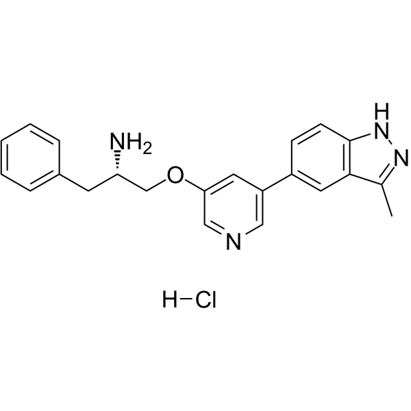
CC1=C2C=C(C=CC2=NN1)C3=CC(=CN=C3)OC[C@H](CC4=CC=CC=C4)N.Cl
|
| InChi Key |
HLNHYVLLEFHBJD-FYZYNONXSA-N
|
| InChi Code |
InChI=1S/C22H22N4O.ClH/c1-15-21-11-17(7-8-22(21)26-25-15)18-10-20(13-24-12-18)27-14-19(23)9-16-5-3-2-4-6-16;/h2-8,10-13,19H,9,14,23H2,1H3,(H,25,26);1H/t19-;/m0./s1
|
| 化学名 |
(2S)-1-[5-(3-methyl-2H-indazol-5-yl)pyridin-3-yl]oxy-3-phenylpropan-2-amine;hydrochloride
|
| 别名 |
A-674563 hydrochloride; A-674563 hcl; A674563 hydrochloride; A674563 hcl; A 674563 hydrochloride; A 674563 hcl
|
| HS Tariff Code |
2934.99.9001
|
| 存储方式 |
Powder -20°C 3 years 4°C 2 years In solvent -80°C 6 months -20°C 1 month 注意: 请将本产品存放在密封且受保护的环境中(例如氮气保护),避免吸湿/受潮。 |
| 运输条件 |
Room temperature (This product is stable at ambient temperature for a few days during ordinary shipping and time spent in Customs)
|
| 溶解度 (体外实验) |
H2O: ~100 mg/mL (~253.2 mM)
DMSO: ~50 mg/mL (~126.6 mM) |
|---|---|
| 溶解度 (体内实验) |
配方 1 中的溶解度: ≥ 2.08 mg/mL (5.27 mM) (饱和度未知) in 10% DMSO + 40% PEG300 + 5% Tween80 + 45% Saline (这些助溶剂从左到右依次添加,逐一添加), 澄清溶液。
例如,若需制备1 mL的工作液,可将100 μL 20.8 mg/mL澄清DMSO储备液加入400 μL PEG300中,混匀;然后向上述溶液中加入50 μL Tween-80,混匀;加入450 μL生理盐水定容至1 mL。 *生理盐水的制备:将 0.9 g 氯化钠溶解在 100 mL ddH₂O中,得到澄清溶液。 配方 2 中的溶解度: ≥ 2.08 mg/mL (5.27 mM) (饱和度未知) in 10% DMSO + 90% (20% SBE-β-CD in Saline) (这些助溶剂从左到右依次添加,逐一添加), 澄清溶液。 例如,若需制备1 mL的工作液,可将 100 μL 20.8 mg/mL澄清DMSO储备液加入900 μL 20% SBE-β-CD生理盐水溶液中,混匀。 *20% SBE-β-CD 生理盐水溶液的制备(4°C,1 周):将 2 g SBE-β-CD 溶解于 10 mL 生理盐水中,得到澄清溶液。 View More
配方 3 中的溶解度: ≥ 2.08 mg/mL (5.27 mM) (饱和度未知) in 10% DMSO + 90% Corn Oil (这些助溶剂从左到右依次添加,逐一添加), 澄清溶液。 配方 4 中的溶解度: 100 mg/mL (253.23 mM) in PBS (这些助溶剂从左到右依次添加,逐一添加), 澄清溶液; 超声助溶 (<60°C). 1、请先配制澄清的储备液(如:用DMSO配置50 或 100 mg/mL母液(储备液)); 2、取适量母液,按从左到右的顺序依次添加助溶剂,澄清后再加入下一助溶剂。以 下列配方为例说明 (注意此配方只用于说明,并不一定代表此产品 的实际溶解配方): 10% DMSO → 40% PEG300 → 5% Tween-80 → 45% ddH2O (或 saline); 假设最终工作液的体积为 1 mL, 浓度为5 mg/mL: 取 100 μL 50 mg/mL 的澄清 DMSO 储备液加到 400 μL PEG300 中,混合均匀/澄清;向上述体系中加入50 μL Tween-80,混合均匀/澄清;然后继续加入450 μL ddH2O (或 saline)定容至 1 mL; 3、溶剂前显示的百分比是指该溶剂在最终溶液/工作液中的体积所占比例; 4、 如产品在配制过程中出现沉淀/析出,可通过加热(≤50℃)或超声的方式助溶; 5、为保证最佳实验结果,工作液请现配现用! 6、如不确定怎么将母液配置成体内动物实验的工作液,请查看说明书或联系我们; 7、 以上所有助溶剂都可在 Invivochem.cn网站购买。 |
| 制备储备液 | 1 mg | 5 mg | 10 mg | |
| 1 mM | 2.5323 mL | 12.6614 mL | 25.3229 mL | |
| 5 mM | 0.5065 mL | 2.5323 mL | 5.0646 mL | |
| 10 mM | 0.2532 mL | 1.2661 mL | 2.5323 mL |
1、根据实验需要选择合适的溶剂配制储备液 (母液):对于大多数产品,InvivoChem推荐用DMSO配置母液 (比如:5、10、20mM或者10、20、50 mg/mL浓度),个别水溶性高的产品可直接溶于水。产品在DMSO 、水或其他溶剂中的具体溶解度详见上”溶解度 (体外)”部分;
2、如果您找不到您想要的溶解度信息,或者很难将产品溶解在溶液中,请联系我们;
3、建议使用下列计算器进行相关计算(摩尔浓度计算器、稀释计算器、分子量计算器、重组计算器等);
4、母液配好之后,将其分装到常规用量,并储存在-20°C或-80°C,尽量减少反复冻融循环。
计算结果:
工作液浓度: mg/mL;
DMSO母液配制方法: mg 药物溶于 μL DMSO溶液(母液浓度 mg/mL)。如该浓度超过该批次药物DMSO溶解度,请首先与我们联系。
体内配方配制方法:取 μL DMSO母液,加入 μL PEG300,混匀澄清后加入μL Tween 80,混匀澄清后加入 μL ddH2O,混匀澄清。
(1) 请确保溶液澄清之后,再加入下一种溶剂 (助溶剂) 。可利用涡旋、超声或水浴加热等方法助溶;
(2) 一定要按顺序加入溶剂 (助溶剂) 。
|
 D-myo-Inositol-1,3,4,5,6-pentaphosphate ammonium salt
 Biotin-AKT3 Recombinant Human Active Protein Kinase
 Biotin-AKT2 Recombinant Human Active Protein Kinase
 AKT1 E17K Recombinant Human Active Protein Kinase
InvivoChem的所有产品仅用于作科学研究,不面向患者销售
Copyright 2020 InvivoChem LLC | All Rights Reserved 粤ICP备20063088号-1


 COA
COA

 463611831
