| 规格 | 价格 | 库存 | 数量 |
|---|---|---|---|
| 10mg |
|
||
| 25mg |
|
||
| 50mg |
|
||
| 100mg |
|
||
| 250mg |
|
||
| 500mg |
|
||
| Other Sizes |
|

| 靶点 |
VEGFR-2 (IC50 = 0.02 μM); VEGFR-1 (IC50 = 0.38 μM); mVEGFR-2 (IC50 = 0.23 μM); VEGFR-3 (IC50 = 0.18 μM); PDGFR-β (IC50 = 1.4 μM)
Vascular Endothelial Growth Factor Receptor 2 (VEGFR2/KDR) (IC50 = 25 nM in recombinant VEGFR2 kinase activity assay; Ki = 18 nM in ATP-competitive binding assay) [1] Fibroblast Growth Factor Receptor 1 (FGFR1) (IC50 = 1200 nM in recombinant FGFR1 kinase activity assay, weak inhibition) [1] Platelet-Derived Growth Factor Receptor β (PDGFRβ) (IC50 = 850 nM in recombinant PDGFRβ kinase activity assay, weak inhibition) [1] |
|---|---|
| 体外研究 (In Vitro) |
NVP-ACC789 是人 VEGFR-1、VEGFR-2(小鼠 VEGFR-2)、VEGFR-3 和 PDGFR-β 的抑制剂,根据数据,IC50 分别为 0.38、0.02 (0.23)、0.18 和 1.4 μM酶促激酶测定。当 VEGFR-2 抑制剂 NVP-ACC789 添加到 VEGF 处理的培养物中时,BME 细胞的数量从 1 μM 减少至基线水平。同样,NVP-ACC789 可显着抑制 bFGF 诱导的 BME 细胞增殖,浓度从 1 μM 抑制至 10 μM,但不能抑制至基础水平。 NVP-ACC789 被发现是 VEGF 诱导的 HUVE 细胞增殖的强抑制剂,IC50 为 1.6 nM。此外,NVP-ACC789完全阻止VEGF引起的BME和BAE细胞侵袭以及VEGF-C引起的BAE细胞侵袭。在这两种细胞类型中,抑制作用都是剂量依赖性的,在 1 μM 时达到最大影响[1]。
NVP-ACC789 (ACC-789)是强效、选择性的ATP竞争性重组人VEGFR2(KDR)激酶抑制剂,IC50为25 nM;对FGFR1(IC50=1200 nM)和PDGFRβ(IC50=850 nM)仅表现出弱抑制活性,对VEGFR2的选择性是这两种受体的40倍以上[1] 在人脐静脉内皮细胞(HUVECs)中,NVP-ACC789 (ACC-789)剂量依赖性抑制VEGF诱导的细胞增殖,IC50为32 nM;抑制碱性成纤维细胞生长因子(bFGF)诱导的增殖的IC50为450 nM;100 nM浓度下可抑制80%的VEGF诱导的HUVEC迁移(Boyden小室实验)和75%的管形成(Matrigel管形成实验),而抑制50%的bFGF诱导迁移和管形成则需要1 μM浓度[1] NVP-ACC789 (ACC-789)在浓度≥50 nM时,可阻断VEGF诱导的HUVEC中VEGFR2及其下游信号分子(ERK1/2、Akt)的磷酸化(蛋白质印迹法检测);浓度≤1 μM时,对bFGF诱导的FGFR1磷酸化无显著影响[1] |
| 体内研究 (In Vivo) |
口服 NVP-ACC789 每天一次,持续六天,以剂量依赖性方式抑制 VEGF 诱导的血管生成。虽然 NVP-ACC789 不具有线性剂量反应曲线,但它确实在一定程度上抑制对 bFGF 的反应[1]。
在鸡胚尿囊膜(CAM)血管生成模型中,局部应用NVP-ACC789 (ACC-789)(0.1-10 μg/卵)可剂量依赖性抑制VEGF诱导的血管形成,ED50为0.8 μg/卵;对bFGF诱导的血管生成也有抑制作用,ED50为5 μg/卵[1] 在小鼠角膜血管生成模型中,结膜下注射NVP-ACC789 (ACC-789)(1-10 μg/眼)可使VEGF诱导的角膜新生血管面积减少60%-90%(血管面积定量),10 μg/眼剂量对bFGF诱导的角膜血管生成的抑制率为40%[1] 在小鼠Matrigel栓血管生成模型中,腹腔注射NVP-ACC789 (ACC-789)(5-20 mg/kg/天)持续7天,可使VEGF/bFGF处理的Matrigel栓血管化程度降低55%-85%(血红蛋白含量检测);20 mg/kg剂量下,栓中血红蛋白水平从溶媒组的12.5 mg/g降至2.1 mg/g[1] |
| 酶活实验 |
在 6 孔板中,将人 VEGFR-2 转染的 CHO 细胞铺板并使其生长至约 80% 汇合。添加连续稀释的 NVP-ACC789 后,将细胞在不含胎牛血清 (FCS) 的培养基中于 37°C 培养两小时。然后,添加 20 ng/mL VEGF。在 37°C 下孵育 5 分钟后,细胞在裂解前进行两次冰冷的磷酸盐缓冲盐水洗涤。 4°C 下离心 10 分钟以去除细胞核。确定裂解物中存在多少蛋白质[1]。
重组VEGFR2(KDR)激酶活性实验:制备重组人VEGFR2胞内激酶域蛋白(785-1354位氨基酸),在含20 mM Tris-HCl(pH 7.5)、10 mM MgCl2和1 mM DTT的激酶反应缓冲液中稀释至终浓度10 nM;将酶与系列浓度的NVP-ACC789 (ACC-789)(10⁻¹²-10⁻⁶ M)及ATP(100 μM,生理浓度)在30℃孵育10分钟;加入VEGFR2特异性生物素化肽底物(100 μM),继续孵育60分钟;用50 mM EDTA终止反应,加入链霉亲和素包被磁珠和抗磷酸化肽抗体,通过酶标仪检测化学发光信号;对抑制曲线进行非线性回归分析,计算IC50值[1] ATP竞争性结合实验:通过胺偶联法将重组VEGFR2激酶域固定在生物传感器芯片上;在含1 mM ATP的结合缓冲液中注入系列浓度的NVP-ACC789 (ACC-789)(10⁻¹²-10⁻⁶ M);利用表面等离子体共振(SPR)实时监测共振单位(RU)的变化;将结合数据拟合至一位点竞争性结合模型,计算Ki值[1] |
| 细胞实验 |
它决定 HUVE 细胞增殖。涂有 1.5% 明胶的 96 孔板每孔接种 5 x 10³ 细胞。然后将细胞在含有 5% 胎牛血清 (FCS) 的内皮细胞生长培养基中孵育 24 小时。更换培养基后,将细胞在必要的基础培养基(1.5% FCS)中再培养 24 小时。完成后,将含有 0.5 ng/mL bFGF 或 50 ng/mL VEGF 的新鲜培养基添加到必需的基础培养基中。 NVP-ACC789 之后立即添加生长因子。在添加 BrdU 标记溶液之前,将细胞再培养 24 小时。 24 小时后,除去标记溶液,固定细胞,使用 TMB 底物和过氧化物酶标记的抗 BrdU 抗体可以看到掺入的 BrdU [1]。
1. HUVEC增殖实验:原代分离人脐静脉内皮细胞(HUVECs),培养至第2-4代;以5×10³个/孔的密度接种于96孔板,无血清培养基培养24小时使细胞同步化;用系列浓度的NVP-ACC789 (ACC-789)(10⁻¹¹-10⁻⁵ M)预处理细胞30分钟,加入VEGF(20 ng/mL)或bFGF(10 ng/mL)孵育72小时;采用BrdU掺入实验评估细胞增殖,检测450 nm处吸光度,计算生长因子诱导增殖的抑制IC50值[1] 2. HUVEC迁移实验:将HUVECs以1×10⁵个/小室的密度接种于Boyden小室(8 μm孔径)上室,培养基为含NVP-ACC789 (ACC-789)(10 nM-1 μM)的无血清培养基;下室加入VEGF(20 ng/mL)或bFGF(10 ng/mL)作为趋化因子;37℃孵育4小时后,甲醇固定细胞,结晶紫染色,在五个随机高倍视野计数迁移细胞数;计算与溶媒处理组相比的迁移抑制百分比[1] 3. HUVEC管形成实验:用Matrigel包被24孔板,37℃孵育30分钟使其聚合;接种HUVECs(2×10⁴个/孔)于含NVP-ACC789 (ACC-789)(10 nM-1 μM)和VEGF/bFGF的培养基中;37℃孵育18小时,光学显微镜下拍摄管结构图像,用图像分析软件定量管长度和分支点数;计算管形成抑制率[1] 4. VEGFR2信号通路的蛋白质印迹实验:无血清饥饿的HUVECs经NVP-ACC789 (ACC-789)(10 nM-1 μM)预处理30分钟后,加入VEGF(20 ng/mL)刺激10分钟;提取总细胞蛋白,经SDS-PAGE分离后转膜,用抗磷酸化VEGFR2(Tyr1175)、总VEGFR2、磷酸化ERK1/2(Thr202/Tyr204)、总ERK1/2、磷酸化Akt(Ser473)、总Akt及GAPDH(内参)抗体孵育;定量条带强度以评估信号通路的抑制程度[1] |
| 动物实验 |
将含有0.8% (w/v) 琼脂和肝素(20 U/mL)的多孔聚四氟乙烯腔室(体积0.5 mL)皮下植入雌性小鼠的背部,腔室中添加或不添加VEGF(2 μg/mL)或bFGF(0.3 μg/mL)。从腔室植入前一天开始,小鼠每天口服NVP-ACC789或赋形剂(5%二甲基亚砜,1% Tween 80水溶液),持续5天。治疗结束后,处死小鼠并取出腔室。小心取出腔室,称量腔室周围生长的血管化组织,并测定血红蛋白水平以确定血液含量。计算血管生成反应的抑制百分比,即组织重量或血容量的增加。剂量反应曲线(抑制百分比与剂量的关系)用于估算EC50值。每个剂量组有六只动物参与每个实验,每个剂量至少在两项独立的研究中进行检验[1]。
1. 鸡绒毛尿囊膜 (CAM) 检测:将受精鸡卵在 37°C 下孵育 3 天,然后在蛋壳上开窗以暴露 CAM;在孵育第 7 天,将浸泡有 NVP-ACC789 (ACC-789)(0.1、1、5、10 μg/卵,溶于 50 μL 10% DMSO + 90% PBS) 或载体的无菌滤片贴于 CAM 上,同时加入 VEGF(50 ng/卵)或 bFGF(100 ng/卵);密封鸡蛋并继续孵育 48 小时;使用立体显微镜观察血管,计数滤膜盘周围绒毛尿囊膜 (CAM) 中的血管分支数量,并计算血管生成抑制百分比 [1] 2. 小鼠角膜血管生成试验:使用 6-8 周龄 C57BL/6 小鼠(雌性,18-22 g);在麻醉下,使用角膜刮刀在角膜基质中创建一个微囊,并将含有 VEGF (50 ng) 或 bFGF (100 ng) 的水凝胶颗粒植入囊内;在颗粒植入后立即通过结膜下注射给予 NVP-ACC789 (ACC-789)(1、5、10 μg/眼,溶于 5 μL 5% DMSO + 95% PBS 中),并每天一次,持续 6 天;在第 7 天,用裂隙灯检查角膜,拍摄新生血管图像,并使用图像分析软件测量血管长入面积[1] 3. 小鼠 Matrigel 塞试验:制备添加了 VEGF (50 ng/mL) 和 bFGF (25 ng/mL) 的 Matrigel;将 NVP-ACC789 (ACC-789) (终浓度 0.1、1、5 μM) 与 Matrigel 混合,或通过腹腔注射(5、10、20 mg/kg/天,溶于 10% DMSO + 40% PEG400 + 50% 生理盐水)给 8 周龄 BALB/c 小鼠(雄性,20-25 g),在皮下植入 Matrigel 塞 (0.5 mL/只小鼠) 后立即给药,并每天一次,持续 7 天;第 8 天,收集 Matrigel 胶塞,在 PBS 中匀浆,并使用比色法测定血红蛋白含量以量化血管化;计算与载体处理组相比的抑制百分比[1] |
| 参考文献 | |
| 其他信息 |
NVP-ACC789 (ACC-789) 是一种合成的小分子 ATP 竞争性 VEGFR2 (KDR) 拮抗剂,被开发为一种潜在的抗血管生成药物,用于治疗实体瘤和血管生成相关疾病(例如,年龄相关性黄斑变性)[1]
NVP-ACC789 (ACC-789) 的抗血管生成机制涉及选择性抑制 VEGFR2 激酶活性和下游信号传导(ERK1/2、Akt),从而阻断 VEGF 诱导的内皮细胞增殖、迁移和管状结构形成——血管生成的关键步骤;在高浓度下,NVP-ACC789 (ACC-789) 对 bFGF 诱导的血管生成也表现出较弱的交叉抑制作用,这可能是由于其间接调节内皮细胞对 bFGF 的反应所致[1]。NVP-ACC789 在浓度高达 10 μM 时对 HUVEC 或其他正常细胞系(例如 NIH 3T3 成纤维细胞)均无明显的细胞毒性(MTT 法检测细胞存活率 >90%),表明其具有良好的抗血管生成治疗安全性[1]。 |
| 分子式 |
C21H17N4BR
|
|---|---|
| 分子量 |
405.29048
|
| 精确质量 |
404.064
|
| 元素分析 |
C, 62.23; H, 4.23; Br, 19.71; N, 13.82
|
| CAS号 |
300842-64-2
|
| 相关CAS号 |
300842-64-2
|
| PubChem CID |
6918616
|
| 外观&性状 |
Light yellow to yellow solid powder
|
| 密度 |
1.441g/cm3
|
| 沸点 |
607.7ºC at 760 mmHg
|
| 闪点 |
321.3ºC
|
| 折射率 |
1.707
|
| LogP |
4.852
|
| tPSA |
53.93
|
| 氢键供体(HBD)数目 |
1
|
| 氢键受体(HBA)数目 |
4
|
| 可旋转键数目(RBC) |
4
|
| 重原子数目 |
26
|
| 分子复杂度/Complexity |
442
|
| 定义原子立体中心数目 |
0
|
| SMILES |
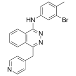
CC1=CC=C(NC2=NN=C(CC3=CC=NC=C3)C4=C2C=CC=C4)C=C1Br
|
| InChi Key |
GXWKSXUPEFVUOO-UHFFFAOYSA-N
|
| InChi Code |
InChI=1S/C21H17BrN4/c1-14-6-7-16(13-19(14)22)24-21-18-5-3-2-4-17(18)20(25-26-21)12-15-8-10-23-11-9-15/h2-11,13H,12H2,1H3,(H,24,26)
|
| 化学名 |
N-(3-bromo-4-methylphenyl)-4-(pyridin-4-ylmethyl)phthalazin-1-amine
|
| 别名 |
ZK-202650; ZK 202650; ZK202650; NVP-ACC789; NVP-ACC 789; NVP-ACC-789; ACC-789; ACC 789; ACC789
|
| HS Tariff Code |
2934.99.9001
|
| 存储方式 |
Powder -20°C 3 years 4°C 2 years In solvent -80°C 6 months -20°C 1 month |
| 运输条件 |
Room temperature (This product is stable at ambient temperature for a few days during ordinary shipping and time spent in Customs)
|
| 溶解度 (体外实验) |
DMSO: ~25 mg/mL (~61.7 mM)
|
|---|---|
| 溶解度 (体内实验) |
配方 1 中的溶解度: ≥ 2.5 mg/mL (6.17 mM) (饱和度未知) in 10% DMSO + 40% PEG300 + 5% Tween80 + 45% Saline (这些助溶剂从左到右依次添加,逐一添加), 澄清溶液。
例如,若需制备1 mL的工作液,可将100 μL 25.0 mg/mL澄清DMSO储备液加入到400 μL PEG300中,混匀;然后向上述溶液中加入50 μL Tween-80,混匀;加入450 μL生理盐水定容至1 mL。 *生理盐水的制备:将 0.9 g 氯化钠溶解在 100 mL ddH₂O中,得到澄清溶液。 配方 2 中的溶解度: ≥ 2.5 mg/mL (6.17 mM) (饱和度未知) in 10% DMSO + 90% Corn Oil (这些助溶剂从左到右依次添加,逐一添加), 澄清溶液。 例如,若需制备1 mL的工作液,可将 100 μL 25.0 mg/mL 澄清 DMSO 储备液加入到 900 μL 玉米油中并混合均匀。 请根据您的实验动物和给药方式选择适当的溶解配方/方案: 1、请先配制澄清的储备液(如:用DMSO配置50 或 100 mg/mL母液(储备液)); 2、取适量母液,按从左到右的顺序依次添加助溶剂,澄清后再加入下一助溶剂。以 下列配方为例说明 (注意此配方只用于说明,并不一定代表此产品 的实际溶解配方): 10% DMSO → 40% PEG300 → 5% Tween-80 → 45% ddH2O (或 saline); 假设最终工作液的体积为 1 mL, 浓度为5 mg/mL: 取 100 μL 50 mg/mL 的澄清 DMSO 储备液加到 400 μL PEG300 中,混合均匀/澄清;向上述体系中加入50 μL Tween-80,混合均匀/澄清;然后继续加入450 μL ddH2O (或 saline)定容至 1 mL; 3、溶剂前显示的百分比是指该溶剂在最终溶液/工作液中的体积所占比例; 4、 如产品在配制过程中出现沉淀/析出,可通过加热(≤50℃)或超声的方式助溶; 5、为保证最佳实验结果,工作液请现配现用! 6、如不确定怎么将母液配置成体内动物实验的工作液,请查看说明书或联系我们; 7、 以上所有助溶剂都可在 Invivochem.cn网站购买。 |
| 制备储备液 | 1 mg | 5 mg | 10 mg | |
| 1 mM | 2.4674 mL | 12.3368 mL | 24.6737 mL | |
| 5 mM | 0.4935 mL | 2.4674 mL | 4.9347 mL | |
| 10 mM | 0.2467 mL | 1.2337 mL | 2.4674 mL |
1、根据实验需要选择合适的溶剂配制储备液 (母液):对于大多数产品,InvivoChem推荐用DMSO配置母液 (比如:5、10、20mM或者10、20、50 mg/mL浓度),个别水溶性高的产品可直接溶于水。产品在DMSO 、水或其他溶剂中的具体溶解度详见上”溶解度 (体外)”部分;
2、如果您找不到您想要的溶解度信息,或者很难将产品溶解在溶液中,请联系我们;
3、建议使用下列计算器进行相关计算(摩尔浓度计算器、稀释计算器、分子量计算器、重组计算器等);
4、母液配好之后,将其分装到常规用量,并储存在-20°C或-80°C,尽量减少反复冻融循环。
计算结果:
工作液浓度: mg/mL;
DMSO母液配制方法: mg 药物溶于 μL DMSO溶液(母液浓度 mg/mL)。如该浓度超过该批次药物DMSO溶解度,请首先与我们联系。
体内配方配制方法:取 μL DMSO母液,加入 μL PEG300,混匀澄清后加入μL Tween 80,混匀澄清后加入 μL ddH2O,混匀澄清。
(1) 请确保溶液澄清之后,再加入下一种溶剂 (助溶剂) 。可利用涡旋、超声或水浴加热等方法助溶;
(2) 一定要按顺序加入溶剂 (助溶剂) 。
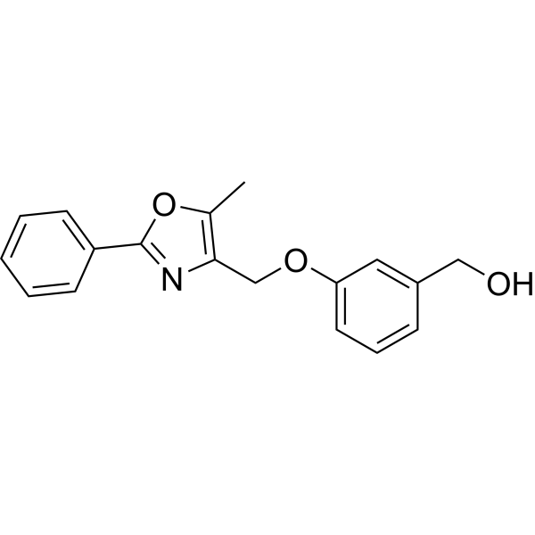 VEGFR2-IN-7
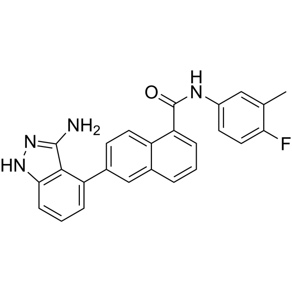 SYHA1813
 VEGFR-2-IN-38
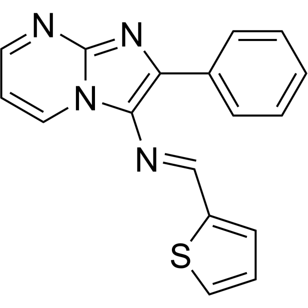
 BHEP
BHEP
InvivoChem的所有产品仅用于作科学研究,不面向患者销售
Copyright 2020 InvivoChem LLC | All Rights Reserved 粤ICP备20063088号-1


 COA
COA
 463611831
