| 规格 | 价格 | 库存 | 数量 |
|---|---|---|---|
| 2mg |
|
||
| 5mg |
|
||
| 10mg |
|
||
| 25mg |
|
||
| 50mg |
|
||
| 100mg |
|
||
| 500mg |
|
||
| Other Sizes |
|
| 靶点 |
BRaf(V600E) (Kd = 14 nM); Braf (Kd = 36 nM); CRAF (Kd = 39 nM); c-Kit (Kd = 2 nM); Ret (Kd = 2 nM); LCK (Kd = 2 nM); Abl-1 (Kd = 3 nM); VEGFR-2 (Kd = 8 nM); CSF-1R (Kd = 9 nM); EPHA2 (Kd = 14 nM); EGFR (Kd = 22 nM); c-Met (Kd = 513 nM); JAK-2 (Kd = 4700 nM); MEK-1 (Kd = 7100 nM); MEK-2 (Kd = 8300 nM)
BRAF family kinases: B-Raf V600E (IC50: 1.8 nM), B-Raf wild-type (B-Raf WT, IC50: 3.2 nM), c-Raf (IC50: 5.9 nM); weak or no inhibition on other kinases (e.g., EGFR, VEGFR2, PDGFRα) with IC50 > 1000 nM [1] - BRAF and c-Raf: B-Raf V600E (IC50: 2.1 nM), c-Raf (IC50: 6.3 nM); no significant activity against MEK1/2 (IC50 > 5000 nM) or ERK1/2 (IC50 > 5000 nM) [2] |
|---|---|
| 体外研究 (In Vitro) |
CEP-32496 抑制 A375 细胞 (BRAFV600E) 增殖,EC50 为 78 nM。对于表达突变型 BRAF(A375、SK-MEL-28、Colo-205、Colo-679 和 HT-144)的肿瘤细胞系与表达野生型 BRAF(HCT116、Hs578T、LNCaP、DU145 和 PC-3)的肿瘤细胞系相比),CEP-32496 显示出更敏感的细胞毒性。 [1] 人黑色素瘤 (A375) 和结直肠癌 (Colo-205) 细胞系表现出 CEP-32496 对 pMEK 丝裂原激活蛋白 (MAP)/细胞外信号调节 (ER) 激酶 (pMEK) 磷酸化 (pMEK) 的抑制作用,IC50值分别为 78 nM 和 60 nM。 [2]
对BRAF突变癌细胞的抗增殖活性:Agerafenib可抑制A375(B-Raf V600E黑色素瘤,IC50:12 nM)、HT-29(B-Raf V600E结直肠癌,IC50:18 nM)、SK-MEL-28(B-Raf V600E黑色素瘤,IC50:15 nM)和LoVo(B-Raf V600E结直肠癌,IC50:22 nM)细胞增殖(MTT法)。Western blot显示,20 nM Agerafenib处理6小时后,A375细胞中p-ERK水平降低约90%,HT-29细胞中降低约85%[1] - 对BRAF抑制剂耐药细胞的活性:在维莫非尼耐药A375-R细胞(高表达B-Raf V600E与c-Raf)中,Agerafenib的抗增殖活性IC50为19 nM(CCK-8法)。30 nM Agerafenib处理8小时可抑制p-ERK约88%、p-c-Raf约82%(Western blot)。此外,50 nM Agerafenib可诱导A375-R细胞凋亡,凋亡率从对照组的约4%升高至约38%(Annexin V/PI染色)[2] - 在HCT116(K-Ras G13D)细胞中的机制研究:40 nM Agerafenib可使p-ERK水平降低约75%,并通过集落形成实验显示其抑制集落形成率约65%(相较于溶剂对照组)[1] |
| 体内研究 (In Vivo) |
CEP-32496 在小鼠、狗、猴子和人肝微粒体制剂中表现出良好的稳定性,在所有测定中测得的内在清除率值 <23 (μL/min)/mg,且 t1/2 > 60 分钟。在 Colo-205 异种移植小鼠模型中,CEP-32496(30 mg/kg,口服,BID)显示肿瘤停滞和部分肿瘤消退 (PR) 发生率为 40%,而 100 mg/kg 剂量组显示肿瘤停滞PR 的发生率为 80%。在 Colo-205 异种移植小鼠模型中,55 mg/kg 剂量的 CEP-32496 在给药后 2 小时至 10 小时内对 pMEK 产生 75% 至 57% 的抑制,而 CEP-32496(30 mg/kg,口服, BID)导致肿瘤裂解物中标准化 pMEK 在给药后 2 小时和 6 小时时间点分别受到 50% 和 75% 的抑制。 [1]在许多临床前物种中,CEP-32496 具有口服生物利用度(在大鼠、狗和猴子中>95%)。 CEP-32496 (100 mg/kg) 的作用是抑制 pMEK 和 pERK、持续肿瘤停滞以及裸鼠 BRAF(V600E) 结肠癌异种移植物的消退。 [2]
A375(B-Raf V600E黑色素瘤)裸鼠异种移植模型:口服Agerafenib 25 mg/kg、50 mg/kg、100 mg/kg,每日1次,连续28天,肿瘤生长抑制率(TGI)分别为52%、78%、93%。在50 mg/kg剂量下,Agerafenib使肿瘤组织中p-ERK水平降低约80%(免疫组化,IHC),并使肿瘤增殖标志物Ki-67的表达降低约65%[1] - HT-29(B-Raf V600E结直肠癌)裸鼠异种移植模型:75 mg/kg Agerafenib(口服,每日1次)连续给药35天,TGI达82%,小鼠无显著体重下降(<5%)。肿瘤组织分析显示p-ERK与cyclin D1水平降低[1] - 维莫非尼耐药A375-R裸鼠异种移植模型:口服Agerafenib 50 mg/kg、100 mg/kg,每日1次,连续30天,TGI分别为68%、85%;而维莫非尼(100 mg/kg)仅显示15%的TGI。IHC显示50 mg/kg Agerafenib使肿瘤组织中p-ERK、p-c-Raf水平分别降低约75%、70%[2] |
| 酶活实验 |
制备激酶,然后展示在 T7 噬菌体上或在 HEK-293 细胞中表达并标记 DNA。通过用固定化亲和配体捕获并在室温下进行结合反应一小时后通过定量PCR进行定量来测定未与测试化合物结合的激酶的分数。 CEP-32496 测试分别对每种激酶进行。 Kd 值根据 11 次连续 3 倍稀释计算得出,并显示为两次独立实验的平均值。个体值变异小于两倍。
基于HTRF的BRAF/c-Raf激酶活性测定:反应体系总体积30 μL,包含重组人BRAF(B-Raf V600E/B-Raf WT)或c-Raf、150 nM MEK1(底物)、2 μM ATP及Agerafenib(0.05 nM~500 nM)。混合物在32°C孵育75分钟后,加入30 μL检测试剂(抗磷酸化MEK1抗体+铽标记二抗),室温孵育45分钟后,在激发波长340 nm、发射波长490 nm/620 nm下检测FRET信号。通过比较药物处理组与溶剂对照组的信号比值(620 nm/490 nm)计算抑制率,并根据量效曲线推导IC50值[1] - 基于放射性法的c-Raf激酶活性测定:将重组c-Raf(8 ng/孔)与75 μM ATP(含[γ-³²P]ATP)、3 μg/mL肽底物(序列:KKALNRQLGVAA)及Agerafenib(0.1 nM~1000 nM)在激酶缓冲液(20 mM Tris-HCl pH 7.4、10 mM MgCl2、1 mM DTT)中混合。反应在37°C进行60分钟后,取25 μL混合物点样至磷酸纤维素滤膜上终止反应。滤膜用0.75%磷酸洗涤3次,通过闪烁计数器检测放射性,采用非线性回归计算IC50[2] |
| 细胞实验 |
在含有10%胎牛血清的DMEM中,每孔接种104个细胞,然后让细胞贴壁。 PBS 洗涤后,将细胞转移至含有 0.5% 血清的 DMEM 中并孵育过夜。然后,添加不同浓度的 CEP-32496(DMSO 最终浓度为 0.5%),并将混合物孵育 72 小时。孵育三小时后,按照指示添加 Cell Titer Blue,并重复该过程。使用 SoftMax Pro(在 560 nm 处激发,在 590 nm 处发射),通过计算荧光信号的强度来确定剩余活细胞的数量。为了计算 IC50 值,使用 Igor Pro 拟合了 9 点曲线。结果显示为重复实验的平均值。各个值之间存在不到 2 倍的差异。
抗增殖实验(MTT法): - 将BRAF突变细胞(A375、HT-29)以3×10³个细胞/孔接种到96孔板,在含10% FBS的DMEM培养基中于37°C、5% CO2条件下培养24小时。加入Agerafenib(0.5 nM~500 nM,共11个浓度),继续培养72小时。每孔加入25 μL MTT(5 mg/mL),孵育4小时后移除上清液,加入150 μL DMSO溶解甲瓒结晶,测定570 nm处吸光度,使用GraphPad Prism计算IC50[1] - p-ERK Western blot实验: - 将A375细胞以2.5×10⁵个细胞/孔接种到6孔板,培养24小时。用Agerafenib(5 nM~100 nM)处理细胞6小时后,用含蛋白酶/磷酸酶抑制剂的RIPA裂解液裂解细胞。通过BCA法测定蛋白浓度,每泳道上样35 μg蛋白进行SDS-PAGE电泳,随后将蛋白转移至PVDF膜,用5% BSA封闭1.5小时,加入抗p-ERK一抗(1:1500)与抗总ERK一抗(1:2000)在4°C下孵育过夜。洗涤后,加入HRP标记二抗(1:5000)孵育1小时,通过ECL法检测信号,使用ImageJ对条带强度进行定量[1] - 凋亡实验(Annexin V/PI染色法): - 将A375-R细胞以3×10⁵个细胞/孔接种到6孔板,用50 nM Agerafenib处理48小时。收集细胞,用冷PBS洗涤2次,重悬于结合缓冲液中。加入5 μL Annexin V-FITC与10 μL PI,避光孵育15分钟后,通过流式细胞术分析凋亡细胞,计算早期(Annexin V+/PI-)与晚期(Annexin V+/PI+)凋亡细胞的百分比[2] - 集落形成实验: - 将HCT116细胞以5×10³个细胞/孔接种到6孔板,培养24小时。加入Agerafenib(10 nM~80 nM),继续培养14天。用4%多聚甲醛固定集落15分钟,0.1%结晶紫染色30分钟,水洗后计数含>50个细胞的集落,相较于对照组计算抑制率[1] |
| 动物实验 |
小鼠:将约 1×10⁶ 个 Colo-205 肿瘤细胞皮下注射到 6 至 8 周龄的无胸腺裸鼠(nu/nu,20-25 g)右侧腹部。待肿瘤平均体积达到 150–200 mm³(植入后 10–12 天)后,将动物随机分配至各治疗组(每组 10 只小鼠)。每组小鼠分别口服阿格拉非尼,剂量为 10、30 或 100 mg/kg,每日两次(BID),剂量根据动物体重调整,每次给药体积为每 20 g 体重 0.1 mL,连续给药 14 天。同时,每组小鼠也给予赋形剂(22% HPβCD)。每周三次使用游标卡尺测量肿瘤体积,并计算体积。
A375 裸鼠异种移植模型: - 将 6×10⁶ 个 A375 细胞(悬浮于 100 μL PBS + 100 μL Matrigel 中)皮下注射到 6-8 周龄、19-23 g 的雌性 BALB/c 裸鼠右侧腹部。当肿瘤体积达到约 120 mm³ 时,将小鼠随机分为 4 组(每组 n=6):载体对照组(10% DMSO + 40% PEG400 + 50% 生理盐水)、Agerafenib 25 mg/kg、50 mg/kg 和 100 mg/kg 组。Agerafenib 溶于载体中,每日口服一次,连续 28 天。每隔2天测量一次肿瘤体积(V = 0.5 × 长 × 宽²)和体重。实验结束时,切除肿瘤进行免疫组化(p-ERK、Ki-67检测)[1] - HT-29裸鼠异种移植模型: - 将5×10⁶个HT-29细胞(100 μL PBS + 100 μL Matrigel)皮下注射到小鼠体内。当肿瘤体积达到约100 mm³时,将小鼠分组(每组n=6):载体组,Agerafenib 75 mg/kg(口服,每日一次)组,持续35天。每隔3天测量一次肿瘤体积和重量;收集肿瘤组织进行蛋白质印迹分析(p-ERK、cyclin D1)[1] - 维莫非尼耐药的A375-R裸鼠异种移植模型: - 将7×10⁶个A375-R细胞皮下注射到6-7周龄的雌性裸鼠体内。当肿瘤体积达到约130 mm³时,将小鼠分为3组(每组n=6):载体组、阿格拉非尼 50 mg/kg组、阿格拉非尼 100 mg/kg组和维莫非尼100 mg/kg组(阳性对照)。所有药物均每日口服一次,持续30天。每2天测量一次肿瘤体积;切除肿瘤组织进行免疫组化分析(p-ERK、pc-Raf)[2] |
| 药代性质 (ADME/PK) |
在SD大鼠(n=3/性别/剂量)中:
- 口服阿格拉非尼(20 mg/kg):血浆峰浓度(Cmax)= 286 ng/mL,达峰时间(Tmax)= 2 小时,半衰期(t1/2)= 5.8 小时,口服生物利用度(F)= 55%,清除率(CL)= 15 mL/min/kg,分布容积(Vd)= 6.2 L/kg [1] - 静脉注射阿格拉非尼(5 mg/kg):Cmax = 312 ng/mL,t1/2 = 5.2 小时,CL = 14 mL/min/kg [1] - 在CD-1小鼠(n=3/性别/剂量)中:口服阿格拉非尼(20 mg/kg)显示Cmax = 215 ng/mL,Tmax = 1.5 小时,t1/2 = 4.9 小时,F = 49% [1] - 人肝微粒体代谢谱:阿格拉非尼主要通过 CYP3A4(约占总代谢的 65%)和 CYP2D6(约占 20%)代谢;CYP1A2、CYP2C9 或 CYP2C19 的代谢不明显 [1] |
| 毒性/毒理 (Toxicokinetics/TK) |
CD-1小鼠急性毒性:单次口服剂量高达300 mg/kg的阿格拉非尼(Agerafenib)未见死亡或严重毒性。小鼠行为正常,体重减轻<8%。肝脏、肾脏、心脏和肺的组织病理学检查未见异常病变[1]
- SD大鼠亚急性毒性:每日一次口服阿格拉非尼(Agerafenib)(50 mg/kg、100 mg/kg),持续28天:血液学参数(白细胞、红细胞、血小板)或血清生化指标(ALT、AST、肌酐、尿素氮)均无显著变化。器官重量(肝脏、肾脏、脾脏)均在正常范围内;未观察到组织病理学毒性[1] - 血浆蛋白结合率:在人血浆中,阿格拉非尼的结合率为94%(平衡透析法);在大鼠和小鼠血浆中,结合率分别为92%和90%[1] |
| 参考文献 | |
| 其他信息 |
阿格拉非尼(Agerafenib)正在临床试验NCT03052569(针对RET基因改变癌症的RXDX-105扩大用药范围)中进行研究。
阿格拉非尼是一种口服的v-raf鼠肉瘤病毒癌基因同源物B1(B-raf)丝氨酸/苏氨酸蛋白激酶抑制剂,具有潜在的抗肿瘤活性。阿格拉非尼特异性地选择性抑制B-raf激酶突变体(V600E)的活性。这抑制了RAF/丝裂原活化蛋白激酶激酶(MEK)/细胞外信号调节激酶(ERK)信号通路的激活,并可能导致表达突变B-raf基因的肿瘤细胞增殖减少。 Raf 基因突变 BRAF V600E,即第 600 位谷氨酸被缬氨酸取代,常见于多种人类肿瘤中,导致 RAF/MEK/ERK 信号通路持续激活,从而调控细胞增殖和存活。 阿格拉非尼是一种强效、选择性的 BRAF 家族激酶(B-Raf V600E、B-Raf WT、c-Raf)抑制剂,旨在靶向 MAPK 信号通路,该通路在 BRAF 突变型癌症(例如黑色素瘤、结直肠癌)中过度激活。与选择性 B-Raf 抑制剂不同,阿格拉非尼对 c-Raf 的活性有助于克服由 c-Raf 上调或二聚化引起的获得性耐药[1]。 黑色素瘤中 BRAF 抑制剂耐药通常是由 c-Raf 过表达或 B-Raf/c-Raf 二聚体形成导致的 MAPK 通路重新激活所驱动的。阿格拉非尼(Agerafenib)对 B-Raf 和 c-Raf 的双重抑制作用使其能够抑制耐药细胞中的通路激活,使其成为 BRAF 抑制剂耐药癌症的潜在治疗药物。耐药异种移植模型中的临床前数据证实了其克服维莫非尼耐药性的疗效[2]。临床前研究表明,阿格拉非尼在啮齿动物中具有良好的口服生物利用度和药代动力学特性,支持其作为口服抗癌药物进行临床开发的潜力[1]。 |
| 分子式 |
C24H22F3N5O5
|
|
|---|---|---|
| 分子量 |
517.46
|
|
| 精确质量 |
517.157
|
|
| 元素分析 |
C, 55.71; H, 4.29; F, 11.01; N, 13.53; O, 15.46
|
|
| CAS号 |
1188910-76-0
|
|
| 相关CAS号 |
Agerafenib hydrochloride;1227678-26-3
|
|
| PubChem CID |
56846693
|
|
| 外观&性状 |
White to off-white solid powder
|
|
| LogP |
5.346
|
|
| tPSA |
127.35
|
|
| 氢键供体(HBD)数目 |
2
|
|
| 氢键受体(HBA)数目 |
11
|
|
| 可旋转键数目(RBC) |
7
|
|
| 重原子数目 |
37
|
|
| 分子复杂度/Complexity |
776
|
|
| 定义原子立体中心数目 |
0
|
|
| SMILES |
FC(C(C([H])([H])[H])(C([H])([H])[H])C1=C([H])C(N([H])C(N([H])C2C([H])=C([H])C([H])=C(C=2[H])OC2C3=C([H])C(=C(C([H])=C3N=C([H])N=2)OC([H])([H])[H])OC([H])([H])[H])=O)=NO1)(F)F
|
|
| InChi Key |
DKNUPRMJNUQNHR-UHFFFAOYSA-N
|
|
| InChi Code |
InChI=1S/C24H22F3N5O5/c1-23(2,24(25,26)27)19-11-20(32-37-19)31-22(33)30-13-6-5-7-14(8-13)36-21-15-9-17(34-3)18(35-4)10-16(15)28-12-29-21/h5-12H,1-4H3,(H2,30,31,32,33)
|
|
| 化学名 |
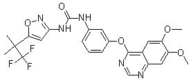
1-[3-(6,7-dimethoxyquinazolin-4-yl)oxyphenyl]-3-[5-(1,1,1-trifluoro-2-methylpropan-2-yl)-1,2-oxazol-3-yl]urea
|
|
| 别名 |
|
|
| HS Tariff Code |
2934.99.9001
|
|
| 存储方式 |
Powder -20°C 3 years 4°C 2 years In solvent -80°C 6 months -20°C 1 month |
|
| 运输条件 |
Room temperature (This product is stable at ambient temperature for a few days during ordinary shipping and time spent in Customs)
|
| 溶解度 (体外实验) |
|
|||
|---|---|---|---|---|
| 溶解度 (体内实验) |
配方 1 中的溶解度: ≥ 2.5 mg/mL (4.83 mM) (饱和度未知) in 10% DMSO + 40% PEG300 + 5% Tween80 + 45% Saline (这些助溶剂从左到右依次添加,逐一添加), 澄清溶液。
例如,若需制备1 mL的工作液,可将100 μL 25.0 mg/mL澄清DMSO储备液加入到400 μL PEG300中,混匀;然后向上述溶液中加入50 μL Tween-80,混匀;加入450 μL生理盐水定容至1 mL。 *生理盐水的制备:将 0.9 g 氯化钠溶解在 100 mL ddH₂O中,得到澄清溶液。 配方 2 中的溶解度: ≥ 2.5 mg/mL (4.83 mM) (饱和度未知) in 10% DMSO + 90% (20% SBE-β-CD in Saline) (这些助溶剂从左到右依次添加,逐一添加), 澄清溶液。 例如,若需制备1 mL的工作液,可将 100 μL 25.0 mg/mL澄清DMSO储备液加入900 μL 20% SBE-β-CD生理盐水溶液中,混匀。 *20% SBE-β-CD 生理盐水溶液的制备(4°C,1 周):将 2 g SBE-β-CD 溶解于 10 mL 生理盐水中,得到澄清溶液。 View More
配方 3 中的溶解度: ≥ 2.5 mg/mL (4.83 mM) (饱和度未知) in 10% DMSO + 90% Corn Oil (这些助溶剂从左到右依次添加,逐一添加), 澄清溶液。 配方 4 中的溶解度: 15% Captisol: 15mg/mL 1、请先配制澄清的储备液(如:用DMSO配置50 或 100 mg/mL母液(储备液)); 2、取适量母液,按从左到右的顺序依次添加助溶剂,澄清后再加入下一助溶剂。以 下列配方为例说明 (注意此配方只用于说明,并不一定代表此产品 的实际溶解配方): 10% DMSO → 40% PEG300 → 5% Tween-80 → 45% ddH2O (或 saline); 假设最终工作液的体积为 1 mL, 浓度为5 mg/mL: 取 100 μL 50 mg/mL 的澄清 DMSO 储备液加到 400 μL PEG300 中,混合均匀/澄清;向上述体系中加入50 μL Tween-80,混合均匀/澄清;然后继续加入450 μL ddH2O (或 saline)定容至 1 mL; 3、溶剂前显示的百分比是指该溶剂在最终溶液/工作液中的体积所占比例; 4、 如产品在配制过程中出现沉淀/析出,可通过加热(≤50℃)或超声的方式助溶; 5、为保证最佳实验结果,工作液请现配现用! 6、如不确定怎么将母液配置成体内动物实验的工作液,请查看说明书或联系我们; 7、 以上所有助溶剂都可在 Invivochem.cn网站购买。 |
| 制备储备液 | 1 mg | 5 mg | 10 mg | |
| 1 mM | 1.9325 mL | 9.6626 mL | 19.3252 mL | |
| 5 mM | 0.3865 mL | 1.9325 mL | 3.8650 mL | |
| 10 mM | 0.1933 mL | 0.9663 mL | 1.9325 mL |
1、根据实验需要选择合适的溶剂配制储备液 (母液):对于大多数产品,InvivoChem推荐用DMSO配置母液 (比如:5、10、20mM或者10、20、50 mg/mL浓度),个别水溶性高的产品可直接溶于水。产品在DMSO 、水或其他溶剂中的具体溶解度详见上”溶解度 (体外)”部分;
2、如果您找不到您想要的溶解度信息,或者很难将产品溶解在溶液中,请联系我们;
3、建议使用下列计算器进行相关计算(摩尔浓度计算器、稀释计算器、分子量计算器、重组计算器等);
4、母液配好之后,将其分装到常规用量,并储存在-20°C或-80°C,尽量减少反复冻融循环。
计算结果:
工作液浓度: mg/mL;
DMSO母液配制方法: mg 药物溶于 μL DMSO溶液(母液浓度 mg/mL)。如该浓度超过该批次药物DMSO溶解度,请首先与我们联系。
体内配方配制方法:取 μL DMSO母液,加入 μL PEG300,混匀澄清后加入μL Tween 80,混匀澄清后加入 μL ddH2O,混匀澄清。
(1) 请确保溶液澄清之后,再加入下一种溶剂 (助溶剂) 。可利用涡旋、超声或水浴加热等方法助溶;
(2) 一定要按顺序加入溶剂 (助溶剂) 。
|
 |
 |
InvivoChem的所有产品仅用于作科学研究,不面向患者销售
Copyright 2020 InvivoChem LLC | All Rights Reserved 粤ICP备20063088号-1


 COA
COA

 463611831
