| 规格 | 价格 | 库存 | 数量 |
|---|---|---|---|
| 5mg |
|
||
| 10mg |
|
||
| 25mg |
|
||
| 50mg |
|
||
| 100mg |
|
||
| 250mg |
|
||
| 500mg |
|
||
| Other Sizes |
|

| 靶点 |
CBFβ-SMHHC
|
|---|---|
| 体外研究 (In Vitro) |
AI-10-49 的 IC50 值为 0.26 μM,可阻断 CBFβ-SMMHC 与 RUNX1 Runt 结构域的结合 [1]。 AI-10-49(1 μM;3、6、12 小时)以特定方式减少 CBFβ-SMMHC 与 RUNX1 的结合[1]。
肝微粒体稳定性的测量表明,AI-10-47降低了代谢不稳定性,因此证明了二价衍生物AI-10-49的合成是合理的(表1)。AI-10-49是强效的(FRET IC50=260nM)(表1)[等温滴定量热法(ITC)测量得出解离常数(KD)=168 nM](图S6),改善了体内药代动力学特性(t½=380min)(图S5),与母体质子化二价化合物AI-4-83(IC50约为3μM)相比,对ME-1细胞生长的抑制活性增强(IC50=0.6 mM)(图1F)(图1E)。请注意,AI-10-49在正常人骨髓细胞中显示出可忽略的活性(IC50>25μM)(图1G),这表明了一个强大的潜在治疗窗口。在11种人类白血病细胞系中,ME-1细胞是唯一对AI-10-49高度敏感的细胞系(图S7)。 在ME-1细胞中评估了AI-10-49在破坏内源性RUNX1与CBFβ-SMMHC结合方面的特异性。AI-10-49有效地将DRUNX1与CBFβ-SMMHC分离,治疗6小时后分离率为90%,而它对CBFβ-RUNX1的结合只有适度的影响(图2A)。AI-10-49不影响RUNX1、CBFβ和CBFβ-SMMHC的稳定性(图S8A)。在inv(16)AML中,CBFβ-SMMHC抑制了RUNX1调控基因RUNX3、CSF1R和CEBPA的表达。先前的研究表明,在CBFβ-SMMHC存在的情况下,RUNX1与靶基因的结合减少,这表明CBFβSMMHC通过阻断RUNX1与靶标DNA位点的结合来抑制RUNX1靶基因(图2B)。与该模型一致,染色质免疫沉淀(ChIP)检测表明,用AI-10-49处理ME-1细胞6小时,RUNX3、CSF1R和CEBPA启动子处的RUNX1占有率分别增加了8倍、2.2倍和8倍,而在对照位点没有观察到富集(图2C和图S8、B和C)。据此,用AI-10-49处理ME-1细胞6或12小时可增加RUNX3、CSF1R和CEBPA的表达,但对对照基因PIN1没有影响(图2D)。在inv(16)阴性的U937细胞中没有观察到这些效应。这些数据确立了AI-10-49在抑制CBFβ-SMMHC与RUNX1结合方面的选择性,并验证了我们使用二价抑制剂实现这种特异性的方法[1]。 为了测试AI-10-49在人类inv(16)白血病治疗中的潜在效用,我们评估了用单价AI-10-47和二价AI-10-49剂量范围治疗48小时的四种原发inv(6)AML细胞样本的存活率。如图3B所示,用浓度为5和10μM的AI-10-49处理后,inv(16)患者细胞的存活率降低(个体剂量反应实验如图S12所示)。请注意,二价AI-10-49比单价化合物AI-10-47更有效,因此概括了在人类inv(16)细胞系ME-1中观察到的效果。相比之下,正常核型AML样本的存活率不受AI-10-49治疗的影响(图3C)。对另外五组AML样本的分析表明,AI-10-49治疗特异性降低了inv(16)白血病细胞的存活率,但对其分化没有明显影响(图S13)。当我们通过评估化合物暴露后的集落形成单位(CFU)来评估AML细胞形成集落的能力时,AI-10-49的特异性也很明显。与正常核型和t(8;21)AML患者样本相比,AI-10-49选择性降低了inv(16)AML细胞形成CFU的能力(图3D)。这种抑制作用是剂量依赖性的(在5和10μM时分别为40%和60%)(图3E),而用AI-10-47处理的AML细胞、具有正常核型的AML细胞(图3F)或CD34+脐带血细胞(图3G)的CFU没有变化。这些研究表明,AI-10-49选择性抑制inv(16)AML母细胞的存活率和CFU能力,而它对具有正常核型的AML母细胞或重要的是对正常人类造血祖细胞的影响可以忽略不计[1]。 |
| 体内研究 (In Vivo) |
AI-10-49(200 mg/kg;每日)可延缓小鼠白血病的进展[1]。
高达90%的inv AML患者在受体酪氨酸激酶途径的成分中存在协同突变,包括N-RAS和c-Kit。我们最近通过结合条件性NrasLSL-G12D和CbfbMYH11等位基因,开发了一种有效的inv AML小鼠模型。为了在体内测试AI-10-49给药的效果,我们用Cbfb+/MYH11:Ras+/G12D白血病细胞移植小鼠,等待5天植入,然后用载体[二甲亚砜(DMSO)]或200mg/kg体重AI-10-49治疗小鼠10天,并评估其对疾病潜伏期的影响。如图3A所示,载体治疗的小鼠死于白血病的中位潜伏期为33.5天,而AI-10-49治疗的小鼠存活时间明显更长(中位潜伏期=61天;P=0.7×10-6)。因此,用AI-10-49进行短暂治疗可以减少体内白血病的扩大。尽管我们没有评估长期暴露后的毒性,但在服用AI-10-49 7天后,我们没有观察到毒性证据[1]。 |
| 酶活实验 |
FRET检测。[1]
如前所述,Cerulean Runt结构域被表达和纯化。Venus CBFβ-SMMHC是通过将6xHis标签和Venus插入NdeI和NcoI位点之间的pET22b载体中,并在NcoI和BamHI位点之间插入CBFβ-SSMHC(CBFβ/SMMHC构建体包含369个氨基酸,CBFβ为1-166,MYH11为166-369(氨基酸1526-1730))构建的。融合蛋白通过标准镍亲和层析纯化,柱上苯并酶处理以去除残留的DNA污染物。使用前,将蛋白质透析到FRET缓冲液(25mM Tris-HCl,pH 7.5,150mM KCl,2mM MgCl2)中。蛋白质浓度分别通过天蓝色和金星在433和513 nm处的紫外吸光度测定。Cerulean Runt结构域和Venus CBFβ-SMMHC以1:1混合,在96孔黑色COSTAR板中达到10nM的最终浓度。将化合物的DMSO溶液加入至DMSO的最终浓度为5%(v/v),然后在室温下在黑暗中孵育平板一小时。使用PHERAstar微孔板读数器测量荧光(激发波长为433 nm,发射波长为474和525 nm)。对于IC50测定,绘制525nm和474nm处的荧光强度比与化合物浓度的对数,并使用Origin7.0将所得曲线拟合为S形曲线。进行了三次独立的测量,并使用它们的平均值和偏差进行IC50数据拟合。[1] 蛋白质核磁共振波谱[1] 所有核磁共振实验均在30°C下在配备低温探头的布鲁克800 MHz仪器上进行。所有NMR样品均在50 mM磷酸钾、0.1 mM EDTA、0.1 mM NaN3、1 mM DTT和5%(v/v)D2O中制备,最终pH值为7.5。15N1 H HSQC实验使用500µM样品,13C1 H HSQC试验在1 mM样品上进行。所有核磁共振数据均使用NMRPipe和Sparky进行处理。加权化学位移变化(百万分之几)通过以下方程式计算:�HN |+(|∆�N|/4.69)。[1] 等温滴定量热法[1] 将由MYH11(MYH11氨基酸1526-1730)的CBFß残基1-166和166-369组成的369个氨基酸CBFß-SMMHC构建体克隆到具有N端6xHis标签和Tev蛋白酶位点的修饰pET22b载体中,并在30°C下用1mM IPTG诱导的Terrific Broth培养基在Rosetta2(DE3)细胞中表达8小时。CBFß-SMMHC通过Niaffinity色谱纯化,然后进行苯并酶处理和Tev蛋白酶消化过夜。然后将融合蛋白第二次通过Ni-NTA柱,然后进行Q-Sepharose离子交换层析以去除残留的核酸。通过使用Sephacryl S300柱的尺寸排阻色谱实现了额外的纯化。将纯化的CBFß-SMMHC透析到1 L ITC缓冲液(12.5 mM KPi(pH 6.5)、150 mM NaCl、2 mM MgCl2、1 mg/mL NaN3、1 mM DTT和0.25%DMSO)中4小时。将AI-10-49单独加入10 mL ITC缓冲液中,使终浓度达到2µM。使用DSS作为标准,在布鲁克600 MHz NMR光谱仪上使用1H NMR验证了AI-10-49的浓度。所有ITC测量均在30°C下在微量热系统上进行。CBFß-SMMHC和AI-10-49样品脱气20分钟,并在量热池中向400 nM CBFßSMMHC溶液中注射8µL 2µMAI-10-49。ITC数据中一致观察到双相转变,表明有多个过程导致了观察到的热量。因此,由于观察到的热量相对较小,无法使用热量计上可用的标准软件分析这些数据,该软件使用热值来确定KD。相反,使用Origin 7.5对数据进行分析,并在校正稀释焓以得出表观KD值后,将其拟合到单点S形结合曲线。重复测量三次,并将值报告为平均值±标准偏差。 |
| 细胞实验 |
蛋白质印迹分析[1]
细胞类型: ME-1 细胞 测试浓度: 1 μM 孵育时间: > 3、6 小时 实验结果: RUNX1 与 CBFβ-SMMHC 有效解离。 RT-PCR[1] 细胞类型: ME-1 和 U937 细胞 测试浓度: 1 μM 孵育持续时间:6、12 小时 实验结果:RUNX3、CSF1R 和 CEBPA 表达增加。 |
| 动物实验 |
动物/疾病模型:小鼠(Cbfb+/MYH11:Ras+/G12D白血病细胞)[1]
剂量:200 mg/kg 给药途径:每日 实验结果:体内白血病细胞增殖减少,且存活时间显著延长。 小鼠白血病移植研究[1] 如前所述,在CD45.2 C57BL/6小鼠中生成携带Cbfb+/MYH11和Nras+/G12D致癌等位基因的白血病细胞。简而言之,将2×10³个Cbfb+/MYH11;Nras+/G12D白血病细胞移植到22只经亚致死剂量照射的6至8周龄CD45.1 C57BL/6雌性小鼠体内。在初步试验中,根据既定的实验条件选择每组小鼠的数量,以达到统计效力。移植后第5天,将小鼠随机分为两组,分别腹腔注射50 µL DMSO或溶于DMSO的AI-10-49(200 mg/kg),连续注射10天。为确定白血病潜伏期的中位数,由多人对小鼠进行观察。一旦发现疾病症状,包括活动减少、梳理毛发减少、弓背、爪子苍白(贫血)和体温过低,即对小鼠实施安乐死。安乐死时,提取外周血和脾细胞,并按先前所述方法进行分析。通过测量外周血中白细胞总数和c-kit(+)阳性细胞群的数量来分析白血病负荷。 |
| 药代性质 (ADME/PK) |
对 AI-4-57(AI-10-49 的类似物)的药代动力学性质分析表明,该化合物在小鼠血浆中的半衰期较短(t½ = 37 分钟)(图 S5),且甲氧基上的甲基脱除是其主要代谢产物。三氟甲氧基 (CF3O) 取代已被证明反应活性较低 (18, 19),因此我们合成了带有该取代基的 AI-10-47。FRET 测量结果表明,该取代基实际上增强了单价化合物的活性(表 1)。肝微粒体稳定性测定表明,AI-10-47 降低了代谢风险,因此合成二价衍生物AI-10-49是合理的(表 1)。AI-10-49 具有强效活性(FRET IC50=260nM)(表 1)[等温滴定量热法 (ITC) 测定解离常数 (KD) = 168 nM](图 S6),体内药代动力学性质得到改善(t½ = 380min)(图 S5),并且与母体质子化二价化合物 AI-4-83(IC50 为 ~3 μM)(图 1E)相比,对 ME-1 细胞生长的抑制活性增强(IC50 = 0.6 mM)(图 1F)。值得注意的是,AI-10-49在正常人骨髓细胞中活性极低(IC50 > 25 μM)(图1G),这表明其具有良好的潜在治疗窗口。在11种人白血病细胞系中,ME-1细胞是唯一对AI-10-49高度敏感的细胞系(图S7)。[1]
药代动力学研究[1] 研究开始前,小鼠禁食至少3小时,并可自由饮水。动物饲养于12小时光照/12小时黑暗循环、温度72-74°C、相对湿度30-50%的环境中。腹腔注射给药时,将24-28克雄性C57BL/6小鼠进行人工固定,并在胸骨和耻骨之间中点、略偏离小鼠中线的位置进行腹腔注射。每次注射均使用1毫升注射器和27号针头。根据预定时间点采集动物血液。动物用异氟烷麻醉,并通过心脏穿刺采集血液。血液立即转移至1.5毫升肝素化微量离心管中,并在4000转/分钟下离心10分钟。然后将血浆转移至干净的离心管中并冷冻保存。由于放血致死,动物未从麻醉中苏醒,并通过开胸手术确保其在麻醉状态下死亡。该方法符合美国兽医协会(AVMA)关于安乐死的指南中关于使用放血作为安乐死手段的建议。使用PK Solutions 2.0软件对受试化合物的血浆浓度-时间数据进行非房室药代动力学分析。 [1] AI-10-49– 采用由岛津SCL-10Avp控制器、SIL-10A自动进样器、LC-10ADvp泵、SPD-10Avp检测器和CTO-10Avp柱温箱组成的液相色谱系统进行高效液相色谱分析。数据采集、峰积分和计算均使用LabSolutions软件完成。采用4.6 mm × 13 mm × 150 mm Atlantis T3 5 μm色谱柱进行分离,流动相为A相和B相,乙腈/水/三氟乙酸的比例分别为5/95/0.1和95/5/0.1,梯度洗脱程序为:4分钟内B相比例从50%升至95%,95% B相保持2分钟,95% B相从0.5分钟降至50%,最后50% B相保持4.5分钟。流速为 1 mL/min,温度为 45°C。进样量为 40 μL,检测波长为 325 nm。定量分析采用空白血浆配制的外部标准品,线性范围为 25 – 750 ng/mL (R² > 0.995)。血浆样品提取步骤如下:取 100 μL 样品,加入 0.5 mL 甲基叔丁基醚 (MtBE),并加入 10 μL AI-10-49 加标溶液 (10X),涡旋混匀,室温静置 5 min。将两相混合物涡旋混匀 5 min,然后以 12,000 rpm 离心 5 min。取 450 μL MtBE 层转移至干净的离心管中,蒸干。将所得残渣用 100 µL 50/50/0.1 乙腈/水/三氟乙酸溶液重悬,涡旋振荡后,以 12,000 rpm 离心 5 分钟。取 90 µL 上清液转移至带有聚丙烯插片的自动进样器小瓶中进行分析。 |
| 毒性/毒理 (Toxicokinetics/TK) |
尽管我们尚未评估长期暴露后的毒性,但在连续7天给予AI-10-49后,我们未观察到任何毒性迹象(图S9至S11)。
毒理学研究[1] 我们评估了AI-10-49每日给药7天后的累积毒性。这些研究不包括最大耐受剂量或长期(>30天)毒性。6周龄C57BL/6雌性小鼠每日腹腔注射DMSO或200mg/kg AI-10-49,持续7天(每组n=3至4)。我们分析了小鼠的外观,以评估毒性迹象,包括梳理毛发、活动能力和体重。末次注射4小时后,我们使用流式细胞术分析外周血细胞,并使用血细胞计数器进行定量。随后,我们对小鼠实施安乐死,以收集组织样本。通过流式细胞术分析骨髓造血祖细胞,包括造血干细胞和多系祖细胞[LSK+= Lin(-), kit(+), Sca1(+)]、髓系祖细胞[CMP=Lin(-)Sca1(-)kit(+)CD34(+)CD16/32(-)]、粒细胞/单核细胞祖细胞[GMP=Lin(-)Sca1(-)kit(+)CD34(+)CD16/32(+)]和巨核细胞/红系祖细胞[MEP=Lin(-)Sca1(-)kit(+)CD34(-)CD16/32(- )]。将骨髓、肝脏、肺、脾脏、肠道和脑制成石蜡切片,并用苏木精和伊红染色,以分析组织结构。 |
| 参考文献 | |
| 其他信息 |
急性髓系白血病 (AML) 是成人白血病中最常见的类型。在染色体倒位 inv(16)(p13q22) 的 AML 中表达的转录因子融合体 CBFβ-SMMHC(核心结合因子 β 与平滑肌肌球蛋白重链)会与野生型 CBFβ 竞争结合转录因子 RUNX1,从而扰乱造血过程中 RUNX1 的活性,并诱发 AML。目前,采用非选择性细胞毒性化疗治疗 inv(16) AML 可获得良好的初始疗效,但长期生存率有限。本文报道了一种蛋白质-蛋白质相互作用抑制剂 AI-10-49 的研发,该抑制剂可选择性地与 CBFβ-SMMHC 结合,并破坏其与 RUNX1 的结合。AI-10-49 可恢复 RUNX1 的转录活性,具有良好的药代动力学特性,并能延缓小鼠白血病的进展。用 AI-10-49 治疗原发性 inv(16) AML 患者的原始细胞可诱导选择性细胞死亡。这些数据表明,直接抑制致癌的 CBFβ-SMMHC 融合蛋白可能是治疗 inv(16) AML 的有效方法,并为其他癌症的转录因子靶向治疗提供了支持。[1]
|
| 分子式 |
C30H22F6N6O5
|
|
|---|---|---|
| 分子量 |
660.5233
|
|
| 精确质量 |
660.155
|
|
| 元素分析 |
C, 54.55; H, 3.36; F, 17.26; N, 12.72; O, 12.11
|
|
| CAS号 |
1256094-72-0
|
|
| 相关CAS号 |
|
|
| PubChem CID |
49806644
|
|
| 外观&性状 |
Off-white to yellow solid powder
|
|
| 密度 |
1.5±0.1 g/cm3
|
|
| 沸点 |
790.3±70.0 °C at 760 mmHg
|
|
| 闪点 |
431.8±35.7 °C
|
|
| 蒸汽压 |
0.0±2.8 mmHg at 25°C
|
|
| 折射率 |
1.611
|
|
| LogP |
7
|
|
| tPSA |
129.29
|
|
| 氢键供体(HBD)数目 |
2
|
|
| 氢键受体(HBA)数目 |
15
|
|
| 可旋转键数目(RBC) |
12
|
|
| 重原子数目 |
47
|
|
| 分子复杂度/Complexity |
913
|
|
| 定义原子立体中心数目 |
0
|
|
| SMILES |
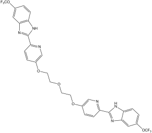
FC(OC1C([H])=C([H])C2=C(C=1[H])N([H])C(C1C([H])=C([H])C(=C([H])N=1)OC([H])([H])C([H])([H])OC([H])([H])C([H])([H])OC1=C([H])N=C(C([H])=C1[H])C1=NC3C([H])=C([H])C(=C([H])C=3N1[H])OC(F)(F)F)=N2)(F)F
|
|
| InChi Key |
WJBSSBFGPKTMQQ-UHFFFAOYSA-N
|
|
| InChi Code |
InChI=1S/C30H22F6N6O5/c31-29(32,33)46-17-1-5-21-25(13-17)41-27(39-21)23-7-3-19(15-37-23)44-11-9-43-10-12-45-20-4-8-24(38-16-20)28-40-22-6-2-18(14-26(22)42-28)47-30(34,35)36/h1-8,13-16H,9-12H2,(H,39,41)(H,40,42)
|
|
| 化学名 |
6-(trifluoromethoxy)-2-[5-[2-[2-[6-[6-(trifluoromethoxy)-1H-benzimidazol-2-yl]pyridin-3-yl]oxyethoxy]ethoxy]pyridin-2-yl]-1H-benzimidazole
|
|
| 别名 |
|
|
| HS Tariff Code |
2934.99.9001
|
|
| 存储方式 |
Powder -20°C 3 years 4°C 2 years In solvent -80°C 6 months -20°C 1 month |
|
| 运输条件 |
Room temperature (This product is stable at ambient temperature for a few days during ordinary shipping and time spent in Customs)
|
| 溶解度 (体外实验) |
|
|||
|---|---|---|---|---|
| 溶解度 (体内实验) |
配方 1 中的溶解度: ≥ 2.5 mg/mL (3.78 mM) (饱和度未知) in 10% DMSO + 40% PEG300 + 5% Tween80 + 45% Saline (这些助溶剂从左到右依次添加,逐一添加), 澄清溶液。
例如,若需制备1 mL的工作液,可将100 μL 25.0 mg/mL澄清DMSO储备液加入到400 μL PEG300中,混匀;然后向上述溶液中加入50 μL Tween-80,混匀;加入450 μL生理盐水定容至1 mL。 *生理盐水的制备:将 0.9 g 氯化钠溶解在 100 mL ddH₂O中,得到澄清溶液。 配方 2 中的溶解度: ≥ 2.5 mg/mL (3.78 mM) (饱和度未知) in 10% DMSO + 90% Corn Oil (这些助溶剂从左到右依次添加,逐一添加), 澄清溶液。 例如,若需制备1 mL的工作液,可将 100 μL 25.0 mg/mL 澄清 DMSO 储备液添加到 900 μL 玉米油中并混合均匀。 View More
配方 3 中的溶解度: 10% DMSO +90%PEG400: 30mg/mL 1、请先配制澄清的储备液(如:用DMSO配置50 或 100 mg/mL母液(储备液)); 2、取适量母液,按从左到右的顺序依次添加助溶剂,澄清后再加入下一助溶剂。以 下列配方为例说明 (注意此配方只用于说明,并不一定代表此产品 的实际溶解配方): 10% DMSO → 40% PEG300 → 5% Tween-80 → 45% ddH2O (或 saline); 假设最终工作液的体积为 1 mL, 浓度为5 mg/mL: 取 100 μL 50 mg/mL 的澄清 DMSO 储备液加到 400 μL PEG300 中,混合均匀/澄清;向上述体系中加入50 μL Tween-80,混合均匀/澄清;然后继续加入450 μL ddH2O (或 saline)定容至 1 mL; 3、溶剂前显示的百分比是指该溶剂在最终溶液/工作液中的体积所占比例; 4、 如产品在配制过程中出现沉淀/析出,可通过加热(≤50℃)或超声的方式助溶; 5、为保证最佳实验结果,工作液请现配现用! 6、如不确定怎么将母液配置成体内动物实验的工作液,请查看说明书或联系我们; 7、 以上所有助溶剂都可在 Invivochem.cn网站购买。 |
| 制备储备液 | 1 mg | 5 mg | 10 mg | |
| 1 mM | 1.5140 mL | 7.5698 mL | 15.1396 mL | |
| 5 mM | 0.3028 mL | 1.5140 mL | 3.0279 mL | |
| 10 mM | 0.1514 mL | 0.7570 mL | 1.5140 mL |
1、根据实验需要选择合适的溶剂配制储备液 (母液):对于大多数产品,InvivoChem推荐用DMSO配置母液 (比如:5、10、20mM或者10、20、50 mg/mL浓度),个别水溶性高的产品可直接溶于水。产品在DMSO 、水或其他溶剂中的具体溶解度详见上”溶解度 (体外)”部分;
2、如果您找不到您想要的溶解度信息,或者很难将产品溶解在溶液中,请联系我们;
3、建议使用下列计算器进行相关计算(摩尔浓度计算器、稀释计算器、分子量计算器、重组计算器等);
4、母液配好之后,将其分装到常规用量,并储存在-20°C或-80°C,尽量减少反复冻融循环。
计算结果:
工作液浓度: mg/mL;
DMSO母液配制方法: mg 药物溶于 μL DMSO溶液(母液浓度 mg/mL)。如该浓度超过该批次药物DMSO溶解度,请首先与我们联系。
体内配方配制方法:取 μL DMSO母液,加入 μL PEG300,混匀澄清后加入μL Tween 80,混匀澄清后加入 μL ddH2O,混匀澄清。
(1) 请确保溶液澄清之后,再加入下一种溶剂 (助溶剂) 。可利用涡旋、超声或水浴加热等方法助溶;
(2) 一定要按顺序加入溶剂 (助溶剂) 。
|
|---|
|
 Cetiedil
 Tinlarebant
 Amelenodor (NX-13)
 Mebufotenin
InvivoChem的所有产品仅用于作科学研究,不面向患者销售
Copyright 2020 InvivoChem LLC | All Rights Reserved 粤ICP备20063088号-1


 COA
COA


 463611831
