| 规格 | 价格 | 库存 | 数量 |
|---|---|---|---|
| 5mg |
|
||
| 10mg |
|
||
| 25mg |
|
||
| 50mg |
|
||
| 100mg |
|
||
| 250mg |
|
||
| 500mg |
|
||
| Other Sizes |
|
| 靶点 |
human LPA1 ( pIC50 = 0.98 μM ); mouse LPA1 ( pIC50 = 0.73 μM )
LPA1 receptor (IC50 = 0.025 µM for human LPA1, IC50 = 0.023 µM for mouse LPA1) Selective against LPA2, LPA3, LPA4, and LPA5 receptors (IC50 > 5 µM) [1] |
|---|---|
| 体外研究 (In Vitro) |
AM095 抑制稳定转染人或小鼠 LPA1 的 CHO 细胞中 LPA 诱导的钙流。 AM095 拮抗人或小鼠 LPA1 转染的 CHO 细胞中 LPA 诱导的钙流的 IC50 分别为 0.025 和 0.023 μM[1]。与赋形剂对照相比,AM095 在 10 μM 时可将 LPA 诱导的血管舒张作用降低约 90%[2]。 AM095 抑制过表达小鼠 LPA1 (IC50=778 nM) 和人 A2058 黑色素瘤细胞 (IC50=233 nM) 的 CHO 细胞的 LPA 驱动的趋化性[3]。
AM095 是一种强效、选择性的 LPA1 拮抗剂,在表达人或小鼠 LPA1 受体的 CHO 细胞中,能抑制 LPA 诱导的钙流,IC50 分别为 0.025 µM 和 0.023 µM。在浓度高达 5 µM 时,对其他 LPA 受体无明显活性。[1] |
| 体内研究 (In Vivo) |
LPA1 与 AM095 的药理拮抗作用可显着减轻博来霉素诱导的真皮纤维化[1]。 AM095 具有较高的口服生物利用度和中等的半衰期,并且在口服和静脉给药后在大鼠和犬中测试的剂量下具有良好的耐受性。 AM095 剂量依赖性地减少 LPA 刺激的组胺释放。 AM095 减弱博莱霉素诱导的支气管肺泡灌洗液中胶原蛋白、蛋白质和炎症细胞浸润的增加。 AM095 可减少小鼠单侧输尿管梗阻模型中的肾纤维化[3]。
在博来霉素诱导的小鼠皮肤纤维化模型中,口服 AM095(30 mg/kg,每日两次)能显著减轻皮肤厚度和羟脯氨酸含量(胶原蛋白标志物)的增加。无论是预防性(从博来霉素起始时开始)还是治疗性(博来霉素开始后 7 或 14 天开始)给药方案均显示出疗效。[1] |
| 酶活实验 |
在测定中,使用 hLPA1/CHO 和 mLPA1/CHO 细胞。将蛋白酶抑制剂、10 mM HEPES、pH 7.4、1 mM 二硫苏糖醇和约 20 mL 冰冷的膜缓冲液添加到 hLPA1/CHO 或 mLPA1/CHO 细胞的细胞沉淀中。对细胞进行超声处理,并将细胞裂解物在 4°C 下以 2000 rpm 离心 10 分钟。 4°C、25,000 rpm 进一步离心上清液 70 分钟。使用 Potter-Elvehjem 组织研磨机,将膜沉淀重悬于 5 mL 冰冷的膜缓冲液中并均质化。使用 Bradford 蛋白质检测试剂盒可计算最终蛋白质浓度。加入 25 至 40 μg hLPA1/CHO 或 mLPA1/CHO 膜和 0.1 nM [35S]-GTPηS 缓冲液(50 mM HEPES、0.1 mM NaCl、10 mM MgCl22 的冷缓冲液洗涤 3 次。 30°C 下孵育 30 分钟。板干燥后,使用 Packard TopCount NXT 微孔板闪烁计数器测量 cpm。
使用瞬时或稳定表达人或小鼠 LPA 受体的 CHO、HEK 或 B103 细胞进行钙流实验。细胞负载钙敏感染料,与待测化合物孵育 30 分钟,然后用 LPA 刺激。使用荧光酶标仪检测钙动员,绘制抑制曲线以计算 IC50 值。[1] |
| 细胞实验 |
在体外,AM095是一种强效的LPA受体拮抗剂,因为它抑制了GTPγS与过表达重组人或小鼠LPA的中国仓鼠卵巢(CHO)细胞膜的结合,IC值分别为0.98和0.73μM,并且没有表现出LPA激动作用。在功能测定中,AM095抑制了过表达小鼠LPA 8321(IC 832=778 nM)和人A2058黑色素瘤细胞(IC 833=233 nM)的CHO细胞的LPA驱动的趋化性[3]。
使用稳定表达人或小鼠 LPA1 的 CHO 细胞通过钙流实验评估拮抗活性。细胞接种于 96 孔板,血清饥饿后负载染料,加入化合物处理,再用 LPA 刺激,检测钙动员以确定抑制效果。[1] |
| 动物实验 |
小鼠的左肾分别接受单侧输尿管梗阻(UUO)或假手术。简而言之,手术方法是在左上腹做纵向切口,暴露左肾。找到肾动脉后,将一根6/0丝线插入肾动脉和输尿管之间。为确保输尿管完全结扎,将丝线缠绕在输尿管周围并打结三次。缝合皮肤,将肾脏放回腹腔,并用缝合钉闭合切口。健康对照组的肾脏为对侧(右)肾。在UUO手术前1至4小时,以及手术后根据需要,分别灌胃给予AM095(30 mg/kg)或溶剂(水)。小鼠经二氧化碳吸入麻醉8天后,取出肾脏并切成两半,进行组织病理学和生化检查,以评估纤维化情况。将肾脏样本固定于 10% 中性缓冲福尔马林溶液中,并用 Masson 三色染色法染色,以测量纤维化程度。为了进行胶原蛋白含量的生化分析,将肾脏的另一半冷冻于 -80°C。
野生型 (WT)、LPA₁ 基因敲除 (KO) 和 LPA₂ 基因敲除 (LPA₂-KO) 小鼠每天皮下注射一次博来霉素或磷酸盐缓冲液 (PBS),持续 28 天。测定博来霉素注射组和 PBS 注射组皮肤的真皮厚度、胶原蛋白含量以及 α-平滑肌肌动蛋白 (α-SMA) 或磷酸化 Smad2 阳性细胞的数量。在另一组实验中,将一种新型选择性 LPA₁ 拮抗剂AM095或载体单独灌胃给予接受 28 天博来霉素或 PBS 注射的 C57BL/6 小鼠。在注射博来霉素和PBS的同时或之后7天或14天,分别开始给予AM095或载体治疗,并持续至实验结束。测定注射部位皮肤的真皮厚度和胶原蛋白含量。[1] C57Bl/6小鼠皮下注射博来霉素(10 µg/ml,100 µl),每日一次,持续28天,以诱导真皮纤维化。AM095(30 mg/kg)或载体(无菌水)通过灌胃给药,工作日每日两次,周末每日一次。治疗开始时间可以是博来霉素注射开始时(预防性治疗),也可以是注射后7天或14天(治疗性治疗)。在第28天采集皮肤样本进行组织学和生化分析。[1] |
| 药代性质 (ADME/PK) |
在体内实验中,我们证实了AM095:1)具有较高的口服生物利用度和中等的半衰期,并且在大鼠和犬经口服和静脉给药后,在所测试的剂量范围内耐受性良好;2)呈剂量依赖性地降低LPA刺激的组胺释放;3)减弱博来霉素诱导的支气管肺泡灌洗液中胶原蛋白、蛋白质和炎症细胞浸润的增加;4)在小鼠单侧输尿管梗阻模型中减轻肾纤维化。尽管AM095具有抗纤维化活性,但它对大鼠切口和切除伤口后的正常伤口愈合没有影响。这些数据表明,AM095是一种LPA₁受体拮抗剂,在啮齿动物模型中具有良好的口服暴露量和抗纤维化活性。 [3]在C57Bl/6小鼠中,口服AM095(0和8小时,30 mg/kg)后,Cmax为6200 nM (28 µg/ml),Cmin为170 nM (0.08 µg/ml),AUC为118.7 µg·hr/ml。在整个24小时内,血浆浓度均高于LPA1的IC50。[1]
|
| 参考文献 |
|
| 其他信息 |
目的:硬皮病(系统性硬化症[SSc])的特征是进行性多器官纤维化。我们近期发现溶血磷脂酸(LPA)与肺纤维化的发病机制有关。本研究旨在探讨LPA及其两种受体LPA₁和LPA₂在SSc小鼠模型皮肤纤维化中的作用。方法:将野生型(WT)、LPA₁敲除(KO)和LPA₂敲除小鼠皮下注射博来霉素或磷酸盐缓冲液(PBS),每日一次,持续28天。分别检测博来霉素注射组和PBS注射组皮肤的真皮厚度、胶原蛋白含量以及α-平滑肌肌动蛋白(α-SMA)或磷酸化Smad2阳性细胞的数量。在另一项实验中,我们通过灌胃法向C57BL/6小鼠施用新型选择性LPA₁拮抗剂AM095或载体,这些小鼠此前已连续28天接受博来霉素或PBS注射。AM095或载体处理与博来霉素和PBS注射同时开始,或在注射后7天或14天开始,并持续至实验结束。我们测定了注射部位皮肤的真皮厚度和胶原蛋白含量。
结果:LPA₁-KO小鼠对博来霉素诱导的真皮厚度和胶原蛋白含量增加表现出显著的抵抗力,而LPA₂-KO小鼠与野生型小鼠一样易感。在LPA₁-KO小鼠中,博来霉素诱导的真皮α-SMA+和磷酸化Smad2+细胞的增加被消除。使用 AM095 进行 LPA₁ 的药理学拮抗,无论采用预防方案还是两种治疗方案,均能显著减轻博来霉素诱导的皮肤纤维化。 结论:这些结果表明,LPA/LPA₁ 通路抑制可能成为系统性硬化症 (SSc) 的一种有效新疗法,并且 LPA₁ 是皮肤纤维化中一个有吸引力的药理学靶点。[1] 溶血磷脂酸 (LPA) 已被证实参与多种心血管功能,但其在血管张力控制中的潜在作用尚不明确。本文研究表明,LPA (18:1) 和 VPC31143(LPA1-3 受体的合成激动剂)均能舒张完整的小鼠胸主动脉,且二者的最大舒张效应 (Emax) 值相似(分别为苯肾上腺素诱导预收缩的 53.9% 和 51.9%),但 LPA 和 VPC31143 诱导血管舒张的半数有效浓度 (EC50) 不同(分别为 400 nM 和 15 nM)。机械去除内皮或基因敲除内皮型一氧化氮合酶 (eNOS) 不仅减弱了 LPA 或 VPC31143 的血管舒张作用,而且使其转变为血管收缩。新鲜分离的小鼠主动脉内皮细胞表达 LPA1、LPA2、LPA4 和 LPA5 转录本。 LPA1,3拮抗剂Ki16425、LPA1拮抗剂AM095以及LPA1基因敲除(而非LPA2基因敲除)均能消除LPA诱导的血管舒张。沃特曼宁或MK-2206对磷脂酰肌醇3激酶-蛋白激酶B/Akt通路的抑制作用未能影响LPA的作用。然而,U73122或依地福辛对磷脂酶C (PLC)的药理学抑制(而非PLCε基因敲除)能消除LPA诱导的血管舒张,表明除PLCε以外的其他PLC酶介导了该反应。总之,本研究确定LPA是一种内皮依赖性血管舒张物质,其作用机制涉及LPA1、PLC和eNOS。[2] AM095是一种新型的、口服生物利用度高的选择性LPA1拮抗剂,可减轻博来霉素诱导的硬皮病小鼠模型中的皮肤纤维化。它能减少肌成纤维细胞的积累和Smad2的磷酸化,提示其抑制了TGF-β通路激活。该研究表明LPA1是治疗硬皮病等纤维化疾病的潜在药物靶点。[1] |
| 分子式 |
C₂₇H₂₄N₂O₅
|
|---|---|
| 分子量 |
456.49
|
| 精确质量 |
456.168
|
| 元素分析 |
C, 71.04; H, 5.30; N, 6.14; O, 17.52
|
| CAS号 |
1228690-36-5
|
| 相关CAS号 |
AM095; 1345614-59-6
|
| PubChem CID |
46213949
|
| 外观&性状 |
White to off-white solid powder
|
| 密度 |
1.3±0.1 g/cm3
|
| 沸点 |
641.9±55.0 °C at 760 mmHg
|
| 闪点 |
342.0±31.5 °C
|
| 蒸汽压 |
0.0±2.0 mmHg at 25°C
|
| 折射率 |
1.629
|
| LogP |
4.94
|
| tPSA |
105.15
|
| 氢键供体(HBD)数目 |
2
|
| 氢键受体(HBA)数目 |
6
|
| 可旋转键数目(RBC) |
8
|
| 重原子数目 |
34
|
| 分子复杂度/Complexity |
666
|
| 定义原子立体中心数目 |
1
|
| SMILES |
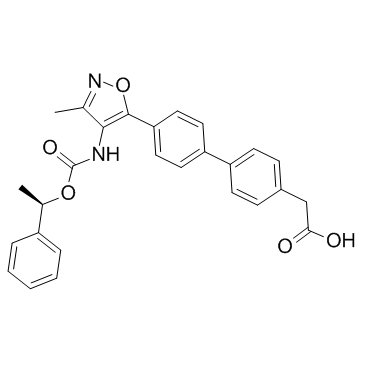
O=C(O)CC1=CC=C(C2=CC=C(C3=C(NC(O[C@@H](C4=CC=CC=C4)C)=O)C(C)=NO3)C=C2)C=C1
|
| InChi Key |
LNDDRUPAICPXIN-GOSISDBHSA-N
|
| InChi Code |
InChI=1S/C27H24N2O5/c1-17-25(28-27(32)33-18(2)20-6-4-3-5-7-20)26(34-29-17)23-14-12-22(13-15-23)21-10-8-19(9-11-21)16-24(30)31/h3-15,18H,16H2,1-2H3,(H,28,32)(H,30,31)/t18-/m1/s1
|
| 化学名 |
2-[4-[4-[3-methyl-4-[[(1R)-1-phenylethoxy]carbonylamino]-1,2-oxazol-5-yl]phenyl]phenyl]acetic acid
|
| 别名 |
AM095; AM 095; AM-095; AM095 free acid
|
| HS Tariff Code |
2934.99.9001
|
| 存储方式 |
Powder -20°C 3 years 4°C 2 years In solvent -80°C 6 months -20°C 1 month |
| 运输条件 |
Room temperature (This product is stable at ambient temperature for a few days during ordinary shipping and time spent in Customs)
|
| 溶解度 (体外实验) |
DMSO: ~67.3 mg/mL (~147.4 mM)
|
|---|---|
| 溶解度 (体内实验) |
配方 1 中的溶解度: ≥ 2.25 mg/mL (4.93 mM) (饱和度未知) in 10% DMSO + 90% Corn Oil (这些助溶剂从左到右依次添加,逐一添加), 澄清溶液。
例如,若需制备1 mL的工作液,可将100 μL 22.5 mg/mL 澄清 DMSO 储备液加入900 μL 玉米油中,混合均匀。 请根据您的实验动物和给药方式选择适当的溶解配方/方案: 1、请先配制澄清的储备液(如:用DMSO配置50 或 100 mg/mL母液(储备液)); 2、取适量母液,按从左到右的顺序依次添加助溶剂,澄清后再加入下一助溶剂。以 下列配方为例说明 (注意此配方只用于说明,并不一定代表此产品 的实际溶解配方): 10% DMSO → 40% PEG300 → 5% Tween-80 → 45% ddH2O (或 saline); 假设最终工作液的体积为 1 mL, 浓度为5 mg/mL: 取 100 μL 50 mg/mL 的澄清 DMSO 储备液加到 400 μL PEG300 中,混合均匀/澄清;向上述体系中加入50 μL Tween-80,混合均匀/澄清;然后继续加入450 μL ddH2O (或 saline)定容至 1 mL; 3、溶剂前显示的百分比是指该溶剂在最终溶液/工作液中的体积所占比例; 4、 如产品在配制过程中出现沉淀/析出,可通过加热(≤50℃)或超声的方式助溶; 5、为保证最佳实验结果,工作液请现配现用! 6、如不确定怎么将母液配置成体内动物实验的工作液,请查看说明书或联系我们; 7、 以上所有助溶剂都可在 Invivochem.cn网站购买。 |
| 制备储备液 | 1 mg | 5 mg | 10 mg | |
| 1 mM | 2.1906 mL | 10.9531 mL | 21.9063 mL | |
| 5 mM | 0.4381 mL | 2.1906 mL | 4.3813 mL | |
| 10 mM | 0.2191 mL | 1.0953 mL | 2.1906 mL |
1、根据实验需要选择合适的溶剂配制储备液 (母液):对于大多数产品,InvivoChem推荐用DMSO配置母液 (比如:5、10、20mM或者10、20、50 mg/mL浓度),个别水溶性高的产品可直接溶于水。产品在DMSO 、水或其他溶剂中的具体溶解度详见上”溶解度 (体外)”部分;
2、如果您找不到您想要的溶解度信息,或者很难将产品溶解在溶液中,请联系我们;
3、建议使用下列计算器进行相关计算(摩尔浓度计算器、稀释计算器、分子量计算器、重组计算器等);
4、母液配好之后,将其分装到常规用量,并储存在-20°C或-80°C,尽量减少反复冻融循环。
计算结果:
工作液浓度: mg/mL;
DMSO母液配制方法: mg 药物溶于 μL DMSO溶液(母液浓度 mg/mL)。如该浓度超过该批次药物DMSO溶解度,请首先与我们联系。
体内配方配制方法:取 μL DMSO母液,加入 μL PEG300,混匀澄清后加入μL Tween 80,混匀澄清后加入 μL ddH2O,混匀澄清。
(1) 请确保溶液澄清之后,再加入下一种溶剂 (助溶剂) 。可利用涡旋、超声或水浴加热等方法助溶;
(2) 一定要按顺序加入溶剂 (助溶剂) 。
|
|
InvivoChem的所有产品仅用于作科学研究,不面向患者销售
Copyright 2020 InvivoChem LLC | All Rights Reserved 粤ICP备20063088号-1


 463611831
