| 规格 | 价格 | 库存 | 数量 |
|---|---|---|---|
| 5g |
|
||
| 10g |
|
||
| 25g |
|
||
| 50g |
|
||
| 100g |
|
||
| Other Sizes |
|

| 靶点 |
Influenza A viruses; ion channels NMDA, M2; CDK2; Bcl-2; Bax
|
|---|---|
| 体外研究 (In Vitro) |
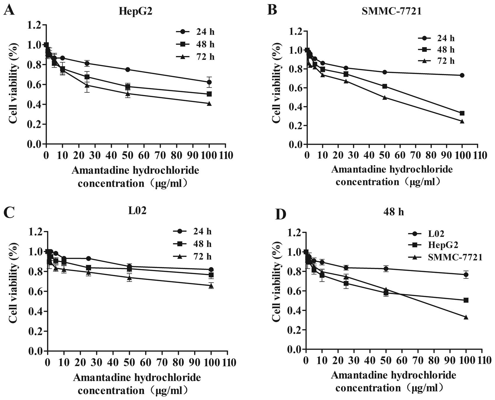
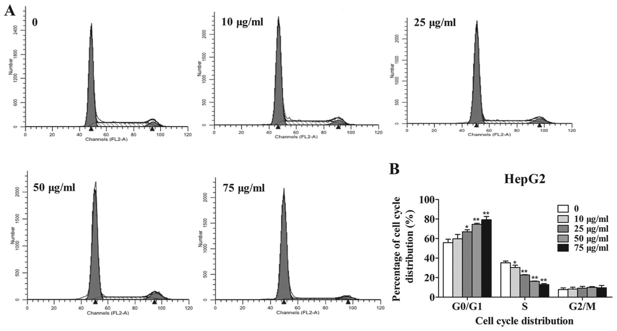
盐酸金刚烷胺 (0-500 μM,26 小时) 抑制 SARS-CoV-2 复制,IC50 浓度在 83 和 119 μM 之间[4]。 盐酸金刚烷胺 (0-100 μg/mL,24-72 h) 显着抑制 HepG2。盐酸金刚烷胺 (0-75 μg/mL,48 h) 将细胞周期阻滞在 G0/G1 期并诱导细胞的肿胀[6]。 盐酸金刚烷胺 (0-75 μg) 和 SMMC-7721 细胞的肿胀[6]。 /mL,48 h) 可降低细胞周期相关基因和蛋白(cyclin D1、cyclin E 和 CDK2),减少 Bcl-2 并增加 Bax 蛋白和 mRNA 水平[6]。 Cell Viability Assay[4] Cell Line: Vero E6细胞浓度:500 µM、100 µM、20 µM、4 µM 和 8 nM 孵育时间:26 小时 结果:10 ℃感染 26 小时后,导致上清液中病毒核酸浓度依赖性减少(IC50=83 µM) -500 µM。感染后 26 小时,导致细胞质中病毒核酸浓度依赖性减少 (IC50=119 µM)。细胞增殖测定[6] 细胞系:人 HCC 细胞系(HepG2 和 SMMC-7721)和正常肝细胞(L02 细胞) 浓度:0、1、2、5、10、25、50 和 100 µg/mL 孵育时间:24、48 和 72 小时 结果:在 HepG2 和 SMMC-7721 细胞中以时间和剂量依赖性方式抑制细胞增殖。细胞周期分析[6] 细胞系:HepG2 和 SMMC-7721 细胞 浓度:0、10、25、50 和 75 µg/mL 孵育时间:48 小时 结果:G0 期 HepG2 和 SMMC-7721 细胞数量显着增加/G1期呈剂量依赖性,并显着减少S期HepG2细胞的数量。细胞凋亡分析[6] 细胞系:HepG2 和 SMMC-7721 细胞 浓度:0、10、25、50 和 75 µg/mL 孵育时间:48 小时 结果:凋亡 HepG2 和 SMMC-7721 细胞的百分比显着增加(早期-和晚期细胞凋亡)以剂量依赖性方式。 Western Blot 分析[6] 细胞系:HepG2 和 SMMC-7721 细胞 浓度:0、10、25、50 和 75 µg/mL 孵育时间:48 h 结果:显示 cyclin D1、cyclin E 和 CDK2 下调,并显示HepG2 和 SMMC-7721 细胞中 Bcl-2 水平降低,Bax 水平升高。 RT-PCR[6] 细胞系:HepG2 和 SMMC-7721 细胞 浓度:0、10、25、50 和 75 µg/mL 孵育时间:48 小时 结果:显示 Bax 增加,Bcl-2 基因减少。
金刚烷胺(0-500 µM,26 小时)抑制 SARS-CoV-2 复制的 IC50 值范围为 83 至 119 µM [4]。金刚烷胺(0-100 µg/mL,24-72 小时)强烈抑制 HepG2 和 SMMC-7721 细胞生长 [6]。金刚烷胺(0-75 µg/mL,48 小时)会导致细胞凋亡并使细胞周期停止在 G0/G1 期 [6]。金刚烷胺(0-75 µg/mL,48 小时)可降低 Bcl-2,增加 Bax 蛋白和 mRNA 水平,并降低细胞周期相关基因和蛋白(细胞周期蛋白 D1、细胞周期蛋白 E 和 CDK2)[6]。 自2019年底严重急性呼吸系统综合征冠状病毒2型疫情开始以来,寻找保护性疫苗和药物治疗已成为应对全球卫生紧急情况的强制性措施。旅行限制、社交距离和口罩是合适的应对措施,但可能无法控制疫情,因为人们会无意中或在一定程度的限制严重程度或持续时间内不遵守规定。即使疫苗获得批准,对抗严重急性呼吸系统综合征冠状病毒2型的抗病毒药物的需求也将持续存在。然而,迄今为止,尚未有明确证据表明任何重新使用的抗病毒药物对严重急性呼吸系统综合征冠状病毒2型有疗效金刚烷胺已被批准为抗甲型流感的抗病毒药物,对严重急性呼吸系统综合征冠状病毒2型的抗病毒活性已通过类比得出,但没有数据。我们在体外测试了金刚烷胺对感染严重急性呼吸系统综合征冠状病毒2型的Vero E6细胞的疗效。事实上,金刚烷胺在两个单独的实验中抑制了严重急性呼吸系统综合征冠状病毒2型的复制,IC50浓度在83至119µM之间。尽管这些IC50浓度在全身给药后高于治疗性金刚烷胺水平,但通过吸入或鼻内滴注局部给药可能会导致气道上皮中金刚烷胺浓度充足,而不会产生高全身暴露。然而,需要在其他模型中进行进一步的研究来证明这一假设。[4] 肝细胞癌(HCC)是全球最具侵袭性的恶性肿瘤之一,近年来与病毒感染相关的发病率有所增加金刚烷胺是一种三环对称胺,可以有效预防丙型肝炎病毒。然而,其抗肿瘤特性尚不清楚。本研究探讨了金刚烷胺对肿瘤细胞存活率、细胞周期调控和凋亡的影响。MTT法检测HepG2和SMMC-7721细胞(HCC细胞系)的生长情况。流式细胞术用于研究细胞周期调控和凋亡。还进行了逆转录定量聚合酶链式反应和蛋白质印迹分析,以检测细胞周期和凋亡相关基因和蛋白质的表达,包括细胞周期蛋白E、细胞周期蛋白D1、细胞周期素依赖性激酶2(CDK2)、B细胞淋巴瘤2(Bcl-2)和Bax。我们的研究结果表明,金刚烷胺以剂量和时间依赖的方式显著抑制HepG2和SMMC-7721细胞的增殖,并将细胞周期阻滞在G0/G1期。金刚烷胺降低了细胞周期相关基因和蛋白质(细胞周期蛋白D1、细胞周期蛋白E和CDK2)的水平,并显著诱导了细胞凋亡。金刚烷胺治疗还降低了Bcl-2,增加了Bax蛋白和mRNA水平。此外,金刚烷胺治疗后,两种HCC细胞系的Bcl-2/Bax比值较低。总的来说,这些结果强调了金刚烷胺在抑制HCC细胞增殖和诱导凋亡方面的作用,主张将其作为一种新型的肿瘤抑制治疗候选药物[6]。 |
| 体内研究 (In Vivo) |
盐酸金刚烷胺(25 mg/kg,IP,每天一次,持续3天)抑制调节引起的神经调节和学习记忆障碍[5]。 动物模型:Fischer 344大鼠(4月龄,雄性,290-330 g,每组15只大鼠)[5] 剂量:25 mg/kg 给药方式:IP,每日一次,连续3天(手术前15分钟第一次给药) 结果:抑制手术引起的神经炎症和学习记忆障碍,增加GDNF(胶质细胞)线源性神经营养因子)与海马体中的神经胶质原纤维酸性蛋白(星形细胞标记物)共定位。
金刚烷胺(25 mg/kg,IP,每天一次,持续 3 天)可减少手术引起的神经炎症以及学习和记忆缺陷 [5]。 在训练后1天或8天进行测试时,手术增加了在巴恩斯迷宫中识别目标框的时间[22(中位数)(11-66)(四分位数间距)对照组对158(29-180)手术组,n=15,P=0.022),并减少了恐惧条件测试中与背景相关的冷冻行为。这些影响被金刚烷胺和侧脑室GDNF减弱。金刚烷胺增加了与星形胶质细胞标志物胶质纤维酸性蛋白共定位的GDNF。海马。侧脑室注射抗GDNF抗体而不是变性抗体阻断了金刚烷胺对认知的影响。手术诱导的神经炎症被金刚烷胺抑制。脂多糖增加了C8-B4细胞白细胞介素1β的产生。这种作用被GDNF抑制[5]。 金刚烷胺减轻了手术引起的学习记忆障碍[5] 随着对照组大鼠、仅接受麻醉的大鼠、只接受Amantadine金刚烷胺的大鼠和接受手术加金刚烷胺的大鼠训练时间的增加,巴恩斯迷宫试验4天训练期间识别目标框的时间缩短了。这四组大鼠在第4天的时间明显短于第1天。这种效果在单独手术后的大鼠身上并不明显。手术对训练课程中识别目标框所需的时间有显著影响[F(1,28)=5.625,P=0.025]。金刚烷胺可消除这种作用[F(1,28)=0.840,P=0.367;与对照组相比]。金刚烷胺或麻醉对训练期间识别目标框的时间没有显著影响[F(1,28)=0.063,P=0.804;F(1,14)=0.074,P+0.790](图1和图2)。当在训练课程后1天对大鼠进行测试时,接受手术的大鼠识别目标框的时间比对照组大鼠长。金刚烷胺可以减轻这种延长。在训练课程结束8天后进行测试时,也出现了类似的变化模式。然而,无论测试是在训练后1天还是8天进行,单独使用麻醉和金刚烷胺都不会影响识别目标框的时间(图1B和2B)。 与对照组大鼠相比,手术组大鼠(而非仅麻醉组或金刚烷胺组大鼠)在恐惧条件反射测试中的情境相关冷冻行为较少。金刚烷胺消除了这种手术效果(图1C)。对照组、接受金刚烷胺治疗的大鼠、接受手术的大鼠和接受手术加金刚烷胺的大鼠在音调相关的冷冻行为方面没有差异(图1C和2C)。 金刚烷胺减轻了手术引起的神经炎症[5] 术后6小时和24小时,海马中Iba-1(一种小胶质细胞标志物)、IL-1β和IL-6的表达显著增加。Amantadine金刚烷胺消除了这些增加(图3和图4)。同样,手术后10天,海马齿状回区域的Iba-1表达也增加,这种增加被金刚烷胺阻断(图5)。这些结果表明,手术诱导了金刚烷胺抑制的神经炎症。 金刚烷胺增加了抑制小胶质细胞活化的GDNF的表达[5] 金刚烷胺/Amantadine显著增加了海马中的GDNF(图7)。GDNF主要与星形胶质细胞标志物GFAP共定位,但与Iba-1不共定位(图7A和7B)。一些GDNF似乎位于神经元标记物NeuN周围(图7C)。手术也增加了GFAP,但这种增加不受海马中金刚烷胺的影响(图7A和7E)。 抗GDNF抗体可抑制金刚烷胺诱导的术后学习记忆障碍的减弱[5] 与对照组大鼠相似,仅抗体组和手术加金刚烷胺/Amantadine加煮沸抗体组的大鼠找到目标框的时间缩短,训练次数增加。这两组大鼠在训练第4天的时间比训练第1天的时间短。对于手术加金刚烷胺加抗GDNF抗体组的大鼠来说,这种效果并不明显。研究发现,抗GDNF抗体对训练期间识别目标框的时间有显著影响[F(1,14)=19.009,P<0.001;与对照组相比)(图9A)。训练后第1天识别目标框所需的时间在对照组大鼠、接受抗体的大鼠、接受手术加金刚烷胺加抗-GDNF抗体或接受手术加金刚烷基胺加煮沸抗体的大白鼠之间没有差异。然而,接受手术加金刚烷胺加抗-DDNF抗体的大老鼠在训练后第8天需要比对照组大白鼠或接受手术加金刚烷胺加煮沸抗体的老鼠更长的时间来识别目标框(图9B)。 同样,在恐惧条件测试中,接受手术加金刚烷胺加抗GDNF抗体的大鼠也比对照组大鼠或接受手术加金刚烷胺加煮沸抗体的大白鼠有更少的与环境相关的冷冻行为。然而,三组之间与音调相关的冷冻行为没有差异(图9C)。 |
| 酶活实验 |
S-蛋白-ACE2结合试验[4]
使用严重急性呼吸系统综合征冠状病毒2型刺突:ACE2抑制剂筛选试剂盒测试化合物抑制严重急性呼吸系综合征冠状病毒2中刺突蛋白(S蛋白)与ACE2结合的能力。简而言之,将严重急性呼吸系统综合征冠状病毒2型刺突蛋白以1µg/mL的磷酸盐缓冲盐水包被到96微孔板上。去除未结合的蛋白质,并阻断孔中的非特异性结合位点。然后,去除阻断溶液,将稀释的化合物和对照样品加入孔中。将包被的刺突蛋白与化合物预孵育后,加入His标记的ACE2蛋白,并与化合物一起孵育,以允许与刺突蛋白结合。洗涤和阻断后,用与辣根过氧化物酶(HRP)偶联的抗His抗体检测结合的ACE2蛋白。使用化学发光HRP底物进行检测,并在微量滴定板读数器中读取发光强度。将含有稀释化合物的每个样品的发光信号除以不存在任何抑制剂时的发光,并将所得值与化合物浓度绘制成图。 RT-PCR读数抗病毒活性测定(第一次实验)[4] 将指数增长的Vero E6细胞以每孔8×10⁴个细胞的密度接种到48孔板中,并孵育过夜。取出培养基,用严重急性呼吸系统综合征冠状病毒2型(hCoV-19/意大利/INMI1 isl/2020,MOI为0.01,在含有不同抑制剂浓度的300µL培养基中)感染细胞三次。将Amantadine金刚烷胺溶解在无菌水中,并用培养基进一步稀释至500µM、100µM、20µM、4µM和8 nM的浓度。将雷德西韦溶解在DMSO中,用培养基稀释至50µM、10µM、2µM、0.4µM和80 nM的剂量。雷德西维尔MOCK对照品含有不同量的DMSO。 用核衣壳蛋白读数进行抗病毒活性测定(第二次实验)[4] 将指数增长的Vero E6细胞在完全培养基中以最佳密度接种到96孔板中;24小时后,细胞以0.01 moi(多重感染)感染严重急性呼吸系统综合征冠状病毒2型(病毒株INMI1),然后暴露于不同浓度的药物(0-0.1-1-10-100-300μM的Amantadine/金刚烷胺)72小时。在培养基中进行药物稀释。检查每个浓度点的复制品。在潜伏期结束时,通过ELISA(定量严重急性呼吸系综合征冠状病毒-2核蛋白)和细胞保护试验(通过倒置显微镜检查毒性效应)检查抗病毒活性。 |
| 细胞实验 |
细胞活力测定[4]
细胞类型: Vero E6 细胞 测试浓度: 500 µM、100 µM、20 µM、4 µM 和 8 nM 孵育持续时间: 26 小时 实验结果:导致病毒浓度依赖性减少 (IC50=83 µM) 26 感染后上清液中的核酸浓度为10-500 µM。导致感染后 26 小时细胞质中病毒核酸浓度依赖性减少 (IC50=119 µM)。 细胞增殖测定[6] 细胞类型:人 HCC 细胞系(HepG2 和 SMMC-7721)和正常肝细胞(L02 细胞) 测试浓度:0、1、2、5、10、25、50 和 100 µg/mL 孵育时间:24、48 和 72 小时 实验结果:在HepG2和SMMC-7721细胞中以时间和剂量依赖性方式抑制细胞增殖。 细胞周期分析[6] 细胞类型: HepG2 和 SMMC-7721 细胞 测试浓度: 0、10、25、 50 和 75 µg/mL 孵育持续时间:48 小时 实验结果:HepG2 和 SMMC- 数量显着增加7721细胞以剂量依赖性方式处于G0/G1期,并且数量急剧减少 |
| 动物实验 |
Fischer 344 大鼠(4 月龄,雄性,290-330 g,每组 15 只)
25 mg/kg 给药途径:腹腔注射,每日一次,连续 3 天(首次给药于术前 15 分钟) 动物/疾病模型: Fischer 344 大鼠(4 月龄,雄性,290-330 g,每组 15 只)[5] 剂量: 25 mg/kg 给药途径: 腹腔注射,每日一次,连续 3 天(首次给药于术前 15 分钟) 实验结果: 抑制手术诱导的神经炎症和学习记忆障碍,增加与海马胶质纤维酸性蛋白共定位的 GDNF(胶质细胞系衍生的神经元)的表达。 (星形胶质细胞标志物)营养因素)。 四个月龄、体重290-330克的雄性Fischer 344大鼠被随机分为四组:1)对照组(未接受手术或任何药物治疗),2)金刚烷胺组,3)手术组(暴露右侧颈动脉),4)手术+金刚烷胺组。每组15只大鼠。在第二个实验中,大鼠被分为四组:5)对照组,6)抗GDNF抗体组,7)手术+金刚烷胺+煮沸的抗GDNF抗体组,8)手术+金刚烷胺+抗GDNF抗体组。每组8只大鼠。在第三个实验中,大鼠被随机分为四组:7)对照组,8)仅麻醉组,9)手术+GDNF组。每组8只大鼠。将GDNF和抗GDNF抗体脑室内注射。一周后,开始对这些大鼠进行Barnes迷宫测试和恐惧条件反射测试。将大鼠随机分为三组:1)对照组,2)手术组,3)手术加金刚烷胺组(每组n=6),分别于术后6小时、24小时或10天处死,用于Western blotting和免疫组织化学分析。 金刚烷胺溶于生理盐水,腹腔注射,剂量为25 mg/kg/天,连续三天,首次给药于术前15分钟。仅注射金刚烷胺组采用类似方法,但不进行手术和麻醉。金刚烷胺的剂量参考既往研究[5]。 |
| 药代性质 (ADME/PK) |
吸收、分布和排泄
金刚烷胺口服后在胃肠道吸收良好。 主要以原形经肾小球滤过和肾小管分泌从尿液中排出。 3~8 L/kg [健康受试者] 0.2~0.3 L/hr/kg 0.10±0.04 L/hr/kg [健康老年男性] 从胃肠道迅速且几乎完全吸收。 金刚烷胺可分布到乳汁中。 排泄:肾脏;>90%以原形经肾小球滤过和肾小管分泌从尿液中排出。酸性尿液中排泄速率迅速增加。透析:仅少量(约 4%)通过血液透析从血液中清除。 分布于唾液、泪膜和鼻分泌物中;在动物中,组织(尤其是肺)浓度高于血清浓度。可穿过胎盘和血脑屏障;分布于母乳中。一名患者的脑脊液浓度为相应血浆浓度的 52%。VolD - 4.4 ± 0.2 L/kg(肾功能正常)。 5.1 ± 0.2 L/kg(肾功能衰竭)。 有关金刚烷胺(共7种代谢物)的更多吸收、分布和排泄(完整)数据,请访问HSDB记录页面。 代谢/代谢物 未发现明显的代谢,但已鉴定出微量的乙酰代谢物。 已在人尿中鉴定出8种金刚烷胺代谢物。其中一种代谢物,即N-乙酰化化合物,在人尿中被定量,占给药剂量的5-15%。在12名健康志愿者中,有5名志愿者在服用200 mg金刚烷胺后,血浆中乙酰金刚烷胺的浓度占同期金刚烷胺血浆浓度的80%。在其余七名志愿者的血浆中未检测到乙酰金刚烷胺。 虽然检测到微量的乙酰代谢物,但未发现明显的代谢。金刚烷胺口服后可从胃肠道良好吸收。其抗帕金森病作用机制尚未完全阐明,但似乎是通过促进脑细胞神经末梢释放多巴胺并刺激去甲肾上腺素反应来实现的。其抗病毒机制似乎与此无关。该药物干扰一种病毒蛋白M2(一种离子通道),病毒颗粒在通过内吞作用进入细胞后需要M2蛋白才能“脱壳”。代谢物经尿液排泄 (A308)。 消除途径:主要以原形经肾小球滤过和肾小管分泌经尿液排泄。 半衰期:平均半衰期为 10 至 14 小时,但肾功能损害会导致半衰期显著延长至 7 至 10 天。 生物半衰期 平均半衰期为 10 至 14 小时,但肾功能损害会导致半衰期显著延长至 7 至 10 天。 在 24 名健康成年男性志愿者口服单粒 100 mg 盐酸金刚烷胺软胶囊后,测定了金刚烷胺的药代动力学。……半衰期为 17 ± 4 小时(范围:10 至 25 小时)。在其他研究中,19名健康志愿者的金刚烷胺血浆半衰期平均为16±6小时(范围:9至31小时)。 肾功能正常:11至15小时。老年患者:24至29小时。严重肾功能损害:7至10天。血液透析:24小时。 当肌酐清除率低于40 mL/min/1.73 m²时,消除半衰期增加2至3倍或更多,长期维持性血液透析患者的平均半衰期为8天。 |
| 毒性/毒理 (Toxicokinetics/TK) |
毒性概述
其抗帕金森病作用机制尚未完全阐明,但似乎是通过促进脑细胞神经末梢释放多巴胺,并刺激去甲肾上腺素反应来实现的。它还具有NMDA受体拮抗作用。其抗病毒机制似乎与此无关。该药物会干扰一种病毒蛋白 M2(一种离子通道),病毒颗粒在通过内吞作用进入细胞后,需要这种蛋白才能“脱壳”。 肝毒性 尽管广泛使用,但几乎没有证据表明口服金刚烷胺会导致肝损伤,无论是血清酶升高还是临床上明显的肝病。 可能性评分:E(不太可能导致临床上明显的肝损伤)。 妊娠和哺乳期影响 ◉ 哺乳期用药概述 由于金刚烷胺可能对泌乳产生负面影响,因此哺乳期最好避免使用。 ◉ 对母乳喂养婴儿的影响 截至修订日期,未找到相关的已发表信息。 ◉ 对泌乳和母乳的影响 金刚烷胺是一种多巴胺激动剂。临床研究表明,每日 2 或 3 次服用 100 毫克金刚烷胺可降低服用吩噻嗪类、氟哌啶醇和洛沙平等多巴胺能抗精神病药物的患者血清催乳素水平,并减少溢乳。[1][2] 目前尚无关于金刚烷胺对哺乳期母亲乳汁分泌影响的研究报道。对于已建立泌乳的母亲,其催乳素水平可能不会影响其哺乳能力。 蛋白结合 在浓度范围为 0.1 至 2.0 μg/mL 时,约 67% 与血浆蛋白结合。 毒性数据 LD50:800 mg/kg(口服,大鼠) LD50:700 mg/kg(口服,小鼠) 相互作用 分别研究了金刚烷胺和利巴韦林的抗甲型流感活性以及联合用药的抗甲型流感活性。在雪貂气管纤毛上皮中,药物联合使用可协同延缓病毒诱导的细胞病变效应。 不建议同时服用酒精和金刚烷胺,因为这可能会增加中枢神经系统副作用的风险,例如头晕、头昏眼花、体位性低血压或意识混乱。 同时服用抗胆碱能药物或其他具有抗胆碱能活性的药物;三环类抗抑郁药;其他抗运动障碍药;抗组胺药;或吩噻嗪类药物可能会增强抗胆碱能样副作用,尤其是意识混乱、幻觉和噩梦;可能需要调整这些药物或金刚烷胺的剂量。此外,应告知患者及时报告任何胃肠道问题,因为同时服用阿片类药物可能导致麻痹性肠梗阻。 同时服用含阿片类和抗胆碱能药物的止泻药可能会增强金刚烷胺的抗胆碱能样副作用;虽然使用常用剂量的含阿片类和抗胆碱能药物的止泻药不太可能发生显著的相互作用,但如果滥用这些药物,则可能发生显著的相互作用。 有关金刚烷胺(共10种)的更多药物相互作用(完整)数据,请访问HSDB记录页面。 |
| 参考文献 | |
| 其他信息 |
根据州或联邦政府的标签要求,盐酸金刚烷胺可能引起发育毒性。
盐酸金刚烷胺是金刚烷胺的盐酸盐,金刚烷胺是一种合成的三环胺,具有抗病毒、抗帕金森病和抗痛觉过敏活性。金刚烷胺似乎通过干扰病毒M2蛋白跨膜结构域的功能来发挥其抗甲型流感病毒的作用,从而阻止感染性病毒核酸释放到宿主细胞中;此外,该药物还能阻止病毒复制过程中的病毒组装。金刚烷胺通过刺激纹状体多巴胺能神经末梢释放多巴胺并抑制其突触前重吸收来发挥其抗帕金森病作用。该药物可能通过抑制N-甲基-D-天冬氨酸(NMDA)受体介导的乙酰胆碱刺激发挥一定的抗胆碱能作用,从而产生抗痛觉过敏作用。 一种用于预防或治疗甲型流感的抗病毒药物。它还可用作抗帕金森病药物,用于治疗锥体外系反应和带状疱疹后神经痛。其治疗运动障碍的机制尚不完全清楚,但可能反映了多巴胺合成和释放的增加,以及可能对多巴胺摄取的抑制。 另见:金刚烷胺(具有活性部分)。 药物适应症 治疗帕金森病和帕金森综合征。 治疗用途 抗帕金森病药物;抗病毒药物;多巴胺类药物 金刚烷胺用于治疗多发性硬化症相关的某些疲劳症状,包括精力下降、幸福感降低、注意力不集中、记忆力减退以及问题解决能力下降。/未包含在美国或加拿大产品标签中/ 金刚烷胺适用于治疗特发性帕金森综合征(震颤麻痹;震颤性麻痹)、脑炎后帕金森综合征、药物引起的锥体外系反应、一氧化碳中毒导致的神经系统损伤后的症状性帕金森综合征以及老年人脑动脉硬化相关的帕金森综合征。 /美国产品标签包含/ 金刚烷胺适用于预防和治疗由甲型流感病毒株引起的呼吸道感染,适用于高危人群(包括患有肺部或心血管疾病的患者、老年人、以及居住在疗养院和其他长期护理机构且患有慢性疾病的居民)、高危患者的病房密切接触者、免疫功能低下患者、从事重要公共服务岗位的人员(例如警察、消防员、医务人员)、流感疫苗禁忌的高危人群以及重症甲型流感病毒感染患者。它对迄今为止测试过的所有甲型流感病毒株均有效,包括俄罗斯株、巴西株、德克萨斯株、伦敦株等。在产生保护性抗体之前,它可以与灭活甲型流感疫苗同时作为化学预防药物使用。然而,必须强调的是,每年为高危人群接种疫苗是降低流感影响的最重要措施。目前尚无严格对照研究检验金刚烷胺能否预防高危人群甲型流感并发症。服用利曼他定(金刚乙胺)的患者中曾报告出现耐药性甲型流感病毒株;这些耐药株显然也是通过家庭接触传播的。利曼他定与金刚烷胺具有相似的化学结构、抗菌谱和作用机制,且耐药病毒株对金刚烷胺和利曼他定存在交叉耐药性。/美国产品标签包含/ 有关金刚烷胺(共6种)的更多治疗用途(完整)数据,请访问HSDB记录页面。 药物警告 猪流感(H1N1)病毒包含独特的基因片段组合,此前在美国或其他地区的猪流感或人流感病毒中均未曾报道过。 H1N1病毒对金刚烷胺和利巴韦林耐药,但对奥司他韦或扎那米韦耐药。服用金刚烷胺的患者中,虽有自杀未遂(部分患者死亡)的罕见报道,其中许多患者接受该药短期疗程用于流感预防或治疗。生产商表示,这些自杀未遂的发生率和病理生理机制尚不清楚。有精神疾病史或无精神疾病史的患者均有自杀意念或自杀未遂的报道。金刚烷胺可能会加重有精神疾病史或药物滥用史患者的精神状态。有自杀倾向的患者可能出现异常的精神状态,包括定向障碍、意识混乱、抑郁、人格改变、躁动、攻击性行为、幻觉、妄想、其他精神病性反应、嗜睡或失眠。 服用金刚烷胺的患者曾有发生神经阻滞剂恶性综合征(NMS)的报道,该病与药物剂量减少或停药有关。NMS 可能致命,需要立即启动强化对症和支持治疗。当金刚烷胺剂量减少或停药时,应密切观察患者;对于同时接受抗精神病药物治疗的患者,此项预防措施尤为重要。 恶心是金刚烷胺最常见的不良反应之一,据报道,5-10% 服用常规剂量药物的患者会出现恶心。服用金刚烷胺的患者中,1-5%报告出现厌食、便秘、腹泻和口干,高达1%报告出现呕吐。也有报告出现腹部不适或吞咽困难。金刚烷胺和利曼他定的胃肠道不良反应发生率相当。 有关金刚烷胺(共19条)的更多药物警告(完整)数据,请访问HSDB记录页面。 药效学 金刚烷胺是一种抗病毒药物,也具有抗帕金森病作用,通常在左旋多巴疗效下降时(可能是由于耐受性)与左旋多巴联合使用。它与类似药物利曼他定一样,是金刚烷的衍生物。金刚烷胺治疗帕金森病和药物引起的锥体外系反应的作用机制尚不明确。研究表明,金刚烷胺可增加动物脑内多巴胺的释放,且不具有抗胆碱能活性。 目前,日本有三种获批的抗流感病毒药物:金刚烷胺、扎那米韦和奥司他韦。这些抗病毒药物可用于控制和预防流感,但不能替代疫苗接种。金刚烷胺是一种抗病毒药物,对甲型流感病毒有效,但对乙型流感病毒无效。甲型流感病毒感染者接受金刚烷胺治疗后,在治疗早期可能排出敏感病毒,之后可能排出耐药病毒,尤其是在治疗5-7天后。即使出现耐药病毒,这些患者仍可从治疗中获益。金刚烷胺敏感性筛查采用酶联免疫吸附试验、噬斑减少试验和TCID50/0.2 ml滴定法。与耐药性相关的分子变化已被确定为单核苷酸变化,导致M2蛋白跨膜区四个关键位点(氨基酸26、27、30和31)之一的相应氨基酸发生替换。聚合酶链式反应(PCR)-限制性片段长度多态性分析方法非常有效。在养老院暴发疫情期间,耐药病毒曾出现,而金刚烷胺不仅用于治疗流感病毒感染,还用于治疗帕金森病。应采取措施尽可能减少服用抗病毒药物进行治疗或化学预防的人员与未服用抗病毒药物的人员之间的接触。[1] 目的:探讨金刚烷胺如何从抗流感药物转变为抗帕金森病药物。方法:回顾了1966年至今金刚烷胺使用的历史文献。结果:金刚烷胺最初是作为一种抗病毒药物引入和使用的。一位帕金森病(PD)患者在服用金刚烷胺治疗流感后,发现其症状有所缓解。这一发现引起了人们的兴趣,并促成了几项重要的研究,最终发现了金刚烷胺的新适应症。结论:多年来,金刚烷胺作为治疗流感的药物已不再常用;然而,它已成为早期帕金森病症状治疗的常用药物之一,也是治疗运动障碍的一种选择。[2] 人们已使用多种动物模型(最常用的是小鼠)、兔子或猴子来筛选对正痘病毒感染有效的化合物。牛痘病毒感染的治疗已在多种模型中得到充分研究,包括皮肤或眼睛划痕感染模型,以及静脉、腹腔、脑内或鼻内接种病毒模型。牛痘病毒已被用于鼻内或气溶胶感染研究,以评估其对致命性呼吸道感染的治疗效果。兔痘病毒、猴痘病毒和天花病毒在化疗实验中的应用不如其他病毒广泛。回顾过去50年的文献,已发现多种化合物可有效治疗一种或多种此类感染,包括硫代氨基脲类、核苷和核苷酸类似物、干扰素、干扰素诱导剂以及其他不相关的化合物。最具潜力的抗正痘病毒药物是无环核苷酸(S)-1-(3-羟基-2-膦酰基甲氧基丙基)胞嘧啶(西多福韦,HPMPC)和1-[((S)-2-羟基-2-氧代-1,4,2-二氧杂磷杂环庚烷-5-基)甲基]胞嘧啶(环状HPMPC),以及无环核苷类似物2-氨基-7-[(1,3-二羟基-2-丙氧基)甲基]嘌呤(S2242)。其他一些在致死性感染模型中研究不足但值得进一步关注的化合物类别包括与甲硫氨酰脒相关的硫代氨基脲类化合物,以及腺苷-N(1)-氧化物和1-(苄氧基)腺苷的类似物。[3] 背景:术后认知功能障碍是一种与不良预后相关的临床疾病。我们研究了金刚烷胺在减轻手术引起的认知障碍方面的有效性,以及胶质细胞系衍生神经营养因子 (GDNF) 在此作用中的作用。方法:对4月龄雄性Fischer 344大鼠进行右侧颈动脉暴露术,术中采用静脉麻醉。部分大鼠在术前15分钟腹腔注射25 mg/kg/天的金刚烷胺,连续3天;或在手术结束时脑室内注射GDNF或抗GDNF抗体。一周后,开始对大鼠进行巴恩斯迷宫和恐惧条件反射测试。术后6小时、24小时或10天采集海马组织进行生化分析。C8-B4细胞(一种小胶质细胞系)先用1 ng/ml GDNF预处理30分钟,再用5 ng/ml脂多糖处理2小时。[5] 总之,金刚烷胺抑制Vero E6细胞系统中的病毒复制。在本研究中,由于上述局限性,未能证实金刚烷胺对病毒刺突蛋白与靶细胞上ACE2结合存在功能性干扰。该问题源于预测金刚烷胺与SARS-CoV-2受体结合域(RBD)中的Tyr489和Phe456紧密结合; SARS-CoV-2 RBD(Arg319–Phe541残基)与ACE2的N端肽酶结构域(Ser19–Asp615残基)相互作用,这可能提示金刚烷胺具有潜在的抗病毒作用机制,但我们的数据并不支持这一计算机模拟假设。抑制病毒孔蛋白作为另一种作用机制需要在未来的研究中进行分析。在最近发表的一篇预印本中,金刚烷胺抑制了重组SARS-CoV-2病毒孔蛋白E和推定的SARS-CoV-2病毒孔蛋白Orf10。作者在非洲爪蟾卵母细胞模型中观察到,10 µM 金刚烷胺可抑制蛋白 E 离子通道介导的电流达 77%,这似乎比我们在更复杂的真核细胞培养模型中发现的 IC50 值(83–119 µM)对病毒整体复制的抑制作用更强;这些数据表明,病毒孔蛋白抑制剂值得进一步研究。最后,迄今为止,金刚烷胺似乎也对已知的 SARS-CoV-2 突变体有影响,因为在从印度患者身上采集的 SARS-CoV-2 突变株中,蛋白 E 或 Orf10 几乎没有或根本没有突变。B 1.1.7 株既没有蛋白 E 突变,也没有 Orf10 突变。然而,单个氨基酸的替换即可降低小分子药物的疗效,正如多年前甲型流感病毒的情况一样。 |
| 分子式 |
C10H18CLN
|
|
|---|---|---|
| 分子量 |
187.7
|
|
| 精确质量 |
187.112
|
|
| 元素分析 |
C, 63.99; H, 9.67; Cl, 18.89; N, 7.46
|
|
| CAS号 |
665-66-7
|
|
| 相关CAS号 |
Amantadine; 768-94-5; Amantadine sulfate; 31377-23-8
|
|
| PubChem CID |
64150
|
|
| 外观&性状 |
White to off-white solid powder
|
|
| 密度 |
1.067g/cm3
|
|
| 沸点 |
225.7ºC at 760 mmHg
|
|
| 熔点 |
>300 °C(lit.)
|
|
| 闪点 |
96ºC
|
|
| 折射率 |
1.558
|
|
| LogP |
3.416
|
|
| tPSA |
26.02
|
|
| 氢键供体(HBD)数目 |
2
|
|
| 氢键受体(HBA)数目 |
1
|
|
| 可旋转键数目(RBC) |
0
|
|
| 重原子数目 |
12
|
|
| 分子复杂度/Complexity |
144
|
|
| 定义原子立体中心数目 |
0
|
|
| SMILES |
Cl[H].N([H])([H])C12C([H])([H])C3([H])C([H])([H])C([H])(C([H])([H])C([H])(C3([H])[H])C1([H])[H])C2([H])[H]
|
|
| InChi Key |
WOLHOYHSEKDWQH-UHFFFAOYSA-N
|
|
| InChi Code |
InChI=1S/C10H17N.ClH/c11-10-4-7-1-8(5-10)3-9(2-7)6-10;/h7-9H,1-6,11H2;1H
|
|
| 化学名 |
adamantan-1-amine;hydrochloride
|
|
| 别名 |
|
|
| HS Tariff Code |
2934.99.9001
|
|
| 存储方式 |
Powder -20°C 3 years 4°C 2 years In solvent -80°C 6 months -20°C 1 month 注意: 请将本产品存放在密封且受保护的环境中,避免吸湿/受潮。 |
|
| 运输条件 |
Room temperature (This product is stable at ambient temperature for a few days during ordinary shipping and time spent in Customs)
|
| 溶解度 (体外实验) |
|
|||
|---|---|---|---|---|
| 溶解度 (体内实验) |
配方 1 中的溶解度: 6.88 mg/mL (36.65 mM) in PBS (这些助溶剂从左到右依次添加,逐一添加), 澄清溶液; 超声助溶。 (<60°C).
请根据您的实验动物和给药方式选择适当的溶解配方/方案: 1、请先配制澄清的储备液(如:用DMSO配置50 或 100 mg/mL母液(储备液)); 2、取适量母液,按从左到右的顺序依次添加助溶剂,澄清后再加入下一助溶剂。以 下列配方为例说明 (注意此配方只用于说明,并不一定代表此产品 的实际溶解配方): 10% DMSO → 40% PEG300 → 5% Tween-80 → 45% ddH2O (或 saline); 假设最终工作液的体积为 1 mL, 浓度为5 mg/mL: 取 100 μL 50 mg/mL 的澄清 DMSO 储备液加到 400 μL PEG300 中,混合均匀/澄清;向上述体系中加入50 μL Tween-80,混合均匀/澄清;然后继续加入450 μL ddH2O (或 saline)定容至 1 mL; 3、溶剂前显示的百分比是指该溶剂在最终溶液/工作液中的体积所占比例; 4、 如产品在配制过程中出现沉淀/析出,可通过加热(≤50℃)或超声的方式助溶; 5、为保证最佳实验结果,工作液请现配现用! 6、如不确定怎么将母液配置成体内动物实验的工作液,请查看说明书或联系我们; 7、 以上所有助溶剂都可在 Invivochem.cn网站购买。 |
| 制备储备液 | 1 mg | 5 mg | 10 mg | |
| 1 mM | 5.3277 mL | 26.6383 mL | 53.2765 mL | |
| 5 mM | 1.0655 mL | 5.3277 mL | 10.6553 mL | |
| 10 mM | 0.5328 mL | 2.6638 mL | 5.3277 mL |
1、根据实验需要选择合适的溶剂配制储备液 (母液):对于大多数产品,InvivoChem推荐用DMSO配置母液 (比如:5、10、20mM或者10、20、50 mg/mL浓度),个别水溶性高的产品可直接溶于水。产品在DMSO 、水或其他溶剂中的具体溶解度详见上”溶解度 (体外)”部分;
2、如果您找不到您想要的溶解度信息,或者很难将产品溶解在溶液中,请联系我们;
3、建议使用下列计算器进行相关计算(摩尔浓度计算器、稀释计算器、分子量计算器、重组计算器等);
4、母液配好之后,将其分装到常规用量,并储存在-20°C或-80°C,尽量减少反复冻融循环。
计算结果:
工作液浓度: mg/mL;
DMSO母液配制方法: mg 药物溶于 μL DMSO溶液(母液浓度 mg/mL)。如该浓度超过该批次药物DMSO溶解度,请首先与我们联系。
体内配方配制方法:取 μL DMSO母液,加入 μL PEG300,混匀澄清后加入μL Tween 80,混匀澄清后加入 μL ddH2O,混匀澄清。
(1) 请确保溶液澄清之后,再加入下一种溶剂 (助溶剂) 。可利用涡旋、超声或水浴加热等方法助溶;
(2) 一定要按顺序加入溶剂 (助溶剂) 。
A Safety and Efficacy Study of ADS-5102 in Patients With Multiple Sclerosis and Walking Impairment
CTID: NCT03567057
Phase: Phase 3 Status: Completed
Date: 2022-01-18
|
|
|
|
|
|
 SRI-45949
 Thiethylperazine-d3
 MLS6357
 Dopamine D3 receptor antagonist-3
InvivoChem的所有产品仅用于作科学研究,不面向患者销售
Copyright 2020 InvivoChem LLC | All Rights Reserved 粤ICP备20063088号-1


 COA
COA


 463611831
