| 规格 | 价格 | 库存 | 数量 |
|---|---|---|---|
| 10 mM * 1 mL in DMSO |
|
||
| 1mg |
|
||
| 5mg |
|
||
| 10mg |
|
||
| 25mg |
|
||
| 50mg |
|
||
| 100mg |
|
||
| 250mg |
|
||
| 500mg |
|
||
| 1g |
|
||
| Other Sizes |
|

| 靶点 |
JNK; DNA synthesis
Eukaryotic elongation factor 2 (eEF2) (IC₅₀ = 0.02 μM for inhibiting eEF2 activity, measured by [³H]-leucine incorporation assay); also activates JNK1/2 and p38α/β MAPKs (EC₅₀ = 0.5 μM for inducing JNK phosphorylation in HeLa cells) [1] - Tumor cell protein synthesis (IC₅₀ = 0.08–0.15 μM in human cancer cell lines, including HeLa, A549, and MCF-7) [3] |
|---|---|
| 体外研究 (In Vitro) |
Anisomycin (3 μM) 可减少 MDA16 和 MDA-MB-468 细胞蛋白质合成以及 MDA-MB-468 细胞集落形成。在 MDA-MB-468 培养物中,茴香霉素会增加凋亡细胞的数量,但在 MDA16 培养物中则不然。在 MDA-MB-468 细胞中,anisomycin 激活 JNK 磷酸化。 [2] Anisomycin 以浓度和时间依赖性方式抑制 U251 和 U87 细胞的细胞生长,IC50 (48 h) 值分别为 0.233 和 0.192 μmol/L。在 U251 和 U87 细胞中,anisomycin (4 μM) 分别诱导 21.5% 和 25.3% 的细胞凋亡比例,并激活 p38 MAPK 和 JNK,同时灭活 ERK1/2。在 U251 和 U87 细胞中,anisomycin (4 μM) 以时间依赖性方式降低 PP2A/C 亚基的水平。 [3]茴香霉素以浓度依赖性方式抑制EAC细胞的增殖。 [4]
目的:探讨樟柳霉素对胶质瘤细胞的体外作用及其相关机制。 方法:对U251和U87人胶质母细胞瘤细胞系进行检测。使用CCK-8细胞活力测定分析细胞的生长。使用流式细胞术检测细胞凋亡。使用蛋白质印迹检测蛋白质和磷酸化激酶的表达。 结果:用茴香霉素(0.01-8μmol/L)处理U251和U87细胞以时间和浓度依赖的方式抑制细胞生长(48小时的IC(50)值分别为0.233±0.021和0.192±0.018μmol/L)。茴香霉素(4μmol/L)分别导致U251和U87细胞凋亡率为21.5%±2.2%和25.3%±3.1%。在这两种细胞系中,樟柳霉素(4μmol/L)激活了p38 MAPK和JNK,并灭活了ERK1/2。然而,无论是p38 MAPK抑制剂SB203580(10μmol/L)还是JNK抑制剂SP600125(10μmol/L)都不能阻止樟柳素诱导的细胞死亡。另一方面,茴香霉素(4μmol/L)以时间依赖的方式降低了两种细胞系中PP2A/C亚基(催化亚基)的水平。用PP2A抑制剂冈田酸(100nmol/L)处理这两种细胞系会导致明显的细胞死亡。 结论:茴香霉素通过下调PP2A催化亚基诱导胶质瘤细胞死亡。樟柳霉素对PP2A/C表达的调控为进一步研究其在胶质瘤治疗中的作用提供了线索。[3] 蛋白质合成抑制:Anisomycin (NSC-76712, AI 3-50846, Flagecidin, Wuningmeisu C) 强效抑制HeLa细胞蛋白质合成,0.1 μM处理2小时可使[³H]-亮氨酸掺入量减少≥80%。该效应与eEF2磷酸化(p-eEF2)上调相关(Western blot检测),表明eEF2失活 [1] - MAPK通路激活:在HeLa和NIH/3T3细胞中,Anisomycin(0.2–1 μM)可剂量依赖性诱导JNK1/2磷酸化(p-JNK1/2)达≥90%,p38α/β磷酸化(p-p38α/β)达≥85%(Western blot检测),作用时间30分钟内即可出现;对ERK1/2磷酸化无影响(1 μM时变化≤5%) [1, 2] - 抗增殖活性:在人癌细胞系(A549、MCF-7、HepG2)中,Anisomycin 通过72小时MTT实验抑制细胞活力,IC₅₀分别为0.08 μM(A549)、0.12 μM(MCF-7)、0.15 μM(HepG2)。正常人成纤维细胞(NHF)的IC₅₀ >1 μM,显示肿瘤细胞选择性 [3] - 诱导凋亡:在A549细胞中,Anisomycin(0.1 μM,48小时)可使凋亡细胞比例从溶媒组的2.5%升至38.6%(Annexin V/PI染色)。Western blot显示切割型caspase-3、切割型PARP及Bax上调,Bcl-2下调 [4] - 神经保护作用:在谷氨酸损伤的PC12细胞中,Anisomycin(0.01–0.05 μM)可减少40–50%的细胞死亡(MTT实验),并抑制≥60%的caspase-3激活(比色法检测) [2] |
| 体内研究 (In Vivo) |
腹腔注射茴香霉素 (5 mg/kg) 可显着减缓艾利希腹水癌 (EAC) 的生长,导致 EAC 接种后 90 天小鼠存活率为 60%。 [4]
本研究旨在探讨茴香霉素在体内治疗肿瘤的潜力及其作用机制。结果显示,瘤周注射樟柳霉素显著抑制了艾氏腹水癌(EAC)的生长,导致接种EAC后90天约60%的小鼠存活。肿瘤组织中浸润淋巴细胞的增强明显优于阿霉素。EAC细胞的生长抑制率随着茴香霉素浓度的增加而增强,随后凋亡率也增加。160ng/ml茴香霉素诱导的总凋亡率高于500ng/ml阿霉素诱导的凋亡率。EAC细胞中也观察到DNA断裂和纳米结构变化。在激活胱天蛋白酶-8和胱天蛋白酶-9后,樟柳霉素处理的EAC细胞中胱天蛋白酶-3 mRNA、胱天蛋白酶3和切割的胱天蛋白酶-3蛋白的水平以剂量和时间依赖的方式增加,最终引发PARP切割。切割的半胱氨酸天冬氨酸蛋白酶-3、切割的胱天蛋白酶-8和切割的胱天蛋白酶-9蛋白主要位于细胞的细胞核中。这些结果表明,樟柳霉素通过胱天蛋白酶信号有效地抑制EAC细胞的体外和体内生长,明显优于阿霉素的作用。这表明了苯甲醚霉素治疗癌症的潜力。 肿瘤生长抑制:携带A549异种移植瘤(100–120 mm³)的雌性裸鼠(6–8周龄),接受Anisomycin(5 mg/kg、10 mg/kg,腹腔注射,每日1次)或溶媒(5% DMSO/95%生理盐水)处理21天。10 mg/kg剂量使肿瘤体积减少72%(平均体积:220±25 mm³ vs 溶媒组785±60 mm³),肿瘤重量减少68%(0.3±0.04 g vs 溶媒组0.94±0.08 g)。免疫组化显示肿瘤中Ki-67(增殖标志物)减少,切割型caspase-3(凋亡标志物)增加 [3] - 缺血性神经保护疗效:在大脑中动脉栓塞(MCAO)诱导缺血的大鼠模型中,Anisomycin(2 mg/kg,静脉注射,缺血后1小时给药)在缺血后72小时可减少约45%的脑梗死体积(TTC染色),并改善神经功能缺损评分(从3.2±0.3降至1.5±0.2) [2] |
| 酶活实验 |
在 6 孔板中,每孔接种 500,000 个细胞并孵育过夜。随后,将测试化合物或作为载体对照的 DMSO 与细胞一起以 1% v/v 的终浓度孵育 1 小时。为了标记正在发育的多肽链,添加嘌呤霉素(终浓度:18 μM),然后将细胞再孵育 10 分钟。通过在没有嘌呤霉素的情况下孵育细胞,可以确定背景标记。 HBSS 洗涤、刮取细胞收获并 300 g 离心 5 分钟后,分离细胞。使用 0.5 mL 含有磷酸酶抑制剂的 50 mM DTT 重悬细胞,然后将其在 95°C 下孵育 10 分钟。然后立即将样品冷冻在液氮中并保存在-20°C直至吸干。将样品(每个样品 20-30 g 蛋白质)印迹到 PVDF 膜上。抗磷酸-Thr183/Tyr185-JNK 抗体在封闭并在 4 °C 下孵育过夜后施加到膜上。红外扫描仪用于检测用于标记一抗的二抗。抗磷酸-JNK 抗体荧光信号强度经过背景调整和上样标准化。
抑制蛋白质合成本身不会增强应激激活的蛋白激酶(SAPKs;也称为cJun NH2末端激酶[JNKs])。然而,蛋白质合成抑制剂樟柳霉素是SAPKs/JNKs的强效激活剂。这种激活的机制尚不清楚。我们提供的证据表明,为了激活SAPK/JNK1,茴香霉素需要在与药物接触时具有翻译活性的核糖体,这表明茴香霉素诱导的SAPK/JNK2信号传导的核糖体起源。为了支持这一观点,我们发现氨基己糖嘧啶核苷抗生素与28S rRNA中的同一区域结合,该区域是茴香霉素的靶位点,也是SAPK/JNK1的强效激活剂。抗生素与28S rRNA的结合通过改变关键区域的结构相互作用来干扰分子的功能。因此,我们假设28S rRNA中的这种改变可能作为激活SAPK/JNK1的识别信号。为了验证这一假设,我们使用了两种核糖毒酶,蓖麻毒素A链和α-沙霉素,这两种酶都能催化28S rRNA中的序列特异性RNA损伤。与我们的假设一致,蓖麻素A链和β-沙霉素是SAPK/JNK1及其激活剂SEK1/MKK4的强激动剂,并诱导了即刻早期基因c-fos和c-jun的表达。与茴香霉素的情况一样,在接触蓖麻毒素B链或α-沙星时具有活性的核糖体能够启动从受损的28S rRNA到SAPK/JNK1[1]的信号转导。 蛋白质合成抑制实验([³H]-亮氨酸掺入法):HeLa细胞以5×10⁴细胞/孔接种于24孔板,过夜孵育。用系列浓度Anisomycin(0.001–1 μM)处理细胞1小时,再加入[³H]-亮氨酸(1 μCi/孔)孵育2小时。用10%三氯乙酸(TCA)沉淀蛋白质,收集于玻璃纤维滤膜上,通过闪烁计数器测量放射性,根据放射性相对于溶媒组的剂量反应曲线计算IC₅₀ [1] - JNK激酶活性实验(免疫复合物激酶法):NIH/3T3细胞用Anisomycin(0.5 μM)处理30分钟后,用RIPA缓冲液裂解。通过抗JNK抗体和蛋白A/G珠免疫沉淀JNK,将珠子与反应缓冲液(25 mM Tris-HCl pH 7.5、10 mM MgCl₂、1 mM DTT、10 μM ATP、[γ-³²P]ATP)及GST-c-Jun(底物)在30°C孵育40分钟。用SDS缓冲液终止反应,通过放射自显影检测磷酸化GST-c-Jun,量化条带强度以评估JNK活性 [1] |
| 细胞实验 |
EAC 细胞以 10,000 个细胞/孔的密度接种在 96 孔板中,并使用 200 µL 培养基进行测定。将不同浓度的茴香霉素施用于细胞 48 小时。使用阿霉素 (500 ng/mL) 作为阳性对照。将 MTT 以 0.5 mg/mL 的浓度添加到每个孔中。 4小时后,将MTT还原的甲臜产物溶解在DMSO中,并使用680型酶标仪在570 nm处进行吸光度测量。
细胞活力测定[3] 根据制造商的说明,使用细胞计数试剂盒-8(CCK-8)进行细胞存活率测定。将细胞铺在200μL培养基中的96孔板上,培养基中含有不同浓度的茴香霉素。然后将细胞在37°C的含95%空气和5%二氧化碳的加湿培养箱中培养。48小时后,将CCK-8溶液加入每个孔中,并在培养箱中孵育1小时。使用酶联免疫吸附测定板读数器在450nm下进行吸光度测量。 流式细胞术检测细胞凋亡[3] 将细胞置于10cm培养皿中,使其粘附8小时,然后在37°C下暴露于茴香霉素48小时。48小时后,通过胰蛋白酶收集细胞,离心(3500 r/min,5分钟),用PBS洗涤两次。将细胞固定在1 mL 70%乙醇中,离心(3500r/min,5 min)造粒,用PBS冲洗两次。然后,在室温下用膜联蛋白V-FITC和碘化丙啶孵育细胞15分钟,然后用FACSAria III流式细胞仪进行分析。 细胞活力测定(MTT法):癌细胞(A549、MCF-7、HepG2)以5×10³细胞/孔接种于96孔板,过夜孵育。加入系列浓度Anisomycin(0.001–1 μM),在37°C(5% CO₂)下培养72小时。每孔加入10 μL MTT试剂(5 mg/mL)孵育4小时,用DMSO溶解甲臜晶体,在570 nm处测定吸光度,通过非线性回归计算IC₅₀ [3] - MAPK磷酸化Western blot检测:PC12细胞血清饥饿24小时后,用Anisomycin(0.01–0.5 μM)处理30分钟,用含蛋白酶/磷酸酶抑制剂的RIPA缓冲液裂解。裂解物(20 μg蛋白)经SDS-PAGE分离后转移至PVDF膜,用抗p-JNK1/2(Thr183/Tyr185)、抗p-p38α/β(Thr180/Tyr182)、抗总JNK/p38及抗β-肌动蛋白抗体孵育,通过密度测定法量化条带强度 [2] - 凋亡测定(Annexin V/PI法):A549细胞(2×10⁵/孔,6孔板)用Anisomycin(0.1 μM)或溶媒处理48小时。收集细胞,用PBS洗涤后,与Annexin V-FITC和PI共染,通过流式细胞术分析,计数凋亡细胞(Annexin V⁺/PI⁻ + Annexin V⁺/PI⁺)比例 [4] - Caspase-3活性测定:谷氨酸损伤的PC12细胞用Anisomycin(0.01–0.05 μM)处理24小时后裂解,使用比色试剂盒(底物:Ac-DEVD-pNA)检测caspase-3活性,在405 nm处测定吸光度,活性相对于溶媒组归一化 [2] |
| 动物实验 |
Balb/c小鼠(4-5周龄),雌雄均有
84、99、116、136或160 mg/kg;每只小鼠0.2 mL 经小鼠尾静脉注射 A549异种移植瘤研究:将5×10⁶个A549细胞(悬浮于100 μL PBS/Matrigel,1:1)皮下注射到雌性裸鼠右侧腹部。当肿瘤体积达到100-120 mm³时,将小鼠随机分为3组(每组n=8):(1)载体组(5% DMSO/95%生理盐水,腹腔注射,每日一次);(2)茴香霉素5 mg/kg组(腹腔注射,每日一次);(3)茴香霉素10 mg/kg组(腹腔注射,每日一次)。每周测量两次肿瘤体积(体积 = 长 × 宽² × 0.5)。21 天后,处死小鼠;称量肿瘤重量,并用 10% 福尔马林固定,用于免疫组化染色(Ki-67、cleaved caspase-3)[3] - MCAO 缺血模型:雄性 SD 大鼠(250–300 g)通过将尼龙缝线插入大脑中动脉进行 MCAO。缺血 1 小时后,将大鼠随机分为 2 组(每组 n=6):(1)载体组(5% DMSO/95% 生理盐水,静脉注射);(2)茴香霉素组(2 mg/kg,静脉注射)。缺血 72 小时后,处死大鼠;取出脑组织进行 TTC 染色以测量梗死体积,并使用 5 分制评分标准评估神经功能缺损评分[2] |
| 药代性质 (ADME/PK) |
血浆药代动力学:雄性SD大鼠(每时间点n=3)经静脉注射给予茴香霉素(10 mg/kg,溶剂对照)。分别于给药后0.083、0.25、0.5、1、2、4、6、8和12小时采集血样(50 μL)。采用高效液相色谱-紫外检测法(HPLC-UV)测定血浆药物浓度。关键参数:末端半衰期(T₁/₂)= 2.8 ± 0.3小时;AUC₀₋∞ = 15.6 ± 1.8 μg·h/mL;清除率 (CL) = 18.5 ± 2.1 mL/h/kg [3]
- 口服生物利用度:未见口服给药数据或生物利用度计算的报道 [1, 2, 4] - 组织分布:在大鼠中(10 mg/kg 静脉注射),茴香霉素在肝脏(给药后 1 小时肝血浆比 = 4.2)和肾脏(肾血浆比 = 3.5)中的蓄积量最高,脑渗透性较低(脑血浆比 = 0.2)[3] |
| 毒性/毒理 (Toxicokinetics/TK) |
大鼠口服LD50 72 mg/kg,《抗生素与化疗》,5(490),1955
大鼠腹腔注射LD50 345 mg/kg 行为学:惊厥或对癫痫阈值的影响;肺、胸腔或呼吸:呼吸抑制,《抗生素与化疗》,5(490),1955 大鼠皮下注射LD50 230 mg/kg,《抗生素与化疗》,5(490),1955 大鼠静脉注射LD50 167 mg/kg 行为学:惊厥或对癫痫阈值的影响;肺、胸腔或呼吸:呼吸抑制 抗生素和化疗,5(490),1955 小鼠口服 LD50 148 mg/kg 抗生素和化疗,5(490),1955 急性毒性:在 ICR 小鼠中,单次腹腔注射高达 50 mg/kg 的茴香霉素不会导致死亡,但剂量 ≥75 mg/kg 会导致嗜睡和 30% 的死亡率。在 50 mg/kg 剂量下,血清 ALT 和 AST 略有升高(≤1.5 倍于正常值),但在 48 小时后恢复至基线水平 [3] - 血浆蛋白结合率:茴香霉素在人血浆中的血浆蛋白结合率约为 82%(通过平衡透析法测定)[3] - 体外对正常细胞的细胞毒性:正常人成纤维细胞 (NHF) 和外周血单核细胞 (PBMC) 在茴香霉素浓度高达 0.5 μM 时(处理 72 小时)存活率 >90%,表明其对正常细胞的毒性较低 [4] |
| 参考文献 | |
| 其他信息 |
(-)-茴香霉素是一种从多种链霉菌属细菌中分离得到的抗生素。它通过抑制肽基转移酶或80S核糖体系统来干扰蛋白质和DNA的合成。它具有抗寄生虫、DNA合成抑制剂、蛋白质合成抑制剂、抗肿瘤、抗菌、细菌代谢产物和抗冠状病毒等多种活性。它是一种单羟基吡咯烷类有机氮杂环抗生素。
茴香霉素(有时也称为鞭毛霉素)是一种从灰链霉菌(Streptomyces griseolus)中分离得到的抗生素。该药物通过抑制细菌蛋白质和DNA的合成发挥作用。 据报道,在粘毛链霉菌(Streptomyces hygrospinosus)和链霉菌属中也发现了茴香霉素。 一种从多种链霉菌属细菌中分离得到的抗生素。它通过抑制肽基转移酶或80S核糖体系统来干扰蛋白质和DNA的合成。 在临床化合物筛选中,茴香霉素被鉴定为一种能够杀死高表达外排泵ABCB1的乳腺癌细胞(MDA16细胞,源自三阴性乳腺癌细胞系MDA-MB-468)的药物。我们发现MDA16细胞的死亡机制不依赖于caspase,而MDA-MB-468细胞的死亡机制为细胞凋亡。细胞死亡与蛋白质合成或JNK激活之间均无相关性,而此前有研究表明蛋白质合成或JNK激活与茴香霉素诱导的细胞死亡有关。此外,不抑制蛋白质合成或激活JNK的茴香霉素类似物仍然保留了诱导细胞死亡的能力。这些数据表明,核糖体-ANS复合物在JNK未激活的情况下是一种死亡信号,或者ANS通过与尚未鉴定的靶点结合来杀死细胞。[2] 作用机制:茴香霉素发挥双重作用:1)它与60S核糖体亚基不可逆地结合,抑制真核延伸因子2 (eEF2) 的活性并阻断蛋白质合成;2)它通过诱导核糖体毒性应激激活JNK/p38 MAPK通路,导致肿瘤细胞凋亡[1, 3] - 研究应用:它被广泛用作研究蛋白质合成调控和MAPK通路激活的工具化合物。临床上,茴香霉素已评估其抗肿瘤和神经保护潜力,但尚未获准用于临床[2, 4] - 耐药性说明:在长期接受茴香霉素治疗的A549细胞中,低水平的茴香霉素耐药性与eEF2表达增加(约1.8倍)和ABCB1介导的药物外排增强有关[4] |
| 分子式 |
C14H19NO4
|
|
|---|---|---|
| 分子量 |
265.3
|
|
| 精确质量 |
265.131
|
|
| 元素分析 |
C, 63.38; H, 7.22; N, 5.28; O, 24.12
|
|
| CAS号 |
22862-76-6
|
|
| 相关CAS号 |
|
|
| PubChem CID |
253602
|
|
| 外观&性状 |
White to off-white solid powder
|
|
| 密度 |
1.2±0.1 g/cm3
|
|
| 沸点 |
398.7±42.0 °C at 760 mmHg
|
|
| 熔点 |
140-141ºC
|
|
| 闪点 |
194.9±27.9 °C
|
|
| 蒸汽压 |
0.0±1.0 mmHg at 25°C
|
|
| 折射率 |
1.558
|
|
| LogP |
0.42
|
|
| tPSA |
67.79
|
|
| 氢键供体(HBD)数目 |
2
|
|
| 氢键受体(HBA)数目 |
5
|
|
| 可旋转键数目(RBC) |
5
|
|
| 重原子数目 |
19
|
|
| 分子复杂度/Complexity |
302
|
|
| 定义原子立体中心数目 |
3
|
|
| SMILES |
O(C(C([H])([H])[H])=O)[C@]1([H])[C@]([H])(C([H])([H])N([H])[C@]1([H])C([H])([H])C1C([H])=C([H])C(=C([H])C=1[H])OC([H])([H])[H])O[H]
|
|
| InChi Key |
YKJYKKNCCRKFSL-RDBSUJKOSA-N
|
|
| InChi Code |
InChI=1S/C14H19NO4/c1-9(16)19-14-12(15-8-13(14)17)7-10-3-5-11(18-2)6-4-10/h3-6,12-15,17H,7-8H2,1-2H3/t12-,13+,14+/m1/s1
|
|
| 化学名 |
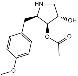
[(2R,3S,4S)-4-hydroxy-2-[(4-methoxyphenyl)methyl]pyrrolidin-3-yl] acetate
|
|
| 别名 |
Wuningmeisu C; NSC 76712; AI3 50846; anisomycin; 22862-76-6; Flagecidin; (-)-Anisomycin; TCMDC-125504; (2R,3S,4S)-4-hydroxy-2-(4-methoxybenzyl)pyrrolidin-3-yl acetate; Upjohn 204t3; (2R,3S,4S)-2-(p-Methoxybenzyl)-3,4-pyrrolidinediol 3-acetate; NSC-76712; AI-350846; NSC76712; AI350846; Flagecidin
|
|
| HS Tariff Code |
2934.99.9001
|
|
| 存储方式 |
Powder -20°C 3 years 4°C 2 years In solvent -80°C 6 months -20°C 1 month |
|
| 运输条件 |
Room temperature (This product is stable at ambient temperature for a few days during ordinary shipping and time spent in Customs)
|
| 溶解度 (体外实验) |
|
|||
|---|---|---|---|---|
| 溶解度 (体内实验) |
配方 1 中的溶解度: ≥ 2.5 mg/mL (9.42 mM) (饱和度未知) in 10% DMSO + 40% PEG300 + 5% Tween80 + 45% Saline (这些助溶剂从左到右依次添加,逐一添加), 澄清溶液。
例如,若需制备1 mL的工作液,可将100 μL 25.0 mg/mL澄清DMSO储备液加入到400 μL PEG300中,混匀;然后向上述溶液中加入50 μL Tween-80,混匀;加入450 μL生理盐水定容至1 mL。 *生理盐水的制备:将 0.9 g 氯化钠溶解在 100 mL ddH₂O中,得到澄清溶液。 配方 2 中的溶解度: ≥ 2.5 mg/mL (9.42 mM) (饱和度未知) in 10% DMSO + 90% (20% SBE-β-CD in Saline) (这些助溶剂从左到右依次添加,逐一添加), 澄清溶液。 例如,若需制备1 mL的工作液,可将 100 μL 25.0 mg/mL澄清DMSO储备液加入900 μL 20% SBE-β-CD生理盐水溶液中,混匀。 *20% SBE-β-CD 生理盐水溶液的制备(4°C,1 周):将 2 g SBE-β-CD 溶解于 10 mL 生理盐水中,得到澄清溶液。 View More
配方 3 中的溶解度: ≥ 2.5 mg/mL (9.42 mM) (饱和度未知) in 10% DMSO + 90% Corn Oil (这些助溶剂从左到右依次添加,逐一添加), 澄清溶液。 配方 4 中的溶解度: 2% DMSO+corn oil: 5mg/mL 1、请先配制澄清的储备液(如:用DMSO配置50 或 100 mg/mL母液(储备液)); 2、取适量母液,按从左到右的顺序依次添加助溶剂,澄清后再加入下一助溶剂。以 下列配方为例说明 (注意此配方只用于说明,并不一定代表此产品 的实际溶解配方): 10% DMSO → 40% PEG300 → 5% Tween-80 → 45% ddH2O (或 saline); 假设最终工作液的体积为 1 mL, 浓度为5 mg/mL: 取 100 μL 50 mg/mL 的澄清 DMSO 储备液加到 400 μL PEG300 中,混合均匀/澄清;向上述体系中加入50 μL Tween-80,混合均匀/澄清;然后继续加入450 μL ddH2O (或 saline)定容至 1 mL; 3、溶剂前显示的百分比是指该溶剂在最终溶液/工作液中的体积所占比例; 4、 如产品在配制过程中出现沉淀/析出,可通过加热(≤50℃)或超声的方式助溶; 5、为保证最佳实验结果,工作液请现配现用! 6、如不确定怎么将母液配置成体内动物实验的工作液,请查看说明书或联系我们; 7、 以上所有助溶剂都可在 Invivochem.cn网站购买。 |
| 制备储备液 | 1 mg | 5 mg | 10 mg | |
| 1 mM | 3.7693 mL | 18.8466 mL | 37.6932 mL | |
| 5 mM | 0.7539 mL | 3.7693 mL | 7.5386 mL | |
| 10 mM | 0.3769 mL | 1.8847 mL | 3.7693 mL |
1、根据实验需要选择合适的溶剂配制储备液 (母液):对于大多数产品,InvivoChem推荐用DMSO配置母液 (比如:5、10、20mM或者10、20、50 mg/mL浓度),个别水溶性高的产品可直接溶于水。产品在DMSO 、水或其他溶剂中的具体溶解度详见上”溶解度 (体外)”部分;
2、如果您找不到您想要的溶解度信息,或者很难将产品溶解在溶液中,请联系我们;
3、建议使用下列计算器进行相关计算(摩尔浓度计算器、稀释计算器、分子量计算器、重组计算器等);
4、母液配好之后,将其分装到常规用量,并储存在-20°C或-80°C,尽量减少反复冻融循环。
计算结果:
工作液浓度: mg/mL;
DMSO母液配制方法: mg 药物溶于 μL DMSO溶液(母液浓度 mg/mL)。如该浓度超过该批次药物DMSO溶解度,请首先与我们联系。
体内配方配制方法:取 μL DMSO母液,加入 μL PEG300,混匀澄清后加入μL Tween 80,混匀澄清后加入 μL ddH2O,混匀澄清。
(1) 请确保溶液澄清之后,再加入下一种溶剂 (助溶剂) 。可利用涡旋、超声或水浴加热等方法助溶;
(2) 一定要按顺序加入溶剂 (助溶剂) 。
|
|
|
|
 JNK-9L
 JNK3 Recombinant Human Active Protein Kinase
 JNK2 Recombinant Human Active Protein Kinase
 JNK-IN-25
InvivoChem的所有产品仅用于作科学研究,不面向患者销售
Copyright 2020 InvivoChem LLC | All Rights Reserved 粤ICP备20063088号-1


 COA
COA


 463611831
