BAY 60-6583

目录号: V12305 纯度: ≥98%
BAY 60-6583 (BAY606583) 是一种新型有效的腺苷 A2B 受体 (EC50 = 3 nM) 激动剂,在心肌缺血模型中具有心脏保护作用。
BAY 60-6583 CAS号: 910487-58-0
产品类别: New1
产品仅用于科学研究,不针对患者销售
规格 价格 库存 数量
1mg
5mg
10mg
50mg
100mg
Other Sizes
点击了解更多
  • 与全球5000+客户建立关系
  • 覆盖全球主要大学、医院、科研院所、生物/制药公司等
  • 产品被大量CNS顶刊文章引用
InvivoChem产品被CNS等顶刊论文引用
产品描述
BAY 60-6583 (BAY606583) 是一种新型强效腺苷 A2B 受体激动剂 (EC50 = 3 nM),在心肌缺血模型中具有心脏保护作用。腺苷 A2B 受体在抗炎反应和预/后适应性心脏保护中发挥重要作用。
BAY 60-6583 (CAS# 910487-58-0) 是一种强效且选择性的腺苷 A2B 受体激动剂。其分子式为 C19H17N5O2S,分子量为 379.43 g/mol。BAY 60-6583 对 A2B 受体的 EC50 为 3 nM。它对 A2B 受体的选择性高于 A1、A2A 和 A3 受体。研究表明,它在心肌缺血模型中具有心脏保护作用。
生物活性&实验参考方法
靶点
BAY 60-6583 targets the adenosine A2B receptor, a G protein-coupled receptor (GPCR). It acts as a partial agonist, binding to the receptor and activating downstream signaling pathways. Its affinity (Ki) for human/mouse/rat A2B receptors is 114/136/100 nM, respectively, as determined by binding assays with [3H]PSB-603.
体外研究 (In Vitro)
在表达重组人A1、A2A或A2B肾上腺素能受体(AR)的CHO细胞中,BAY 60-6583对A1和A2A AR的受体激活EC50值均大于10,000 nM,对A2B AR亚型的EC50值为3 nM[1]。当不使用siRNA时,BAY 60-6583(0-10 μM)的最高激动剂效应为68%,这与存在5、50和500 nM siRNA时的激动剂效应(分别为54%、48%和36%)存在显著差异。在T24细胞中,无论是否存在siRNA,BAY 60-6583的EC50值分别为98±22、102±17、127±31和93±19 nM[3]。在 RAW264.7 前破骨细胞中,BAY 60-6583(5 μM;24 小时)可促进 G1 期细胞的积累,同时减少 G2/M 期细胞的积累[4]。虽然 BAY 60-6583(5 μM;24 小时)不影响 RAW264 细胞中 M-CSF 诱导的 ERK1/2 激活,但它能选择性地抑制 M-CSF 激活的 Akt[4]。体外实验表明,BAY 60-6583 能高效激活 A2B 受体(EC50 = 3 nM)。放射性配体结合实验和功能实验证实了其对 A2B 受体的选择性高于其他腺苷受体亚型。它被用作研究 A2B 受体在各种生理过程中作用的研究工具。
体内研究 (In Vivo)
在缺血性兔心脏模型中,BAY 60-6583(静脉注射;100 mcg/kg)可在再灌注前迅速缩小梗死面积[1]。当用作预处理时,BAY 60-6583(腹腔注射;2 mg/kg)可显著降低脂多糖(LPS)引起的肺损伤。在野生型小鼠中,BAY 60-6583 可降低 IL-6 水平,但在 A2BAR-/- 小鼠中,BAY 60-6583 的给药未能消除这些炎症标志物[2]。肿瘤内注射 BAY 60-6583 可显著增加肿瘤浸润性髓源性抑制细胞(MDSC)的数量;它不影响细胞抑制T细胞增殖或成熟阶段的能力,并且还能促进肿瘤组织内CCL2和IL-10的生成[5]。
体内研究表明,BAY 60-6583在心肌缺血模型中具有心脏保护作用。它是一种用于研究A2B受体激活在心血管疾病和其他疾病中治疗潜力的研究化合物。
酶活实验
BAY 60-6583 的体外受体结合试验包括测定其与 A2B 受体的亲和力。这通常使用放射性配体结合试验,以 [3H]PSB-603 和表达该受体的细胞膜制备物进行。竞争性结合实验用于测定 Ki 值。
细胞实验
细胞周期分析[4]
细胞类型: RAW264.7 前破骨细胞
测试浓度: 5 μM
孵育时间: 48 小时
实验结果: 导致细胞停滞在 G1 期。
蛋白质印迹分析[4]
细胞类型: RAW264.7 前破骨细胞
测试浓度: 5 μM
孵育时间: 48 小时
实验结果: 证实了 M-CSF 介导的 Akt 激活受到抑制,并导致破骨细胞增殖减少。
在表达 A2B 受体的细胞上进行 BAY 60-6583 的体外细胞活性测定。通过其提高 cAMP 水平的能力来衡量其功能活性。测定受体激活的 EC50 值。
动物实验
动物/疾病模型: A2BAR−/− 小鼠与 C57BL/6J 小鼠[1]
剂量: 2 mg/kg
给药途径: 腹腔注射 (ip);2 mg/kg
实验结果: 证实可减轻野生型小鼠的肺部炎症和肺水肿,但在 A2BAR 基因敲除小鼠中未观察到此现象。
BAY 60-6583 的体内动物实验在心肌缺血模型中进行,例如啮齿动物的左前降支 (LAD) 冠状动脉闭塞模型。给予该化合物后,评估其对梗死面积、心脏功能和生存率的影响。
药代性质 (ADME/PK)
BAY 60-6583 已在临床前模型中进行研究,并已确定其药代动力学特性。它通常采用腹腔注射或静脉注射给药。其半衰期和代谢已在动物体内进行研究。它通常以粉末形式储存。
毒性/毒理 (Toxicokinetics/TK)
由于 BAY 60-6583 主要是一种研究用化合物,因此其具体的毒性数据在公共领域并不容易获取。它对 A2B 受体的选择性表明其可能具有良好的安全性。与所有研究用化学品一样,应遵循标准的实验室安全操作规程。
参考文献

[1]. Epithelial-specific A2B adenosine receptor signaling protects the colonic epithelial barrier during acute colitis.Mucosal Immunol. 2015 Nov;8(6):1324-38.

[2]. Signaling through the A2B adenosine receptor dampens endotoxin-induced acute lung injury.J Immunol. 2010 May 1;184(9):5271-9.

[3]. Probing biased/partial agonism at the G protein-coupled A(2B) adenosine receptor.Biochem Pharmacol. 2014 Aug 1;90(3):297-306.

[4]. A2B Adenosine Receptor Stimulation Down-regulates M-CSF-mediated Osteoclast Proliferation. Biomed Sci Letters 2017;23:194-200.

[5]. Characterization of the A2B Adenosine Receptor from Mouse, Rabbit, and Dog. J Pharmacol Exp Ther. 2009 Apr;329(1):2-13.

[6]. Targeting the adenosine A2b receptor in the tumor microenvironment overcomes local immunosuppression by myeloid-derived suppressor cells.Oncoimmunology. 2014 Feb 14;3:e27989. eCollection 2014.

其他信息
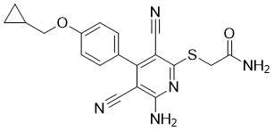
BAY 60-6583 属于氰基吡啶类化合物。其化学名称为 6-氨基-3,5-二氰基-4-(4-羟基苯基)-2-硫代吡啶,其中羟基和硫基上的氢原子分别被环丙基甲基和羧酰胺甲基取代。它具有腺苷 A2B 受体激动剂活性、心脏保护活性和抗炎活性。它是一种氨基吡啶、氰基吡啶、芳基硫醚、单羧酸酰胺、芳香醚和环丙烷类化合物。
BAY 60-6583 是一种用于研究腺苷 A2B 受体的研究化合物。它并非 FDA 批准的药物,仅供研究用途。它是研究 A2B 受体在心血管疾病和其他疾病中作用的重要工具。
*注: 文献方法仅供参考, InvivoChem并未独立验证这些方法的准确性
化学信息 & 存储运输条件
分子式
C19H17N5O2S
分子量
379.43558
精确质量
379.11
CAS号
910487-58-0
PubChem CID
11717831
外观&性状
White to light yellow solid powder
LogP
4.171
tPSA
165.1
氢键供体(HBD)数目
2
氢键受体(HBA)数目
7
可旋转键数目(RBC)
7
重原子数目
27
分子复杂度/Complexity
629
定义原子立体中心数目
0
InChi Key
ZTYHZMAZUWOXNC-UHFFFAOYSA-N
InChi Code
InChI=1S/C19H17N5O2S/c20-7-14-17(12-3-5-13(6-4-12)26-9-11-1-2-11)15(8-21)19(24-18(14)23)27-10-16(22)25/h3-6,11H,1-2,9-10H2,(H2,22,25)(H2,23,24)
化学名
2-[6-amino-3,5-dicyano-4-[4-(cyclopropylmethoxy)phenyl]pyridin-2-yl]sulfanylacetamide
HS Tariff Code
2934.99.9001
存储方式

Powder      -20°C    3 years

                     4°C     2 years

In solvent   -80°C    6 months

                  -20°C    1 month

运输条件
Room temperature (This product is stable at ambient temperature for a few days during ordinary shipping and time spent in Customs)
溶解度数据
溶解度 (体外实验)
DMSO : ~100 mg/mL (~263.55 mM)
溶解度 (体内实验)
配方 1 中的溶解度: ≥ 4.17 mg/mL (10.99 mM) (饱和度未知) in 10% DMSO + 40% PEG300 + 5% Tween80 + 45% Saline (这些助溶剂从左到右依次添加,逐一添加), 澄清溶液。
例如,若需制备1 mL的工作液,可将100 μL 41.7 mg/mL澄清的DMSO储备液加入到400 μL PEG300中,混匀;再向上述溶液中加入50 μL Tween-80,混匀;然后加入450 μL生理盐水定容至1 mL。
*生理盐水的制备:将 0.9 g 氯化钠溶解在 100 mL ddH₂O中,得到澄清溶液。

配方 2 中的溶解度: ≥ 4.17 mg/mL (10.99 mM) (饱和度未知) in 10% DMSO + 90% Corn Oil (这些助溶剂从左到右依次添加,逐一添加), 澄清溶液。
例如,若需制备1 mL的工作液,可将 100 μL 41.7mg/mL澄清DMSO储备液加入900μL玉米油中,混合均匀。

请根据您的实验动物和给药方式选择适当的溶解配方/方案:
1、请先配制澄清的储备液(如:用DMSO配置50 或 100 mg/mL母液(储备液));
2、取适量母液,按从左到右的顺序依次添加助溶剂,澄清后再加入下一助溶剂。以 下列配方为例说明 (注意此配方只用于说明,并不一定代表此产品 的实际溶解配方):
10% DMSO → 40% PEG300 → 5% Tween-80 → 45% ddH2O (或 saline);
假设最终工作液的体积为 1 mL, 浓度为5 mg/mL: 取 100 μL 50 mg/mL 的澄清 DMSO 储备液加到 400 μL PEG300 中,混合均匀/澄清;向上述体系中加入50 μL Tween-80,混合均匀/澄清;然后继续加入450 μL ddH2O (或 saline)定容至 1 mL;

3、溶剂前显示的百分比是指该溶剂在最终溶液/工作液中的体积所占比例;
4、 如产品在配制过程中出现沉淀/析出,可通过加热(≤50℃)或超声的方式助溶;
5、为保证最佳实验结果,工作液请现配现用!
6、如不确定怎么将母液配置成体内动物实验的工作液,请查看说明书或联系我们;
7、 以上所有助溶剂都可在 Invivochem.cn网站购买。
制备储备液 1 mg 5 mg 10 mg
1 mM 2.6355 mL 13.1773 mL 26.3546 mL
5 mM 0.5271 mL 2.6355 mL 5.2709 mL
10 mM 0.2635 mL 1.3177 mL 2.6355 mL

1、根据实验需要选择合适的溶剂配制储备液 (母液):对于大多数产品,InvivoChem推荐用DMSO配置母液 (比如:5、10、20mM或者10、20、50 mg/mL浓度),个别水溶性高的产品可直接溶于水。产品在DMSO 、水或其他溶剂中的具体溶解度详见上”溶解度 (体外)”部分;

2、如果您找不到您想要的溶解度信息,或者很难将产品溶解在溶液中,请联系我们;

3、建议使用下列计算器进行相关计算(摩尔浓度计算器、稀释计算器、分子量计算器、重组计算器等);

4、母液配好之后,将其分装到常规用量,并储存在-20°C或-80°C,尽量减少反复冻融循环。

计算器

摩尔浓度计算器可计算特定溶液所需的质量、体积/浓度,具体如下:

  • 计算制备已知体积和浓度的溶液所需的化合物的质量
  • 计算将已知质量的化合物溶解到所需浓度所需的溶液体积
  • 计算特定体积中已知质量的化合物产生的溶液的浓度
使用摩尔浓度计算器计算摩尔浓度的示例如下所示:
假如化合物的分子量为350.26 g/mol,在5mL DMSO中制备10mM储备液所需的化合物的质量是多少?
  • 在分子量(MW)框中输入350.26
  • 在“浓度”框中输入10,然后选择正确的单位(mM)
  • 在“体积”框中输入5,然后选择正确的单位(mL)
  • 单击“计算”按钮
  • 答案17.513 mg出现在“质量”框中。以类似的方式,您可以计算体积和浓度。

稀释计算器可计算如何稀释已知浓度的储备液。例如,可以输入C1、C2和V2来计算V1,具体如下:

制备25毫升25μM溶液需要多少体积的10 mM储备溶液?
使用方程式C1V1=C2V2,其中C1=10mM,C2=25μM,V2=25 ml,V1未知:
  • 在C1框中输入10,然后选择正确的单位(mM)
  • 在C2框中输入25,然后选择正确的单位(μM)
  • 在V2框中输入25,然后选择正确的单位(mL)
  • 单击“计算”按钮
  • 答案62.5μL(0.1 ml)出现在V1框中
g/mol

分子量计算器可计算化合物的分子量 (摩尔质量)和元素组成,具体如下:

注:化学分子式大小写敏感:C12H18N3O4  c12h18n3o4
计算化合物摩尔质量(分子量)的说明:
  • 要计算化合物的分子量 (摩尔质量),请输入化学/分子式,然后单击“计算”按钮。
分子质量、分子量、摩尔质量和摩尔量的定义:
  • 分子质量(或分子量)是一种物质的一个分子的质量,用统一的原子质量单位(u)表示。(1u等于碳-12中一个原子质量的1/12)
  • 摩尔质量(摩尔重量)是一摩尔物质的质量,以g/mol表示。
/

配液计算器可计算将特定质量的产品配成特定浓度所需的溶剂体积 (配液体积)

  • 输入试剂的质量、所需的配液浓度以及正确的单位
  • 单击“计算”按钮
  • 答案显示在体积框中
动物体内实验配方计算器(澄清溶液)
第一步:请输入基本实验信息(考虑到实验过程中的损耗,建议多配一只动物的药量)
第二步:请输入动物体内配方组成(配方适用于不溶/难溶于水的化合物),不同的产品和批次配方组成不同,如对配方有疑问,可先联系我们提供正确的体内实验配方。此外,请注意这只是一个配方计算器,而不是特定产品的确切配方。
+
+
+

计算结果:

工作液浓度 mg/mL;

DMSO母液配制方法 mg 药物溶于 μL DMSO溶液(母液浓度 mg/mL)。如该浓度超过该批次药物DMSO溶解度,请首先与我们联系。

体内配方配制方法μL DMSO母液,加入 μL PEG300,混匀澄清后加入μL Tween 80,混匀澄清后加入 μL ddH2O,混匀澄清。

(1) 请确保溶液澄清之后,再加入下一种溶剂 (助溶剂) 。可利用涡旋、超声或水浴加热等方法助溶;
            (2) 一定要按顺序加入溶剂 (助溶剂) 。

生物数据图片
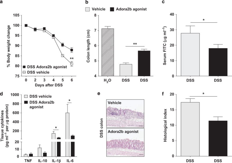
  • Treatment with an Adora2b (A2B adenosine receptor) agonist mediates mucosal protection observed in a model of acute colitis. Gender-, age-, and weight-matched C57BL/6 mice were treated with an Adora2b-specific agonist (BAY 60-6583; 1.2–1.25 mg kg−1 per day) or vehicle (30% Solutol HS15 in 0.9% saline) using a subcutaneous osmotic pump beginning 1 day before administration of DSS (dextran sulfate sodium, 3.5–4%) for 6 days. Mice exposed to DSS were orally gavaged with FITC (fluorescein isothiocyanate)-dextran (0.6 mg g−1 at 80 mg ml−1) 4 h before killing on day 6, serum collection, and blunt dissection of the colon. (a) Daily weight measurements were obtained for each group of mice. (b) Colon lengths were measured upon harvest. (c) Fluorescence measurement determined FITC levels in the serum on day 6 after DSS. (d) Following harvest, cytokines in colonic tissue were measured by Meso Scale. Results are displayed normalized to protein content and represent 4–5 mice per group. (e) Representative colonic histological images from vehicle and Adora2b agonist-treated mice exposed to DSS. Bar = 100 μm; images were acquired at original magnification ×10. [1].Epithelial-specific A2B adenosine receptor signaling protects the colonic epithelial barrier during acute colitis.Mucosal Immunol. 2015 Nov;8(6):1324-38.
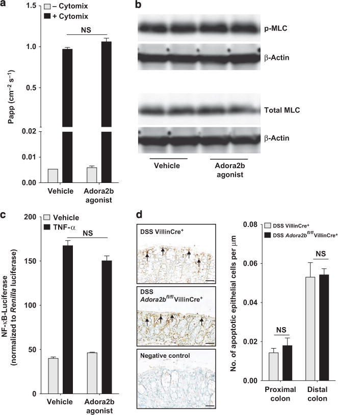
  • Epithelial Adora2b (A2B adenosine receptor) signaling does not affect epithelial barrier breakdown. (a) A combination of tumor necrosis factor-α (TNFα), interleukin-1β (IL-1β), and interferon γ (IFNγ) (all 10 ng ml−1) or media alone was added to the basolateral aspect of polarized T84 intestinal epithelial cells. Vehicle or the Adora2b agonist (BAY 60-6583; 10 μM) was added to both chambers. Following 72 h, FITC (fluorescein isothiocyanate)-labeled dextran (3 kDa) was added to the apical chamber and a flux assay was performed. The permeability of the monolayer was assessed by measuring the concentration of FITC in the basolateral chamber over time, calculated as the apparent permeability (Papp: cm−2 s−1). Results are representative of three independent experiments with 2–4 wells per group. (b) Confluent T84 intestinal epithelial cells were treated on the basolateral and apical aspect with vehicle or Adora2b-specific agonist (BAY 60-6583; 10 μM) for 30 min before cell harvest and total protein extraction. p-MLC (myosin light chain) (Ser19), total MLC, and β-actin levels were determined by Western blot analysis. Results are representative of three independent experiments with 2–3 wells per experiment.[1].Epithelial-specific A2B adenosine receptor signaling protects the colonic epithelial barrier during acute colitis.Mucosal Immunol. 2015 Nov;8(6):1324-38.
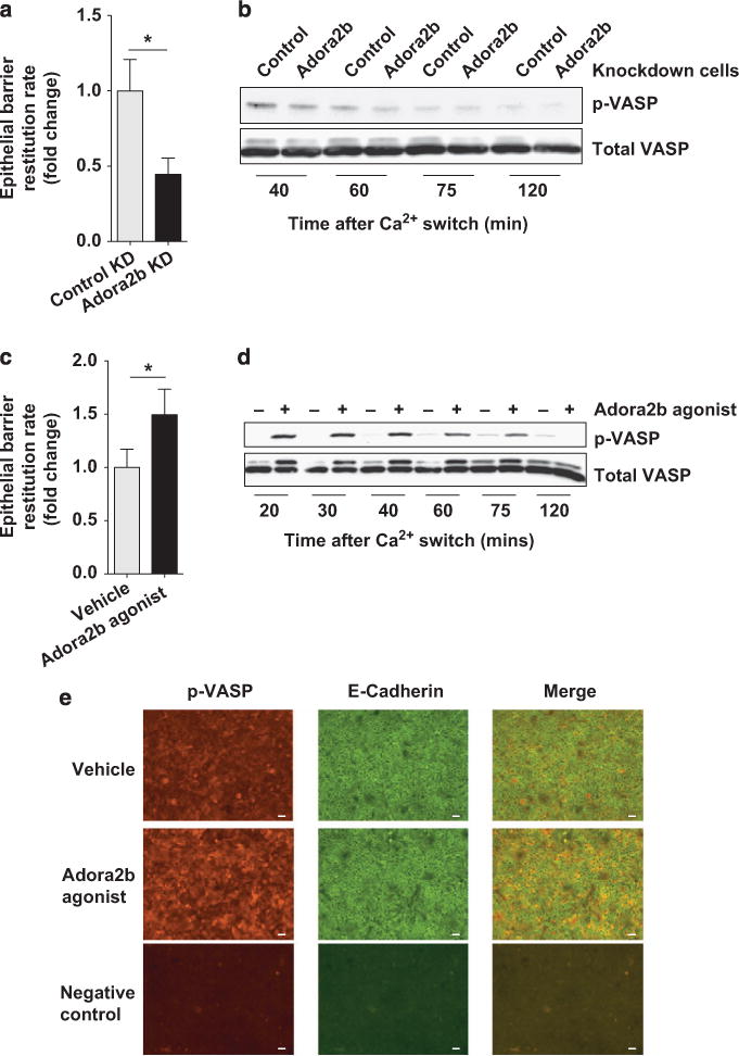
  • Epithelial Adora2b (A2B adenosine receptor) promotes barrier restitution in intestinal epithelial cells. Calcium switch assays were performed using intestinal epithelial cells with a fully competent barrier. Cells were placed in calcium-depleting conditions until barrier function was fully compromised and subsequently allowed to recover in calcium complete media for the time points outlined. (a) Barrier recovery studies were performed with T84 intestinal epithelial cells with constitutive Adora2b knockdown (Adora2b KD) or nonspecific gene KD (control KD) as mentioned. FITC (fluorescein isothiocyanate)-labeled dextran (10 kDa) was added to the apical aspect of the cells at the start of the recovery period. Media from the basolateral aspect were sampled every 15 min for 105 min and fluorescence in the media at each point was measured to analyze rate of barrier restitution. Results are displayed as fold change in the rate of restitution and represent two independent experiments with 4–6 wells per experiment. (b) Constitutive Adora2b or control KD cells were used in calcium switch assays as described above and allowed to recover for the indicated times. Total protein was extracted and phosphorylated vasodilator-stimulated phosphoprotein (p-VASP) (Ser157) and total VASP levels were determined by Western blot analysis. Results are representative of three independent experiments with 2 wells per time point per experiment. (c) T84 intestinal epithelial cells were used in calcium switch assays. Vehicle or the Adora2b-specific agonist (BAY 60-6583; 10 μM) was added to both the basolateral and apical aspects of the cells at the beginning of the recovery period. Barrier restitution rate was determined as described in a. Results are representative of three independent experiments with 4–6 wells per experiment. [1].Epithelial-specific A2B adenosine receptor signaling protects the colonic epithelial barrier during acute colitis.Mucosal Immunol. 2015 Nov;8(6):1324-38.
联系我们