
| 规格 | 价格 | 库存 | 数量 |
|---|---|---|---|
| 10 mM * 1 mL in DMSO |
|
||
| 1mg |
|
||
| 5mg |
|
||
| 10mg |
|
||
| 25mg |
|
||
| 50mg |
|
||
| 100mg |
|
||
| 250mg |
|
||
| 500mg |
|
||
| Other Sizes |
|

| 靶点 |
Topoisomerase I
|
|---|---|
| 体外研究 (In Vitro) |
体外活性:Beta-Lapachone 以剂量依赖性方式抑制 DNA 拓扑异构酶 I 诱导的 DNA 松弛。与 DMSO 对照相比,β-拉帕酮(100 nM 或更高)处理可抑制 >95% 的 Topo I DNA 解旋活性。 β-拉帕酮 (1-5 μM) 可阻断细胞周期的 G0/G1 期,并通过将 Topo I 锁定在 DNA 上并阻断 HL-60 和三种人类前列腺癌(DU-145、PC- 3和LNCaP)细胞。 Beta-Lapachone 通过不同的 MAPK 信号通路促进小鼠 3T3 成纤维细胞和人内皮 EAhy926 细胞的迁移,从而加速体外擦伤伤口的愈合。此外,β-拉帕酮通过非竞争性抑制抑制纯化的重组 IDO1 活性,IC50 为 0.44 μM,β-拉帕酮还表现出优异的细胞内 IDO1 抑制活性保留,IC50 为 1.0 μM,部分依赖于 NQO1 的生物转化。 Beta-lapachone 通过 NQO1 依赖性活性氧 (ROS) 形成和 PARP1 过度激活诱导 NQO1+ 癌细胞程序性坏死。激酶测定:拓扑异构酶I催化活性测定:通过DNA解旋测定来分析酶活性。来自 TopoGEN 的 DNA 拓扑异构酶 I(1 单位,定义为在 37 °C 下 30 分钟内将 0.5 μg 超螺旋 DNA 转化为松弛状态的酶量)与 0.5 μg 6x174 RF DNA 一起孵育,在存在或不存在 Beta-Lapachone,在 20 μL 松弛缓冲液(50 mM Tris (pH 7.5)、50 mM KCI、10 mM MgCl2、0.5 mM 二硫苏糖醇、0.5 mM EDTA、30 μg/mL 牛血清白蛋白)中处理 30 分钟37°C。添加 1% SDS 和蛋白酶 K (50 μg/mL) 终止反应。在 37 °C 下再孵育 1 小时后,通过在 TAE 缓冲液(0.04 M 三乙酸盐、0.001 M EDTA)中的 1% 琼脂糖凝胶中电泳分离产物。电泳后用溴化乙锭对凝胶进行染色。使用 NIH 图像分析系统扫描照相底片。细胞测定:每个细胞系的 IC50 计算由 DNA 量 (IS) 和贴壁依赖性集落形成 (CF) 测定确定。对于 CF 测定,细胞以 500 个活细胞/孔接种在 6 孔板中并孵育过夜,然后用等体积的含有 β-拉帕酮的培养基进行处理,终浓度范围为 0.005 至 50 μM,以半对数增量(对照)用 0.25% DMSO(相当于所使用的最高剂量的 β-lapachonc)处理 4 小时或连续暴露 12 小时。将板(3孔/条件)用结晶紫染色,并计数>50个外观正常的细胞的集落。使用药物剂量计算各种细胞的 IC50 值,其中集落数约为对照的 50%。对于 DNA 测定,使用 CytoFluor 2350 荧光测量系统处理后 8 天收获板进行 IC50 测定。六孔采样包含在每个剂量的 DNA 荧光单位的计算中。使用 β-拉帕酮剂量与荧光单位百分比对照 DNA 的关系图来计算每个 IC50。所有实验至少重复两次,每次一式两份。
|
| 体内研究 (In Vivo) |
β-拉帕酮治疗(50 mg/kg)可有效抑制人卵巢癌异种移植小鼠模型中的体内肿瘤生长,并且β-拉帕酮和紫杉醇的组合可协同诱导细胞凋亡。在正常和糖尿病 (db/db) 小鼠中,β-拉帕酮治疗比仅用载体治疗导致更快的愈合过程。β-lapachone对体内伤口愈合的影响。[3]
为了确定β-拉帕酮是否对伤口愈合有治疗作用,将单独或含有29.8μg/gβ-拉帕酮的软膏涂抹在C57BL/6或db/db小鼠背部的伤口上21天,从受伤第0天到受伤后第21天每5天检查一次伤口愈合情况(图5和图6)。在受伤后第3、7、14或21天切下伤口中心的皮肤组织(约1×1 cm2),并进行苏木精和伊红染色(图6)。使用Imagescope软件测量愈合皮肤下的血管密度(图6E)。显微镜观察显示,db/db小鼠伤口愈合所需的时间明显长于C57BL/6小鼠(图5,A和C),在5至20天内,用含β-lapachone的软膏治疗的C57BL-6或db/db鼠的伤口面积(图5、B和D)明显小于用对照软膏治疗的小鼠(图五,A和C)。与用对照软膏治疗的小鼠相比,C57BL/6和db/db小鼠的β-lapachone治疗伤口面积显著减小(图5E)。与用不含β-拉帕酮的软膏治疗的伤口相比,β-拉帕酮治疗的伤口愈合恢复过程在C57BL/6或db/db小鼠中更快(图6,A-D)。第14天,在C57BL/6小鼠中,用对照软膏治疗的伤口瘢痕组织较厚,真皮出现紊乱(图6A3);然而,在同一天,β-lapachone治疗的伤口的皮肤层完全修复(图6B3)。同样,在db/db小鼠中,瘢痕组织相对较薄,在14天时,β-lapachone治疗的伤口真皮中出现了毛囊(图6D3),但用对照软膏治疗的伤口中的毛囊仅在21天时观察到。 |
| 酶活实验 |
DNA 拓扑异构酶 I 在含有或不含药物(包括 β-Lapachone)的 20 μL 松弛缓冲液(50 mM Tris,pH 7.5)中培养。 (30 μg/mL 牛血清白蛋白、50 mM KCl、10 mM MgCl2、0.5 mM 二硫苏糖醇、0.5 mM EDTA)37°C 30 分钟。添加蛋白酶 K (50 μg/mL) 和 1% SDS 以终止反应。在 37°C 下额外孵育 1 小时后,通过在 TAE 缓冲液(0.04 M 三乙酸盐、0.001 M EDTA)中的 1% 琼脂糖凝胶中电泳分离产物。电泳后,使用溴化乙锭对凝胶进行染色。利用 NIH 图像分析系统扫描底片。
生化测定[4] 纯化的人重组IDO1(hrIDO1)酶在大肠杆菌中表达,并如前所述纯化。IDO1酶测定使用之前发表的磷酸钾缓冲系统进行。25 Ehrlich试剂用于分光光度法检测犬尿氨酸(Kyn),如上所述。25首先混合包括底物和抑制剂在内的试剂,最后加入enyme以在T=0时引发反应。为了确定hrIDO1制剂的酶动力学,在1 mL体积中进行了不同L-Trp浓度(0-400μM)的酶测定,并在多个时间点(0-90分钟)收集了100μL等分试样进行Kyn分析。结果证实,hrIDO1酶遵循之前发表的Michaelis-Menten动力学26,Km为110μM,Vmax为5.9μM/min(补充图S1)。随后,在hrIDO1酶反应中,在固定底物浓度(100μM L-Trp)下,使用不同浓度的抑制剂(0-50μM)测定IC50,或在不同浓度的两种抑制剂(0-800 nM)和底物(0-400μM)进行Ki测定时,评估了β-拉帕酮的抑制活性。反应在100μL体积内进行,并在酶活性在线性范围内时停止15分钟。使用Prism v.5.0进行数据分析和绘图。 |
| 细胞实验 |
MTT 测定用于量化细胞毒性。在添加不同浓度的拓扑替康或 β-拉帕酮前两天,将 IMR-32 和 JCI 细胞以 5.0 × 10⁴(拓扑替康)或 2.5 × 的浓度接种在 96 孔微量滴定板中。 104 (β-lapachone) 细胞/孔/100 µL 培养基。之后,将细胞在37℃的CO2培养箱中保存72小时。细胞增殖试剂盒 I 用于测量细胞增殖。实验中使用了四种不同的培养物。
细胞处理和细胞活力测定。[3] 将100μl培养基中的HS68细胞(103个)、3T3细胞(103)或EAhy926细胞(104个)在37°C下在96孔培养板中在加湿的5%CO2气氛中接种24小时。由于生长速率较低,将HEKn细胞(104)、XB-2细胞(104”和HUVEC(104)接种48小时。对于3-(4,5-二甲基噻唑-2-基)-2,5-二苯基溴化四唑(MTT)试验,在细胞活力试验前24小时向培养基中加入不同浓度的β-lapachone。简而言之,向每个孔中加入10μl MTT(0.5mg/ml),将平板在37°C下孵育4小时。然后将甲赞产物溶解在100μl DMSO中,在37°℃下孵育30分钟,用微孔板读数器测量570nm处的吸光度。 为了测试MAPK抑制剂的作用,将3T3细胞或EAhy926细胞(103个细胞在100μl培养基/孔中)与0、5或10μM ERK抑制剂或p38抑制剂(SB-203580)或0、50或100 nM JNK抑制剂(SP-600125)一起孵育1小时;然后将细胞换成含有相同MAPK抑制剂的培养基,加入或不加入1μMβ-lapachone。使用MTT法测量处理后活细胞的数量。对于所有研究,至少进行了三组独立实验,每组三份。 细胞周期分析。[3] 细胞用1μMβ-lapachone 处理3、6、9、12或24小时,用0.5%胰蛋白酶-EDTA收获,用冷80%乙醇固定。用PBS洗涤三次后,将细胞在37°C下与RNase A(1μg/ml)一起孵育1小时,然后在37°℃下与碘化丙啶(50μg/ml)孵育15分钟。使用FL-2参数通过流式细胞术检测染色细胞,并使用Cell Quest Pro软件分析数据。 免疫荧光染色。 将细胞与1μM的β-lapachone孵育0至24小时,然后在4%的多聚甲醛中固定15分钟。在室温下用10%的正常山羊血清(NGS)封闭1小时后,将细胞在4°C下用抗PCNA单克隆抗体(1:1000)染色过夜,在室温下与罗丹明偶联的二抗和赫斯特染料孵育1小时,并使用徕卡荧光显微镜进行检查和拍照。 蛋白质印迹分析。[3] 用1μMβ-拉帕酮处理0-24小时的细胞用裂解缓冲液(0.25 mM HEPES,pH 7.4,14.9 mM NaCl,10 mM NaF,2 mM MgCl2,0.5%NP-40,0.1 mM PMSF,20μM pepstatin A和20μM亮肽)裂解。然后将裂解物在4°C下以1000 g离心15分钟,收集上清液进行免疫印迹。使用ELISA阅读器通过Bradford测定法测量样品中的蛋白质量。通过10-12%SDS-PAGE从每个样品中分离出约25-50μg蛋白质,然后转移到电泳转移池中的Immobilon-P膜上(在200V下2小时)。所有后续步骤均在室温下进行。用含有0.05%吐温20(PBST)的PBS中的5%脱脂乳将膜封闭1小时,用1%BSA中的抗磷酸化ERK、抗ERK、反磷酸化JNK、抗JNK、反磷酸化p38、抗p38或抗肌动蛋白抗体(1:1000稀释)孵育2小时,用PBST洗涤30分钟,然后用辣根过氧化物酶偶联的二抗孵育1小时。用ECL-Western印迹试剂检测结合抗体,用富士医用X射线胶片检测化学发光。使用Scion软件定量每种蛋白质的量。 刮伤愈合试验。[3] 细胞在24孔培养皿上生长至融合,吸出培养基,加入单独或与ERK抑制剂、p38抑制剂或JNK抑制剂一起含有或不含有1μMβ-lapachone的新培养基。用200-μl一次性塑料移液管尖端在细胞涂层表面刮取一条(约150μm宽)条纹,在37°C下使伤口愈合24小时(内皮细胞)或48小时(成纤维细胞)。通过测量伤口的宽度来评估伤口闭合的平均程度。 Transwell迁移分析。[3] 使用改良的Millicell室(8-μm孔)评估细胞迁移。在0.2 ml培养基中以1×10⁴个细胞/孔的速度接种到上室中的细胞单独用β-拉帕酮或用β-拉帕酮加ERK抑制剂、p38抑制剂或JNK抑制剂处理,并将0.6 ml培养基加入到下室中。在37°C下24小时后,机械去除膜上表面的细胞,固定膜下表面的迁移细胞,并用考马斯亮蓝染色。计数膜下表面上迁移的细胞总数。每个实验进行三次。 |
| 动物实验 |
雄性Balb/c小鼠饲喂商业颗粒饲料,并可自由饮水。适应一周后,将小鼠随机分为五组:对照组、β-拉帕酮组、顺铂组(18 mg/kg,腹腔注射)和β-拉帕酮+顺铂组(18 mg/kg,腹腔注射)。在注射顺铂前两周,β-拉帕酮组小鼠饲喂含药物(0.066)的饲料。注射顺铂三天后,所有小鼠在二氧化碳麻醉下处死。对血液样本进行血清尿素氮(BUN)和肌酐(CRE)分析。用于组织病理学和免疫组织化学(IHC)研究的肾脏被迅速切取一半。剩余的一半保存在低温环境中,直至进行蛋白质印迹实验。伤口活检和伤口愈合情况测量。 [3]
小鼠(C57BL/6 或 db/db)用 2% Rompun 溶液(0.1 ml/20 g 体重;拜耳公司,德国勒沃库森)麻醉。剃除小鼠背部毛发后,用酒精棉签消毒。使用无菌活检穿刺器(直径 6 mm)在肩胛骨下方背部皮肤全层穿刺。该区域的伤口小鼠无法触及,因此可防止其舔舐自身。在伤口处涂抹软膏[100 mg 纯白色凡士林(Vaseline)](对照组)或含有 29.8 μg/g β-拉帕酮的软膏,每 2 天更换一次。从造伤当天开始,每 5 天对每只小鼠的伤口进行数码拍照。所有测量均使用 Scion 软件量化伤口面积。 |
| 参考文献 | |
| 其他信息 |
β-拉帕酮是一种苯并色烯酮,其结构为3,4-二氢-2H-苯并[h]色烯-5,6-二酮,在2位被偕二甲基取代。它从紫葳科植物Tabebuia avellanedae中分离得到,具有抗肿瘤和抗炎活性。它可用作抗肿瘤药、抗炎药和植物代谢产物。它是一种苯并色烯酮,属于邻醌类化合物。
拉帕酮已用于癌症、癌、晚期实体瘤、头颈部肿瘤和鳞状细胞癌的治疗研究。 据报道,β-拉帕酮存在于梓树(Catalpa longissima)、番石榴(Handroanthus guayacan)和其他有相关数据的生物体中。 拉帕酮是一种难溶的邻萘醌,具有潜在的抗肿瘤和放射增敏活性。 β-拉帕酮 (b-lap) 可被 NAD(P)H:醌氧化还原酶-1 (NQO1) 生物活化,产生无效的氧化还原反应,生成高浓度的超氧化物。这些高活性氧 (ROS) 与 DNA 相互作用,导致单链 DNA 断裂,并促使内质网 (ER) 中的钙离子释放。最终,广泛的 DNA 损伤导致 DNA 修复酶——聚(ADP-核糖)聚合酶-1 (PARP-1) 过度激活,同时伴随着 NAD+/ATP 核苷酸水平的快速耗竭。因此,在 NQO1 过表达的肿瘤细胞中,会发生一种不依赖于 caspase 且由内质网应激诱导的 μ-钙蛋白酶介导的细胞死亡。 NQO1是一种黄素蛋白和双电子氧化还原酶,在多种肿瘤中过表达。β-拉帕酮是一种植物产物,已被发现具有多种药理作用。迄今为止,人们对其生化靶点知之甚少。本研究发现,β-拉帕酮能够抑制小牛胸腺和人细胞中拓扑异构酶I的催化活性。但与喜树碱不同,β-拉帕酮并不稳定可裂解复合物,表明其作用机制不同。β-拉帕酮抑制喜树碱诱导的拓扑异构酶I介导的DNA裂解。在加入DNA底物之前,先用β-拉帕酮孵育拓扑异构酶I,可显著增强这种抑制作用。将拓扑异构酶 I 与 DNA 孵育后再加入 β-拉帕酮,可使该酶失去活性;而先用 β-拉帕酮处理 DNA 再加入拓扑异构酶则无影响。这些结果表明 β-拉帕酮与拓扑异构酶 I 直接相互作用,而非与 DNA 底物相互作用。β-拉帕酮不抑制酶与 DNA 底物的结合。在细胞中,β-拉帕酮本身不诱导形成 SDS-K(+) 沉淀复合物,但它能抑制与喜树碱形成复合物。我们推测,β-拉帕酮与拓扑异构酶 I 的直接相互作用不影响酶-DNA 复合物的组装,但会抑制可裂解复合物的形成。[1] β-拉帕酮及其某些衍生物可直接结合并抑制拓扑异构酶 I (Topo I) 的 DNA 解旋活性,形成 DNA-Topo I 复合物,该复合物无法通过 SDS-K+ 检测分离。我们发现β-拉帕酮可以诱导某些细胞凋亡,例如人早幼粒细胞白血病细胞(HL-60)和人前列腺癌细胞(DU-145、PC-3 和 LNCaP),正如 Li 等人(Cancer Res., 55: 0000-0000, 1995)所述。在HL-60细胞中,经≥0.5 μM β-拉帕酮处理4小时后,通过流式细胞术和形态学检查观察到特征性的180-200 bp寡核小体DNA梯状条带和含有DNA片段的凋亡细胞。与等毒性浓度的β-拉帕酮相比,用喜树碱或拓扑替康处理的HL-60细胞产生了更明显的凋亡DNA梯状条带和更多的凋亡细胞,尽管β-拉帕酮是一种更有效的拓扑异构酶I抑制剂。 β-拉帕酮处理(4 小时,1-5 μM)导致 HL-60 细胞和三种不同的前列腺癌细胞(DU-145、PC-3 和 LNCaP)的细胞周期阻滞于 G0/G1 期,S 期和 G2/M 期细胞比例下降,凋亡细胞数量随时间增加。拓扑替康或喜树碱的类似处理(4 小时,1-5 μM)也导致细胞周期阻滞于 S 期并诱导细胞凋亡。因此,β-拉帕酮可阻滞细胞周期于 G0/G1 期,并在 DNA 合成之前或早期诱导细胞凋亡。这些事件与 p53 无关,因为 PC-3 和 HL-60 细胞为 p53 缺失型细胞,LNCaP 为野生型细胞,DU-145 含有突变型 p53,但所有这些细胞在 β-拉帕酮处理后均发生凋亡。有趣的是,β-拉帕酮处理含有野生型p53的前列腺癌细胞(例如LNCaP)并未像喜树碱处理细胞那样诱导p53蛋白的核内表达。与其他拓扑异构酶I抑制剂类似,β-拉帕酮可能通过将拓扑异构酶I锁定在DNA上,阻断复制叉的移动,从而以p53非依赖的方式诱导细胞凋亡。β-拉帕酮及其衍生物以及其他拓扑异构酶I抑制剂,单独使用即可在治疗人类白血病和前列腺癌方面具有潜在的临床应用价值。[2] 伤口愈合障碍是糖尿病患者面临的严重问题。伤口愈合是一个复杂的过程,需要多种细胞类型的协同作用,包括角质形成细胞、成纤维细胞、内皮细胞和巨噬细胞。 β-拉帕酮是从紫葳树(Tabebuia avellanedae)树皮中提取的一种天然化合物,因其在不同浓度和条件下具有抗肿瘤、抗炎和抗肿瘤作用而闻名,但其对伤口愈合的影响尚未得到研究。本研究旨在探讨β-拉帕酮对伤口愈合的影响及其潜在机制。本研究表明,低剂量β-拉帕酮可增强多种细胞的增殖,通过不同的MAPK信号通路促进小鼠3T3成纤维细胞和人内皮细胞EAhy926的迁移,并加速体外擦伤伤口的愈合。在正常小鼠和糖尿病(db/db)小鼠的穿刺伤口上涂抹含或不含β-拉帕酮的软膏后发现,β-拉帕酮治疗组的伤口愈合速度快于仅涂抹溶剂的对照组。此外,β-拉帕酮可诱导巨噬细胞释放VEGF和EGF,这两种细胞因子均有利于多种细胞的生长。我们的研究结果表明,β-拉帕酮能够促进细胞增殖,包括角质形成细胞、成纤维细胞和内皮细胞,并促进成纤维细胞和内皮细胞的迁移,从而加速伤口愈合。因此,我们认为β-拉帕酮可能具有治疗伤口愈合的潜力。[3] β-拉帕酮是一种天然存在的1,2-萘醌类化合物,因其具有肿瘤选择性细胞毒性,已被推进至临床试验阶段。此前,我们曾以相关的1,4-萘醌药效团为基础,开发了一系列强效的吲哚胺2,3-双加氧酶1 (IDO1) 抑制剂。本研究发现,临床候选药物β-拉帕酮具有IDO1抑制活性,这是此前未被发现的特性。基于酶动力学的β-拉帕酮抑制模式为非竞争性抑制,而计算模型预测其与IDO1活性位点的结合方式与其他萘醌衍生物一致。此前已有研究表明,抑制IDO1能够突破致病耐受机制,从而解除免疫系统产生有效抗肿瘤反应的限制。因此,β-拉帕酮具有IDO1抑制活性这一发现,为其作为抗癌药物的潜在用途增添了新的维度,使其区别于其细胞毒性,并提示其细胞毒性和免疫调节作用的联合效应可能产生协同增效作用。[4] 靶向氧化还原酶NAD(P)H:醌氧化还原酶1 (NQO1) 以诱导实体瘤程序性坏死的药物,例如β-拉帕酮,已展现出巨大的潜力,但仍需要更有效的肿瘤选择性化合物。本文报道,脱氧尼波醌以NQO1依赖的方式杀死多种癌细胞,其效力高于β-拉帕酮。脱氧尼波醌的杀伤作用依赖于NQO1依赖的无效氧化还原循环,该循环消耗氧气并产生大量活性氧(ROS)。活性氧(ROS)水平升高会导致广泛的DNA损伤、PARP1过度激活以及严重的NAD+/ATP耗竭,从而刺激Ca2+依赖性程序性坏死,这是这类新型NQO1“生物活化”药物所特有的。NQO1+细胞短期暴露于脱氧尼波醌足以诱导细胞死亡,而基因匹配的NQO1-细胞则不受影响。此外,siRNA介导的NQO1或PARP1敲低可使NQO1+细胞免于短期死亡。用BAPTA-AM(一种胞质Ca2+螯合剂)或过氧化氢酶(一种酶促H2O2清除剂)预处理细胞足以挽救脱氧尼波醌诱导的细胞死亡,这与β-拉帕醌的情况类似。体内研究表明,脱氧尼波醌与β-拉帕醌具有相当的抗肿瘤疗效,但其效力高出6倍。肿瘤组织中观察到PARP1过度激活和ATP显著减少,但在相关的正常肺组织中未观察到。我们的研究结果为去氧尼博醌作为一种强效化疗药物治疗多种难治性实体瘤(如胰腺癌和肺癌)提供了临床前概念验证。[5] 在体外培养的多种人类癌细胞中,用两种低分子量化合物β-拉帕酮和紫杉醇联合治疗后,观察到肿瘤克隆的消融。它们协同诱导培养的卵巢癌、乳腺癌、前列腺癌、黑色素瘤、肺癌、结肠癌和胰腺癌细胞死亡。这种协同作用与给药方案有关;也就是说,紫杉醇必须与β-拉帕酮同时或在其后添加。这种联合疗法对移植到小鼠体内的人类卵巢癌和前列腺癌肿瘤具有异常强的抗肿瘤活性。宿主毒性很小。细胞可在细胞周期检查点启动凋亡,这种机制可清除缺陷细胞以确保基因组的完整性。我们假设,当细胞同时接受激活多个不同细胞周期检查点的药物治疗时,相互冲突的调控信号分子的产生会诱导癌细胞凋亡。β-拉帕酮可导致细胞周期延迟至G1期晚期和S期,而紫杉醇则使细胞停滞于G2/M期。接受这两种药物治疗的细胞在启动凋亡前,会在多个检查点发生延迟。我们的研究结果提示,利用细胞周期检查点处易导致凋亡的“碰撞”机制,或许可以开发出新的抗癌疗法。[6] |
| 分子式 |
C15H14O3
|
|
|---|---|---|
| 分子量 |
242.27
|
|
| 精确质量 |
242.094
|
|
| 元素分析 |
C, 74.36; H, 5.82; O, 19.81
|
|
| CAS号 |
4707-32-8
|
|
| 相关CAS号 |
|
|
| PubChem CID |
3885
|
|
| 外观&性状 |
Brown to red solid powder
|
|
| 密度 |
1.3±0.1 g/cm3
|
|
| 沸点 |
381.4±42.0 °C at 760 mmHg
|
|
| 熔点 |
>110ºC (dec.)
|
|
| 闪点 |
169.7±27.9 °C
|
|
| 蒸汽压 |
0.0±0.9 mmHg at 25°C
|
|
| 折射率 |
1.595
|
|
| LogP |
2.82
|
|
| tPSA |
43.37
|
|
| 氢键供体(HBD)数目 |
0
|
|
| 氢键受体(HBA)数目 |
3
|
|
| 可旋转键数目(RBC) |
0
|
|
| 重原子数目 |
18
|
|
| 分子复杂度/Complexity |
445
|
|
| 定义原子立体中心数目 |
0
|
|
| SMILES |
O1C2C3=C([H])C([H])=C([H])C([H])=C3C(C(C=2C([H])([H])C([H])([H])C1(C([H])([H])[H])C([H])([H])[H])=O)=O
|
|
| InChi Key |
QZPQTZZNNJUOLS-UHFFFAOYSA-N
|
|
| InChi Code |
InChI=1S/C15H14O3/c1-15(2)8-7-11-13(17)12(16)9-5-3-4-6-10(9)14(11)18-15/h3-6H,7-8H2,1-2H3
|
|
| 化学名 |
2,2-dimethyl-3,4-dihydrobenzo[h]chromene-5,6-dione
|
|
| 别名 |
|
|
| HS Tariff Code |
2934.99.9001
|
|
| 存储方式 |
Powder -20°C 3 years 4°C 2 years In solvent -80°C 6 months -20°C 1 month |
|
| 运输条件 |
Room temperature (This product is stable at ambient temperature for a few days during ordinary shipping and time spent in Customs)
|
| 溶解度 (体外实验) |
|
|||
|---|---|---|---|---|
| 溶解度 (体内实验) |
配方 1 中的溶解度: ≥ 2.5 mg/mL (10.32 mM) (饱和度未知) in 10% DMSO + 40% PEG300 + 5% Tween80 + 45% Saline (这些助溶剂从左到右依次添加,逐一添加), 澄清溶液。
例如,若需制备1 mL的工作液,可将100 μL 25.0 mg/mL澄清DMSO储备液加入到400 μL PEG300中,混匀;然后向上述溶液中加入50 μL Tween-80,混匀;加入450 μL生理盐水定容至1 mL。 *生理盐水的制备:将 0.9 g 氯化钠溶解在 100 mL ddH₂O中,得到澄清溶液。 配方 2 中的溶解度: ≥ 2.5 mg/mL (10.32 mM) (饱和度未知) in 10% DMSO + 90% (20% SBE-β-CD in Saline) (这些助溶剂从左到右依次添加,逐一添加), 澄清溶液。 例如,若需制备1 mL的工作液,可将 100 μL 25.0 mg/mL澄清DMSO储备液加入900 μL 20% SBE-β-CD生理盐水溶液中,混匀。 *20% SBE-β-CD 生理盐水溶液的制备(4°C,1 周):将 2 g SBE-β-CD 溶解于 10 mL 生理盐水中,得到澄清溶液。 View More
配方 3 中的溶解度: ≥ 2.5 mg/mL (10.32 mM) (饱和度未知) in 10% DMSO + 90% Corn Oil (这些助溶剂从左到右依次添加,逐一添加), 澄清溶液。 配方 4 中的溶解度: 2.86 mg/mL (11.81 mM) in 20% SBE-β-CD in Saline (这些助溶剂从左到右依次添加,逐一添加), 澄清溶液; 通过加热和超声助溶。 *20% SBE-β-CD 生理盐水溶液的制备(4°C,1 周):将 2 g SBE-β-CD 溶解于 10 mL 生理盐水中,得到澄清溶液。 1、请先配制澄清的储备液(如:用DMSO配置50 或 100 mg/mL母液(储备液)); 2、取适量母液,按从左到右的顺序依次添加助溶剂,澄清后再加入下一助溶剂。以 下列配方为例说明 (注意此配方只用于说明,并不一定代表此产品 的实际溶解配方): 10% DMSO → 40% PEG300 → 5% Tween-80 → 45% ddH2O (或 saline); 假设最终工作液的体积为 1 mL, 浓度为5 mg/mL: 取 100 μL 50 mg/mL 的澄清 DMSO 储备液加到 400 μL PEG300 中,混合均匀/澄清;向上述体系中加入50 μL Tween-80,混合均匀/澄清;然后继续加入450 μL ddH2O (或 saline)定容至 1 mL; 3、溶剂前显示的百分比是指该溶剂在最终溶液/工作液中的体积所占比例; 4、 如产品在配制过程中出现沉淀/析出,可通过加热(≤50℃)或超声的方式助溶; 5、为保证最佳实验结果,工作液请现配现用! 6、如不确定怎么将母液配置成体内动物实验的工作液,请查看说明书或联系我们; 7、 以上所有助溶剂都可在 Invivochem.cn网站购买。 |
| 制备储备液 | 1 mg | 5 mg | 10 mg | |
| 1 mM | 4.1276 mL | 20.6381 mL | 41.2763 mL | |
| 5 mM | 0.8255 mL | 4.1276 mL | 8.2553 mL | |
| 10 mM | 0.4128 mL | 2.0638 mL | 4.1276 mL |
1、根据实验需要选择合适的溶剂配制储备液 (母液):对于大多数产品,InvivoChem推荐用DMSO配置母液 (比如:5、10、20mM或者10、20、50 mg/mL浓度),个别水溶性高的产品可直接溶于水。产品在DMSO 、水或其他溶剂中的具体溶解度详见上”溶解度 (体外)”部分;
2、如果您找不到您想要的溶解度信息,或者很难将产品溶解在溶液中,请联系我们;
3、建议使用下列计算器进行相关计算(摩尔浓度计算器、稀释计算器、分子量计算器、重组计算器等);
4、母液配好之后,将其分装到常规用量,并储存在-20°C或-80°C,尽量减少反复冻融循环。
计算结果:
工作液浓度: mg/mL;
DMSO母液配制方法: mg 药物溶于 μL DMSO溶液(母液浓度 mg/mL)。如该浓度超过该批次药物DMSO溶解度,请首先与我们联系。
体内配方配制方法:取 μL DMSO母液,加入 μL PEG300,混匀澄清后加入μL Tween 80,混匀澄清后加入 μL ddH2O,混匀澄清。
(1) 请确保溶液澄清之后,再加入下一种溶剂 (助溶剂) 。可利用涡旋、超声或水浴加热等方法助溶;
(2) 一定要按顺序加入溶剂 (助溶剂) 。
| NCT Number | Recruitment | interventions | Conditions | Sponsor/Collaborators | Start Date | Phases |
| NCT00622063 | Completed | Drug: ARQ 501 | Cancer | ArQule, Inc., a subsidiary of Merck Sharp & Dohme LLC, a subsidiary of Merck & Co., Inc. (Rahway, NJ USA) |
December 2006 | Phase 1 Phase 2 |
| NCT00075933 | Completed | Drug: ARQ 501 | Cancer | ArQule, Inc., a subsidiary of Merck Sharp & Dohme LLC, a subsidiary of Merck & Co., Inc. (Rahway, NJ USA) |
September 2003 | Phase 1 |
| NCT00524524 | Completed | Drug: ARQ 501 | Advanced Solid Tumors | ArQule, Inc., a subsidiary of Merck Sharp & Dohme LLC, a subsidiary of Merck & Co., Inc. (Rahway, NJ USA) |
August 2007 | Phase 1 |
| NCT00099190 | Completed | Drug: ARQ 501 | Amyotrophic Lateral Sclerosis | ArQule, Inc., a subsidiary of Merck Sharp & Dohme LLC, a subsidiary of Merck & Co., Inc. (Rahway, NJ USA) |
December 2004 | Phase 1 |
| NCT00310518 | Completed | Drug: ARQ 501 | Cancer | ArQule, Inc., a subsidiary of Merck Sharp & Dohme LLC, a subsidiary of Merck & Co., Inc. (Rahway, NJ USA) |
February 2006 | Phase 2 |
 |
|---|
|
|
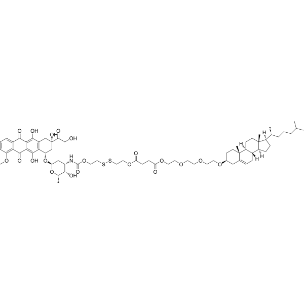 SDOX
SDOX
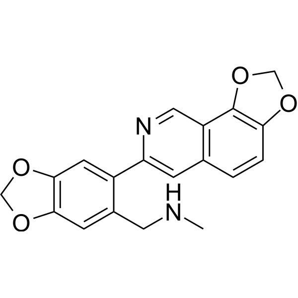 Topoisomerase I/II inhibitor 2
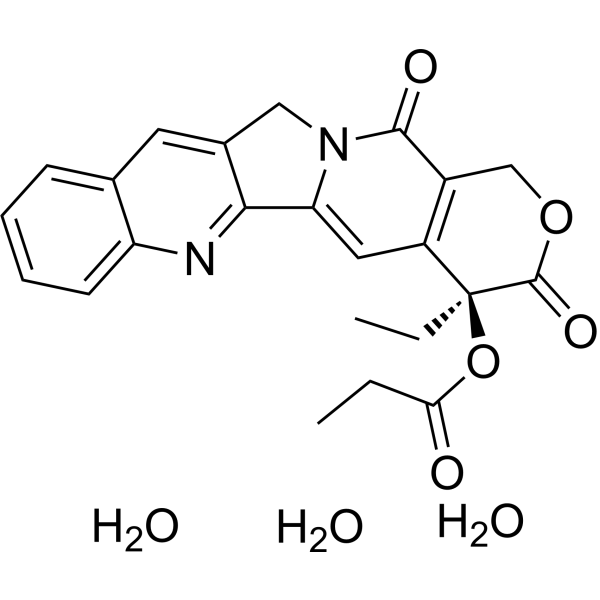 Camptothecin-20(S)-O-propionate hydrate (Camptothecin-20-O-propionate hydrate)
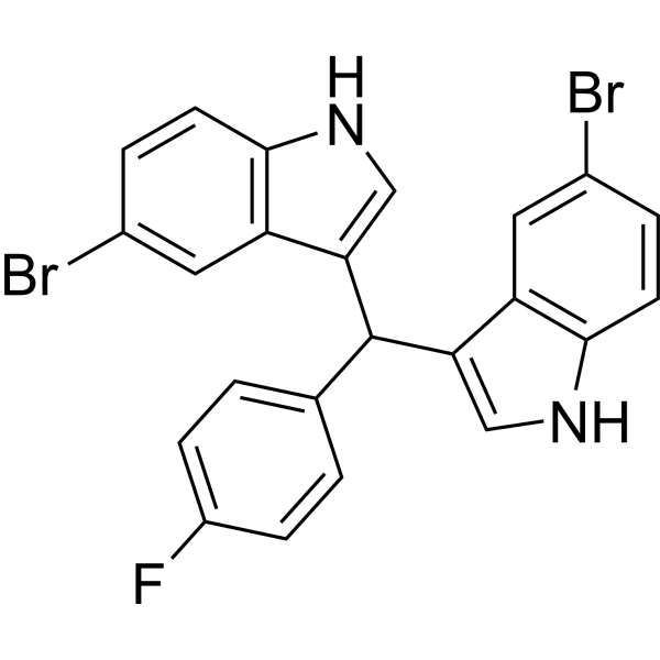 Topoisomerase I inhibitor 9
InvivoChem的所有产品仅用于作科学研究,不面向患者销售
Copyright 2020 InvivoChem LLC | All Rights Reserved 粤ICP备20063088号-1


 COA
COA


 463611831
