| 规格 | 价格 | 库存 | 数量 |
|---|---|---|---|
| 10 mM * 1 mL in DMSO |
|
||
| 1mg |
|
||
| 2mg |
|
||
| 5mg |
|
||
| 10mg |
|
||
| 25mg |
|
||
| 50mg |
|
||
| 100mg |
|
||
| 250mg |
|
||
| Other Sizes |
|
| 靶点 |
PD-1/PD-L1 (IC50 = 18 nM); PD-1/PD-L1 (KD = 8 μM)
Programmed Death Ligand 1 (PD-L1): BMS-202 is a small-molecule inhibitor that binds to PD-L1, blocking its interaction with PD-1 and CD80. It has a dissociation constant (Ki) of 0.7 ± 0.1 μM for PD-L1 (measured by surface plasmon resonance, SPR) and an IC50 of 1.2 ± 0.1 μM for inhibiting PD-L1/PD-1 binding (determined by homogeneous time-resolved fluorescence, HTRF) [1] |
|---|---|
| 体外研究 (In Vitro) |
BMS-202(0-100 μM;4 天;SCC-3 或 Jurkat 细胞)治疗可防止强 PD-L1 阳性 SCC-3 细胞(IC50 为 15 μM)和抗 CD3 抗体激活的 Jurkat 细胞(IC50 10 μM)体外[2]。 BMS-202 特异性诱导 PD-L1 热稳定。 BMS-202 在溶液中引起 PD-L1 二聚化。在同型二聚体的核心,BMS-202 填充了一个大的疏水口袋,促进单体之间的大量额外相互作用。 PD-1/PD-L1 相互作用在生理上由疏水表面介导,BMS-202 与这些表面上的两个 PD-L1 分子相互作用[1]。
PD-L1/PD-1结合抑制:重组人PD-L1(胞外域,ECD)与PD-1(ECD)在BMS-202(0.1~10 μM)存在下孵育。HTRF实验显示BMS-202 浓度依赖性抑制PD-L1/PD-1复合物形成:1 μM时抑制约70%,3 μM时抑制约88%,5 μM时抑制率>95%;同时可阻断PD-L1/CD80结合(IC50=1.5±0.2 μM) [1] - 胶质母细胞瘤细胞抗增殖与促凋亡作用:人胶质母细胞瘤U87MG和U251细胞用BMS-202(0.5~10 μM)处理48小时。MTT实验显示IC50分别为2.5±0.3 μM(U87MG)和2.8±0.2 μM(U251);Annexin V-FITC/PI染色显示凋亡率从对照组的5%升至5 μM BMS-202 处理组的35%(U87MG)和32%(U251)。Western blot显示PD-L1下调(5 μM时降65%)、糖酵解相关蛋白(GLUT1降55%、LDHA降60%)下调,切割型caspase-3升高3.2倍(5 μM)、Bax/Bcl-2比值升高4.0倍(5 μM) [3] - 胶质母细胞瘤细胞代谢重塑:U87MG细胞用BMS-202(2~5 μM)处理24小时后,糖酵解活性降低(胞外酸化率ECAR在5 μM时降40%),氧化磷酸化增强(耗氧率OCR在5 μM时升30%)(Seahorse XF分析仪检测)。RT-PCR证实糖酵解相关基因(HK2降50%、PKM2降55%、LDHA降60%)在5 μM时下调 [3] |
| 体内研究 (In Vivo) |
在人源化 MHC-dKO NOG 小鼠中,BMS-202(20 mg/kg;腹腔注射;每天;持续 9 天;NOG-dKO 小鼠)治疗与对照组相比表现出明显的抗肿瘤作用[2]。
此外,来自荷瘤裸鼠异种移植的数据支持BMS-202在体内显著抑制U251细胞的生长。综上所述,这些体内和体外数据表明,BMS-202在不影响正常胶质细胞的情况下,显著抑制GBM细胞的生长,为其抗肿瘤应用于GBM提供了一个安全的治疗窗口期。 人类化MHC双敲除NOG小鼠的抗肿瘤活性:6~8周龄雌性人类化MHC双敲除NOG小鼠,皮下注射5×10⁶个人非小细胞肺癌(NSCLC)A549细胞(PD-L1阳性)。肿瘤达50~100 mm³时,小鼠分为3组(每组n=6): 1. 溶剂对照组(0.1% DMSO+无菌生理盐水); 2. BMS-202 10 mg/kg组; 3. BMS-202 20 mg/kg组。 BMS-202 每周腹腔注射3次,持续21天。与对照组相比: - 10 mg/kg组:肿瘤体积降45%,重量降40%,21天生存率从30%升至55%; - 20 mg/kg组:肿瘤体积降65%,重量降70%,21天生存率从30%升至70%。 肿瘤免疫组化显示:20 mg/kg组CD8⁺ T细胞浸润增加2.5倍,Foxp3⁺调节性T细胞减少50%,IFN-γ表达升高3.0倍 [2] |
| 酶活实验 |
所有结合研究均在 HTRF 测定缓冲液中进行,该缓冲液由 dPBS 组成,并补充有 0.1%(含 v)牛血清白蛋白和 0.05%(v/v)Tween-20。对于 PD-l-Ig/PD-Ll-His 结合测定,将抑制剂与 PD-Ll-His(最终浓度为 10 nM)在 4 μL 测定缓冲液中预孵育 15 m,然后添加 PD-l- Ig(最终 20 nM)溶于 1 μL 测定缓冲液中,并进一步孵育 15 m。使用来自人类、食蟹猴或小鼠的 PD-L1。 HTRF 检测是使用铕穴酸盐标记的抗 Ig(最终 1 nM)和别藻蓝蛋白 (APC) 标记的抗 His(最终 20 nM)实现的。将抗体在 HTRF 检测缓冲液中稀释,并在结合反应上分配 5 μL。让反应混合物平衡 30 分钟,并使用 En Vision 荧光计获得信号(665 nm/620 nm 比率)。在 PD-1-Ig/PD-L2-His(分别为 20、5 nM)、CD80-His/PD-Ll-Ig(分别为 100、10 nM)和 CD80-His/CTLA4- 之间建立了额外的结合测定。 Ig(分别为 10、5 nM)。
表面等离子体共振(SPR)实验:通过胺偶联法将重组人PD-L1 ECD共价固定于CM5传感芯片。BMS-202 用运行缓冲液(10 mM HEPES pH7.4、150 mM NaCl、0.05% Tween-20)梯度稀释(0.1~10 μM),以30 μL/min流速注入芯片(结合相120秒,解离相300秒)。传感图拟合1:1结合模型,计算结合速率常数(Ka)、解离速率常数(Kd)及Ki(Kd=0.7±0.1 μM) [1] - PD-L1/BMS-202复合物X射线晶体衍射实验:重组PD-L1 ECD(10 mg/mL)与BMS-202(2 mM)4°C孵育过夜,采用悬滴气相扩散法(储液:20% PEG 3350、0.2 M柠檬酸铵pH6.5)20°C培养晶体。同步辐射光源(波长1.0 Å)收集衍射数据,HKL2000处理数据,分子置换法(PDB模板:4ZQK)解析结构,PHENIX精修。结果显示BMS-202 结合于PD-L1同源二聚化界面,与PD-L1的Tyr56、Arg113、Gln66形成氢键 [1] |
| 细胞实验 |
程序性死亡-1/程序性死亡-配体 1 (PD-1/PD-L1) 相互作用在抑制 T 细胞反应中发挥主导作用,特别是在肿瘤微环境中,保护肿瘤细胞免于裂解。据报道,PD-1/PD-L1 抑制剂 2 可阻止 PD-L1 与 PD-1 的相互作用,IC50 值为 18 nM。
胶质母细胞瘤细胞增殖实验(MTT法):U87MG/U251细胞以5×10³个/孔接种于96孔板,过夜培养。加入BMS-202(0.5~10 μM)孵育48小时,每孔加MTT溶液(5 mg/mL)20 μL,孵育4小时后加DMSO 150 μL溶解甲瓒结晶,570 nm测吸光度,非线性回归计算IC50 [3] - 凋亡实验(Annexin V-FITC/PI染色):U87MG细胞以2×10⁵个/孔接种于6孔板,BMS-202(0.5~10 μM)处理72小时。收集细胞,PBS洗涤后,加Annexin V-FITC(5 μL)和PI(10 μL)室温染色15分钟,流式细胞仪分析凋亡(激发光488 nm,FITC发射光530 nm,PI发射光610 nm) [3] - 代谢实验(Seahorse XF法):U87MG细胞以1×10⁴个/孔接种于XF96板,BMS-202(2~5 μM)处理24小时。更换为XF基础培养基(含10 mM葡萄糖、2 mM谷氨酰胺、1 mM丙酮酸),37°C无CO₂孵育1小时。Seahorse XF96分析仪检测ECAR(糖酵解)和OCR(氧化磷酸化),数据以细胞数校正 [3] |
| 动物实验 |
在一项使用人源化MHC双敲除(dKO)NOG小鼠的体内研究中,BMS-202与对照组相比显示出明显的抗肿瘤作用;然而,由于肿瘤部位未观察到淋巴细胞聚集,因此发现其抗肿瘤机制中存在直接的细胞毒性作用。这些结果表明,BMS-202的抗肿瘤作用可能部分是通过直接的非靶向细胞毒性作用介导的,而不仅仅是通过免疫反应机制。此外,本研究中使用的人源化dKO NOG小鼠模型已被证明是筛选PD-1/PD-L1结合小分子抑制剂的有效工具,这些抑制剂可通过免疫反应介导的机制抑制肿瘤生长[2]。
体内治疗性荷瘤异种移植模型(BMS-202)[3] 根据我们之前的研究,将5 × 10~6个U251细胞与基质胶混合,皮下注射到4-6周龄的雄性NOD/SCID裸鼠体内(n = 8)。当肿瘤体积(0.5 × 长 × 宽²)达到100 mm³时,将小鼠随机分为对照组(注射载体)和BMS-202组(腹腔注射20 mg/kg BMS-202,每周两次)。治疗过程停止,直至对照组肿瘤体积达到伦理批准的最大体积 2000 mm³。 人源化 NOG 小鼠非小细胞肺癌异种移植模型:雌性人源化 MHC 双敲除 NOG 小鼠(6-8 周龄)饲养于无特定病原体条件下。将 5×10⁶ 个 A549 细胞(重悬于 0.2 mL PBS 中)皮下注射至小鼠右侧腹部。当肿瘤体积达到 50-100 mm³ 时,将小鼠随机分为 3 组(n=6): - 载体组:0.1% DMSO + 无菌生理盐水,腹腔注射(ip),每周 3 次; - BMS-202 10 mg/kg 组:BMS-202 溶于 0.1% DMSO + 生理盐水(1 mg/mL),腹腔注射,每周 3 次; - BMS-202 20 mg/kg 组:将 BMS-202 溶于 0.1% DMSO + 生理盐水(2 mg/mL),每周腹腔注射 3 次。 每周测量肿瘤体积(长 × 宽² / 2)和体重。21 天后,通过颈椎脱臼处死小鼠:切除肿瘤(称重,用 10% 中性福尔马林固定用于免疫组织化学染色,或冷冻于 -80°C 用于蛋白质/RNA 提取);收集血清,通过 ELISA 法检测细胞因子(IFN-γ、TNF-α)[2] |
| 毒性/毒理 (Toxicokinetics/TK) |
体外正常细胞毒性:将正常人星形胶质细胞 (NHA) 用 BMS-202 (0.5–10 μM) 处理 48 小时。MTT 检测显示所有浓度下细胞存活率均 >90%,表明无显著细胞毒性 [3]
- 体内小鼠安全性:在人源化 NOG 小鼠模型中,BMS-202 (10–20 mg/kg,腹腔注射,21 天) 未引起体重(与对照组相比 ±5%)、器官重量(肝脏、肾脏、脾脏)或血清生化指标(ALT、AST、BUN、肌酐)的显著变化。肝脏和肾脏组织的组织病理学检查未发现炎症、坏死或纤维化 [2] |
| 参考文献 |
|
| 其他信息 |
近年来,利用单克隆抗体靶向PD-1/PD-L1免疫检查点在癌症治疗中取得了前所未有的成果。由于缺乏足够的结构信息,针对该通路的化学抑制剂的研发滞后于抗体的研发。百时美施贵宝公司近期才公开了首个靶向PD-1/PD-L1相互作用的非肽类化学抑制剂。本文研究表明,这些小分子化合物能够直接与PD-L1结合,并有效阻断PD-1的结合。结构研究揭示了一种二聚体蛋白复合物,其中单个小分子能够稳定该二聚体,从而阻断PD-L1与PD-1的相互作用表面。 PD-L1表面的小分子相互作用“热点”为PD-1/PD-L1拮抗剂药物的发现提供了思路。[1]
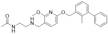
近期,百时美施贵宝(BMS)公司报道了首批PD-1/PD-L1小分子抑制剂,这些抑制剂是基于均相时间分辨荧光(HTRF)筛选PD-1/PD-L1相互作用而开发的。进一步的晶体学和生物物理学研究表明,这些化合物通过诱导PD-L1二聚化来抑制PD-1/PD-L1的相互作用,其中每个二聚体在其界面结合一个稳定剂分子。然而,这些化合物的抗肿瘤免疫学机制仍有待阐明。在本研究中,我们重点关注BMS-202(BMS化合物的代表),并通过体外和体内实验研究了其抗肿瘤活性。体外实验表明,BMS-202 可抑制强阳性 PD-L1 的 SCC-3 细胞(IC50 15 μM)和抗 CD3 抗体激活的 Jurkat 细胞(IC50 10 μM)的增殖。此外,BMS-202 对这些细胞表面 PD-1 或 PD-L1 的表达水平没有调节作用。在利用人源化 MHC 双敲除 (dKO) NOG 小鼠进行的体内研究中,与对照组相比,BMS-202 显示出明显的抗肿瘤作用;然而,由于肿瘤部位未观察到淋巴细胞聚集,因此发现其抗肿瘤机制中存在直接的细胞毒性作用。这些结果提示,BMS-202 的抗肿瘤作用可能部分是通过直接的非靶向细胞毒性作用以及免疫反应机制实现的。此外,本研究中使用的人源化dKO NOG小鼠模型已被证明是筛选PD-1/PD-L1结合小分子抑制剂的有效工具,这些抑制剂可通过免疫反应介导的机制抑制肿瘤生长。[2] 近期,晶体学研究表明,具有甲氧基-1-吡啶化学结构的小分子化合物BMS-202对PD-L1二聚体具有高亲和力。然而,其在胶质母细胞瘤(GBM)中的作用和机制尚不清楚。本研究旨在利用多组学和生物信息学技术,结合大量的体外和体内实验(包括CCK-8实验、流式细胞术、免疫共沉淀、siRNA转染、PCR、蛋白质印迹、细胞迁移/侵袭实验和异种移植治疗实验),研究BMS-202在GBM中的抗肿瘤活性及其潜在机制。我们的研究结果表明,BMS-202在体外和体内均能显著抑制GBM细胞的增殖。此外,它还能在体外有效阻断细胞迁移和侵袭。从机制上讲,它能降低GBM细胞表面PD-L1的表达,并独立于mTOR信号通路阻断PD-L1-AKT-BCAT1轴。综上所述,我们认为BMS-202有望成为GBM患者的潜在治疗药物,其作用机制是通过重塑细胞代谢,从而提高患者的生存率。[3] 作用机制: 1.免疫调节作用(文献1、2):BMS-202与PD-L1的同源二聚体界面和CD80结合位点结合,阻止PD-L1与PD-1(T细胞表面)和CD80(抗原呈递细胞表面)相互作用。这可以缓解 PD-1/PD-L1 介导的免疫抑制,激活 CD8⁺ T 细胞增殖和细胞因子(IFN-γ、TNF-α)分泌,从而杀死肿瘤细胞 [1,2] 2. 代谢重塑(文献 3):在胶质母细胞瘤细胞中,BMS-202 通过下调 GLUT1、LDHA、HK2 和 PKM2 来抑制 Warburg 效应(有氧糖酵解),同时增强氧化磷酸化。这种代谢转变会降低肿瘤增殖所需的ATP生成,并诱导细胞凋亡[3] - 治疗潜力: - BMS-202在体内对PD-L1阳性非小细胞肺癌(NSCLC)和体外对胶质母细胞瘤均显示出疗效,支持其治疗PD-L1过表达实体瘤的潜力[2,3] - 其双重机制(免疫激活+代谢重塑)使其成为与化疗药物或其他免疫疗法联合治疗的理想候选药物[3] - 结构特异性:PD-L1/BMS-202复合物的晶体结构显示出高度结合特异性——BMS-202不与PD-L2(PD-L1的同源物)结合,从而避免脱靶免疫调节[1] |
| 分子式 |
C25H29N3O3
|
|
|---|---|---|
| 分子量 |
419.52
|
|
| 精确质量 |
419.22
|
|
| 元素分析 |
C, 71.57; H, 6.97; N, 10.02; O, 11.44
|
|
| CAS号 |
1675203-84-5
|
|
| 相关CAS号 |
N-deacetylated BMS-202;2310135-18-1
|
|
| PubChem CID |
117951478
|
|
| 外观&性状 |
White to off-white solid powder
|
|
| 密度 |
1.1±0.1 g/cm3
|
|
| 沸点 |
611.4±55.0 °C at 760 mmHg
|
|
| 闪点 |
323.6±31.5 °C
|
|
| 蒸汽压 |
0.0±1.8 mmHg at 25°C
|
|
| 折射率 |
1.575
|
|
| LogP |
3.99
|
|
| tPSA |
72.5Ų
|
|
| 氢键供体(HBD)数目 |
2
|
|
| 氢键受体(HBA)数目 |
5
|
|
| 可旋转键数目(RBC) |
10
|
|
| 重原子数目 |
31
|
|
| 分子复杂度/Complexity |
526
|
|
| 定义原子立体中心数目 |
0
|
|
| SMILES |
O(C1C([H])=C([H])C(=C(N=1)OC([H])([H])[H])C([H])([H])N([H])C([H])([H])C([H])([H])N([H])C(C([H])([H])[H])=O)C([H])([H])C1C([H])=C([H])C([H])=C(C2C([H])=C([H])C([H])=C([H])C=2[H])C=1C([H])([H])[H]
|
|
| InChi Key |
JEDPSOYOYVELLZ-UHFFFAOYSA-N
|
|
| InChi Code |
InChI=1S/C25H29N3O3/c1-18-22(10-7-11-23(18)20-8-5-4-6-9-20)17-31-24-13-12-21(25(28-24)30-3)16-26-14-15-27-19(2)29/h4-13,26H,14-17H2,1-3H3,(H,27,29)
|
|
| 化学名 |
2-[2,6-dichloro-4-(3,5-dimethyl-1,2-oxazol-4-yl)anilino]-N-hydroxybenzamide
|
|
| 别名 |
|
|
| HS Tariff Code |
2934.99.9001
|
|
| 存储方式 |
Powder -20°C 3 years 4°C 2 years In solvent -80°C 6 months -20°C 1 month |
|
| 运输条件 |
Room temperature (This product is stable at ambient temperature for a few days during ordinary shipping and time spent in Customs)
|
| 溶解度 (体外实验) |
|
|||
|---|---|---|---|---|
| 溶解度 (体内实验) |
配方 1 中的溶解度: ≥ 2.5 mg/mL (5.96 mM) (饱和度未知) in 10% DMSO + 40% PEG300 + 5% Tween80 + 45% Saline (这些助溶剂从左到右依次添加,逐一添加), 澄清溶液。
例如,若需制备1 mL的工作液,可将100 μL 25.0 mg/mL澄清DMSO储备液加入到400 μL PEG300中,混匀;然后向上述溶液中加入50 μL Tween-80,混匀;加入450 μL生理盐水定容至1 mL。 *生理盐水的制备:将 0.9 g 氯化钠溶解在 100 mL ddH₂O中,得到澄清溶液。 配方 2 中的溶解度: ≥ 2.5 mg/mL (5.96 mM) (饱和度未知) in 10% DMSO + 90% (20% SBE-β-CD in Saline) (这些助溶剂从左到右依次添加,逐一添加), 澄清溶液。 例如,若需制备1 mL的工作液,可将 100 μL 25.0 mg/mL澄清DMSO储备液加入900 μL 20% SBE-β-CD生理盐水溶液中,混匀。 *20% SBE-β-CD 生理盐水溶液的制备(4°C,1 周):将 2 g SBE-β-CD 溶解于 10 mL 生理盐水中,得到澄清溶液。 View More
配方 3 中的溶解度: ≥ 2.5 mg/mL (5.96 mM) (饱和度未知) in 10% DMSO + 90% Corn Oil (这些助溶剂从左到右依次添加,逐一添加), 澄清溶液。 配方 4 中的溶解度: 4.05 mg/mL (9.65 mM) in 45% PEG300 5% Tween-80 50% Saline (这些助溶剂从左到右依次添加,逐一添加), 澄清溶液; 超声助溶. *生理盐水的制备:将 0.9 g 氯化钠溶解在 100 mL ddH₂O中,得到澄清溶液。 1、请先配制澄清的储备液(如:用DMSO配置50 或 100 mg/mL母液(储备液)); 2、取适量母液,按从左到右的顺序依次添加助溶剂,澄清后再加入下一助溶剂。以 下列配方为例说明 (注意此配方只用于说明,并不一定代表此产品 的实际溶解配方): 10% DMSO → 40% PEG300 → 5% Tween-80 → 45% ddH2O (或 saline); 假设最终工作液的体积为 1 mL, 浓度为5 mg/mL: 取 100 μL 50 mg/mL 的澄清 DMSO 储备液加到 400 μL PEG300 中,混合均匀/澄清;向上述体系中加入50 μL Tween-80,混合均匀/澄清;然后继续加入450 μL ddH2O (或 saline)定容至 1 mL; 3、溶剂前显示的百分比是指该溶剂在最终溶液/工作液中的体积所占比例; 4、 如产品在配制过程中出现沉淀/析出,可通过加热(≤50℃)或超声的方式助溶; 5、为保证最佳实验结果,工作液请现配现用! 6、如不确定怎么将母液配置成体内动物实验的工作液,请查看说明书或联系我们; 7、 以上所有助溶剂都可在 Invivochem.cn网站购买。 |
| 制备储备液 | 1 mg | 5 mg | 10 mg | |
| 1 mM | 2.3837 mL | 11.9184 mL | 23.8368 mL | |
| 5 mM | 0.4767 mL | 2.3837 mL | 4.7674 mL | |
| 10 mM | 0.2384 mL | 1.1918 mL | 2.3837 mL |
1、根据实验需要选择合适的溶剂配制储备液 (母液):对于大多数产品,InvivoChem推荐用DMSO配置母液 (比如:5、10、20mM或者10、20、50 mg/mL浓度),个别水溶性高的产品可直接溶于水。产品在DMSO 、水或其他溶剂中的具体溶解度详见上”溶解度 (体外)”部分;
2、如果您找不到您想要的溶解度信息,或者很难将产品溶解在溶液中,请联系我们;
3、建议使用下列计算器进行相关计算(摩尔浓度计算器、稀释计算器、分子量计算器、重组计算器等);
4、母液配好之后,将其分装到常规用量,并储存在-20°C或-80°C,尽量减少反复冻融循环。
计算结果:
工作液浓度: mg/mL;
DMSO母液配制方法: mg 药物溶于 μL DMSO溶液(母液浓度 mg/mL)。如该浓度超过该批次药物DMSO溶解度,请首先与我们联系。
体内配方配制方法:取 μL DMSO母液,加入 μL PEG300,混匀澄清后加入μL Tween 80,混匀澄清后加入 μL ddH2O,混匀澄清。
(1) 请确保溶液澄清之后,再加入下一种溶剂 (助溶剂) 。可利用涡旋、超声或水浴加热等方法助溶;
(2) 一定要按顺序加入溶剂 (助溶剂) 。
 J Med Chem.2017Jul 13;60(13):5857-5867. |
|---|
|
|
Structural Biology of the Immune Checkpoint Receptor PD-1 and Its Ligands PD-L1/PD-L2.Structure.2017 Aug 1;25(8):1163-1174. |
|---|
New Directions in Designing the Therapeutics Targeting the PD-1/PD-L1 Interaction.Structure.2017 Aug 1;25(8):1163-1174. |
Structural Basis of the PD-1/PD-L1 (PD-L2) Interaction.Structure.2017 Aug 1;25(8):1163-1174. |
InvivoChem的所有产品仅用于作科学研究,不面向患者销售
Copyright 2020 InvivoChem LLC | All Rights Reserved 粤ICP备20063088号-1


 COA
COA


 463611831
