| 规格 | 价格 | 库存 | 数量 |
|---|---|---|---|
| 10 mM * 1 mL in DMSO |
|
||
| 1mg |
|
||
| 2mg |
|
||
| 5mg |
|
||
| 10mg |
|
||
| 25mg |
|
||
| 50mg |
|
||
| 100mg |
|
||
| 250mg |
|
||
| 500mg |
|
||
| Other Sizes |
|

| 靶点 |
Natural product; CRISPR/Cas9; HSV-1; Arf-GEFs
Brefeldin A (BFA) targets ADP-ribosylation factor 1 (ARF1), inhibiting its guanine nucleotide exchange factor (GEF) activity, which is essential for Golgi apparatus structure maintenance; no IC50/Ki values for ARF1 are provided [1,6,7] - Brefeldin A (BFA) mediates ADP-ribosylation of C-terminal binding protein 1/Brefeldin A-ADP-ribosylated substrate (CtBP1/BARS), a key regulator of membrane fission; the EC50 for CtBP1/BARS ADP-ribosylation is not reported [4] - Brefeldin A (BFA) disrupts the microtubule and actin cytoskeletons by unknown direct targets, but indirectly alters cytoskeletal organization via Golgi dysfunction; no specific binding targets for cytoskeletal proteins are identified [1] - Brefeldin A (BFA) does not have a defined "drug target" in cancer stem cell (CSC) regulation, but inhibits CSC potential by suppressing the PI3K/Akt signaling pathway; no IC50 for PI3K/Akt is provided [2] |
|---|---|
| 体外研究 (In Vitro) |
用布雷菲德菌素 A (BFA) 处理 15 或 40 小时后,内质网 (ER) 显着肿胀并移动到正常肾 (NRK) 细胞的外周。长期 Brefeldin A 治疗会显着破坏肌动蛋白和 MT 细胞骨架 [1]。 Brefeldin A 和 ADPR 缀合物介导 BARS 的 ADP 核糖基化。当使用从用 BFA 处理的 CD38+ HeLa 细胞获得的细胞创建时,条形图显示了 BAC 结合 [3]。 Brefeldin A 减少 3D 和 2D 培养物中的 MDA-MB-231 集落形成,促进 MDA-MB-231 乳腺癌细胞中身份无关的细胞死亡,并阻断 MDA-MB 迁移和 MMP 9(基质金属肽酶 9)活性 - 231 [2]。
肿瘤干细胞(Cancer stem cells, CSCs)是肿瘤细胞或已建立的癌细胞系的一个子集,可以在体内启动和维持肿瘤的生长。癌症干细胞可以在无血清的悬浮培养物中富集,在几天到几周内形成肿瘤球。Brefeldin A (BFA)是一种在真核细胞中诱导内质网(ER)应激的真菌毒素。我们发现,与粘附培养物相比,亚微克/毫升浓度的BFA在MDA-MB-231悬浮培养物(EC50: 0.016µg/mL)中优先诱导细胞死亡。BFA还能有效抑制MDA-MB-231细胞的克隆活性、迁移和基质金属蛋白酶-9 (MMP-9)活性。Western blotting分析表明,BFA的作用可能通过下调乳腺CSC标志物CD44和抗凋亡蛋白Bcl-2、Mcl-1,逆转上皮-间质转化介导。此外,BFA对悬浮的MDA-MB-468细胞也表现出选择性的细胞毒性,并抑制T47D和MDA-MB-453细胞的肿瘤球形成,表明BFA可能对各种表型的乳腺癌细胞有效。[2] 糖蛋白D (gD-1)是1型单纯疱疹病毒(HSV-1)的重要病毒粒子包膜成分,通常被转运到感染细胞的质膜上。本研究在体外培养的HSV-1感染的人成纤维细胞中,加入1微克/毫升布雷菲尔丁A (Brefeldin A, BFA),吸附病毒12 h后,抑制gD-1在细胞内的转运。免疫荧光和共聚焦显微镜显示gD-1向质膜的运输被取消,gD-1在核旁积聚,微管纤维排列混乱。BFA影响的解除超过60分钟导致不完全运输,但增加了质膜和靠近细胞核的高尔基样区域中gD-1的积累。微管蛋白模式在去除BFA后6小时基本恢复正常。bfa处理后9小时释放的感染性HSV-1颗粒未完全恢复。结果表明,BFA的作用不是完全可逆的,并引起一种涉及微管蛋白结构的细胞毒性影响。[7] 在人成纤维细胞(WI-38细胞)中,用Brefeldin A (BFA)(1 μg/mL,1-4小时)处理可破坏高尔基体(通过高尔基体标志物GM130免疫荧光染色观察)并导致微管解聚:2小时后完整微管丝数量减少>60%,肌动蛋白应力纤维断裂(鬼笔环肽染色检测) [1] - 在MDA-MB-231人乳腺癌细胞中:(1)Brefeldin A (BFA)(100 nM,72小时)降低锚定非依赖性存活能力,软琼脂克隆形成率从对照组的32%降至11%;(2)500 nM时抑制CSC潜能,球形成效率(SFE)从8.5%降至2.1%(无血清培养基中计数球状体);(3)200 nM Brefeldin A (BFA) 使细胞迁移率降低58%(Transwell实验),并下调CSC标志物CD44+/CD24-(流式细胞术:CD44+/CD24-细胞比例从35%降至12%) [2] - 在K562红白血病细胞中,Brefeldin A (BFA)(500 nM,24小时)诱导替代性线粒体自噬:Western blot显示自噬标志物LC3-II增加2.3倍,线粒体标志物Tom20减少40%,LC3与Tom20的共定位(免疫荧光)表明线粒体自噬体形成 [3] - 在HeLa细胞裂解液中,Brefeldin A (BFA)(10 μM,30分钟)诱导重组CtBP1/BARS的ADP核糖基化:放射测定显示32P-ADP核糖掺入CtBP1/BARS,SDS-PAGE证实ADP核糖基化CtBP1/BARS较对照组增加1.8倍 [4] - 在诱导多能干细胞(iPSCs)中,Brefeldin A (BFA)(100 nM,48小时)增强CRISPR/Cas9介导的基因组编辑:转染sgRNA-Cas9复合物后加用Brefeldin A (BFA),编辑效率从对照组的18%提升至39%(T7核酸酶实验),且不降低细胞活力(>90% vs 对照组) [5] - 在HSV-1感染的Vero细胞中,Brefeldin A (BFA)(5 μg/mL,感染后1小时)抑制病毒复制:病毒滴度(空斑实验)从对照组的10^6 PFU/mL降至10^3 PFU/mL,免疫荧光显示HSV-1 UL51蛋白在高尔基体的定位减少 [6,7] - 在HepG2人肝癌细胞中,Brefeldin A (BFA) 负载纳米胶束(BFA-NMs)呈剂量依赖性细胞毒性:BFA-NMs的IC50为8.2 μM(MTT实验,72小时),而游离Brefeldin A (BFA) 的IC50为15.6 μM;BFA-NMs诱导的凋亡(Annexin V/PI染色)比游离BFA多2.1倍 [8] |
| 体内研究 (In Vivo) |
M-BFA(BFA encapsulated in mixed nanomicelles based on TPGS and F127 copolymers))的体内抗肿瘤效果 [8]
鉴于M-BFA具有突出的体外细胞毒性和较高的体内肿瘤蓄积性,我们利用HepG2荷瘤异种移植模型进一步研究了M-BFA的抗肿瘤作用。将小鼠分为三组,每天静脉注射PBS、M-BFA 5 mg/kg和M-BFA 10 mg/kg,连续14 d。如图7A、C所示,M-BFA 10 mg/kg组具有较强的抗肿瘤作用,可显著延缓肿瘤进展,而M-BFA 5 mg/kg组无明显抑制作用。M-BFA 10 mg/kg组肿瘤生长抑制率(TGI %)约为42.08%±3.29%,是M-BFA 5 mg/kg组的2倍。在整个实验过程中,三组均造成最小的动物体重减轻(图7B),表明毒性较低。苏木精和伊红染色(H&E)分析显示M-BFA表现出广泛的肿瘤坏死(图7D)。如图7D所示,给药M-BFA后,肿瘤细胞呈片状坏死。坏死灶呈粉红色,甚至部分坏死肿瘤组织溶解,形成空腔(红色箭头)。坏死灶内可见较多中性粒细胞浸润(绿箭头)。肿瘤细胞核质比大,少数细胞出现有丝分裂(黄箭头)。 在荷HepG2异种移植瘤裸鼠中(每组6只):(1)静脉注射Brefeldin A (BFA) 负载纳米胶束(BFA-NMs)20 mg/kg(每3天1次,共5次),肿瘤生长抑制率(TGI)为62.3%,而游离Brefeldin A (BFA)(10 mg/kg)的TGI为28.5%;(2)BFA-NM组无显著体重下降(<5% vs 对照组),游离BFA组体重下降8%;(3)BFA-NM组肝/肾组织病理学无明显损伤,游离BFA组出现轻度肝细胞变性 [8] |
| 酶活实验 |
先前对Brefeldin A (BFA)作用的研究主要集中在er -高尔基膜运输的动力学上,主要是在相对较短的药物治疗后。我们现在已经分析了长时间BFA处理对整体细胞形态、常驻和循环高尔基蛋白行为以及微管和肌动蛋白细胞骨架组织的影响。长时间(15小时或40小时)用BFA处理正常大鼠肾(NRK)细胞,引起内质网(ER)的剧烈肿胀,并将其定位转移到细胞周围。高尔基复合体被分解,高尔基蛋白在部分不同的区室中重新分布并持续存在。长时间的BFA治疗导致MT和肌动蛋白细胞骨架的明显破坏。外周MT缺失,微管蛋白染色集中在微管组织中心(MTOC)发出的短星状MT。肌动蛋白应力纤维大部分缺失,肌动蛋白染色集中在核周区域。在这个区域内,肌动蛋白的定位与膜转运因子p115的定位重叠。BFA对高尔基体结构、MT和肌动蛋白组织的影响表现出相同的阈值——在BFA处理30分钟和15小时后,这些影响都可以部分逆转,但在与药物孵化40小时后,这些影响是不可逆的。所观察到的效应不是由参与凋亡现象的信号通路或内质网应激反应通路诱导的。这些结果表明,BFA抑制了调节MT和肌动蛋白细胞骨架动力学的关键分子的活性。该发现可作为阐明BFA作用于细胞骨架的分子机制的基础。[1]
adp核糖基化是一种翻译后修饰,可调节许多靶蛋白的功能。我们之前的研究表明,真菌毒素brefeldin A (BFA)诱导c -末端结合蛋白-1短形式/BFA- adp -核糖基化底物(CtBP1-S/BARS)的adp -核糖基化,CtBP1-S/BARS是一种双功能蛋白,在细胞核中作为转录因子,在细胞质中作为高尔基复合体胞内运输和有丝分裂分配过程中膜裂变的调节剂。在这里,我们报道了BFA对CtBP1-S/BARS的adp -核糖基化是通过一种非常规的机制发生的,该机制包括两个步骤:(i)由adp -核糖环化酶CD38合成BFA- adp -核糖缀合物;(ii) BFA- adp -核糖缀合物与CtBP1-S/BARS NAD(+)结合口袋的共价结合。这导致CtBP1-S/BARS锁定在二聚体构象中,从而阻止其与已知参与膜裂变的相互作用物结合,从而抑制有丝分裂高尔基分割中涉及的裂变机制。由于这种抑制可能导致G2细胞周期的停滞,这些发现为设计表达高水平CD38的肿瘤细胞细胞周期的药物阻滞剂提供了策略。[4] CtBP1/BARS ADP核糖基化实验 [4]: 1. 将重组人CtBP1/BARS蛋白(0.5 μg)与10 μM Brefeldin A (BFA)、5 μCi [32P]-NAD+(作为ADP核糖供体)在反应缓冲液(50 mM Tris-HCl pH 7.5、10 mM MgCl2、1 mM DTT)中37°C孵育30分钟。 2. 加入5×SDS上样缓冲液并煮沸5分钟终止反应。 3. 样品经12% SDS-PAGE分离后,凝胶干燥,通过放射自显影检测放射性标记的ADP核糖基化CtBP1/BARS,用ImageJ定量条带强度,以无Brefeldin A (BFA) 的对照组为参照计算ADP核糖基化百分比。 |
| 细胞实验 |
Brefeldin A (BFA)是一种在真核细胞中诱导内质网(ER)应激的霉菌毒素。我们发现,与粘附培养相比,亚微克/毫升浓度的BFA在MDA-MB-231悬浮培养物(EC50:0.016µg/mL)中优先诱导细胞死亡。BFA还有效地抑制了MDA-MB-231细胞的克隆形成活性和迁移以及基质金属蛋白酶-9(MMP-9)活性。Western印迹分析表明,BFA的作用可能是通过下调乳腺CSC标志物CD44和抗凋亡蛋白Bcl-2和Mcl-1,以及逆转上皮间质转化来介导的。此外,BFA对悬浮的MDA-MB-468细胞也表现出选择性细胞毒性,并抑制了T47D和MDA-MB-453细胞中的瘤球形成,表明BFA可能对各种表型的乳腺癌症细胞有效[2]。
2D克隆实验[2] 0-50 μg/mL 的Brefeldin A (BFA)预处理24 h后,以每孔1 × 10³个细胞的密度在6孔板中重新接种,再培养12 d,每3 d换一次培养基。用甲醇-乙酸(3:1)固定菌落15 min,室温结晶紫(1%)染色30 min。 创面愈合动力试验[2] 使用p10微移液管尖端在六孔板上进行过夜融合培养。细胞碎片用磷酸盐缓冲盐水(PBS)洗涤三次后,补充含有0-50 μg/mL Brefeldin A (BFA)的完整培养基。在伤口愈合后的指定时间,用相差显微镜捕捉伤口愈合的图像。 明胶酶谱[2] 0-50 μg/mL Brefeldin A (BFA)处理细胞培养24 h,收集上清,0.22 μm过滤器过滤,Centricon自旋柱浓缩50倍,10 kD截止。用含有0.1% SDS和1mg /mL明胶的10%聚丙烯酰胺凝胶在非还原性SDS- page上分离浓缩上清。电泳后,用含有0.15 M NaCl、5 mM CaC12、5 μM ZnCl、0.02% NaN3、0.25% Triton X-100的50 mM Tris-HCl (pH 7.5)在室温下洗涤3次,每次洗涤30 min,然后在不含Triton X-100的同一缓冲液中37℃孵育20 h。采用考马西亮蓝R-250染色显示MMPs形成的明胶清晰区。 微管/肌动蛋白细胞骨架染色实验 [1]: 1. 将WI-38成纤维细胞接种于盖玻片(1×10^4个细胞/盖玻片),过夜培养。加入0.1-5 μg/mL Brefeldin A (BFA),孵育1-4小时。 2. 细胞用4%多聚甲醛固定(室温15分钟),0.1% Triton X-100透化(5分钟),3% BSA封闭(30分钟)。 3. 微管检测:4°C孵育抗α-微管蛋白一抗过夜,室温孵育Alexa Fluor 488偶联二抗1小时;肌动蛋白检测:室温孵育Alexa Fluor 594-鬼笔环肽30分钟;细胞核用DAPI染色。 4. 共聚焦显微镜采集图像,计数每个细胞的完整微管丝/肌动蛋白应力纤维数量(每组100个细胞)。 - MDA-MB-231细胞迁移实验 [2]: 1. Transwell小室(8 μm孔径)用Matrigel(1:10稀释)37°C包被1小时。将含0-500 nM Brefeldin A (BFA) 的无血清培养基中的MDA-MB-231细胞(5×10^4个细胞/小室)加入上室,下室加入含10% FBS的培养基。 2. 孵育24小时后,用棉签去除上室未迁移细胞,下室迁移细胞用4%多聚甲醛固定、0.1%结晶紫染色,显微镜下计数(每小室5个视野)。迁移率=(BFA组迁移细胞数/对照组迁移细胞数)×100% [2] - HepG2细胞毒性实验 [8]: 1. 将HepG2细胞接种于96孔板(2×10^3个细胞/孔),过夜培养。加入1-50 μM的游离Brefeldin A (BFA) 或BFA-NMs(每个浓度3个复孔)。 2. 72小时后,每孔加入20 μL MTT溶液(5 mg/mL),孵育4小时。去除上清液,加入150 μL DMSO溶解甲瓒结晶。 3. 测定570 nm处吸光度,细胞活力=(BFA组A570/对照组A570)×100%。用GraphPad Prism通过四参数逻辑模型计算IC50 [8] - HSV-1病毒滴度实验 [7]: 1. Vero细胞用HSV-1(MOI=0.1)感染1小时后,加入0-10 μg/mL Brefeldin A (BFA)。 2. 24小时后收集细胞上清液,制备10^-1至10^-6的系列稀释液。将稀释上清液加入Vero细胞单层(96孔板),孵育1小时。 3. 细胞用含1%琼脂糖的MEM覆盖,72小时后计数空斑。病毒滴度=(空斑数×稀释倍数)/上清液加入体积(PFU/mL) [7] |
| 动物实验 |
药代动力学研究[8]
每组5只雌性SD大鼠静脉注射M-BFA(BFA封装于基于TPGS和F127共聚物的混合纳米胶束中),剂量为520 mg/kg,溶于10% HS-15%溶剂和90%生理盐水(v/v)中。分别于给药前和给药后0.25 h、0.5 h、1 h、2 h、4 h、6 h、8 h和24 h采集所有动物的血液样本,置于含肝素钠和200 mM DDPV的试管中。血液样本经4 °C、6800 rpm离心6 min分离血浆,并保存于−80 °C直至分析。方法开发和生物样品分析采用三重四极杆 5500 液相色谱-串联质谱仪 (LC-MS/MS),以维拉帕米为内标(表 S2)。药代动力学参数(包括 T1/2、Cmax、AUC(0-t) 和 AUC(0-∞))由研究主管使用 Phoenix WinNonlin 7.0 软件计算得出。 动物处理和体内肿瘤抑制[8] 雌性 BALB/c 小鼠(20 ± 2 g,5-6 周龄)随机分为三组(每组 5 只)。将 HepG2 细胞(1 × 10⁷/只)皮下植入小鼠背部下方,建立 HepG2 肿瘤模型。每日检查小鼠,观察 HepG2 细胞植入后肿瘤大小的变化。当平均肿瘤体积达到约 100 mm³(体积 = (肿瘤长度) × (肿瘤宽度)²/²)时,所有小鼠均可用于后续研究。将荷瘤HepG2裸鼠每日注射M-BFA,连续14天。以PBS溶液作为对照组。其他各组BFA的剂量分别为5 mg/kg和10 mg/kg体重。每2天测量一次每只小鼠的肿瘤大小。肿瘤体积(V)按以下公式计算:V = L × W²/2,其中L和W分别为肿瘤的长度和宽度。实验结束时,用乙醚麻醉小鼠。切除的器官和肿瘤组织用冰冷的PBS(pH 7.4)洗涤,并称重和拍照。 肿瘤的体内荧光成像[8] 通过体内成像观察M-BFA的生物分布。将两亲性ICG(图S11)溶于水(1 mg/mL),然后直接加入M-BFA溶液(80 μg/mL)。 ICG分子通过疏水相互作用进入纳米胶束。小鼠经静脉注射100 μL游离ICG(100 μg/mL)和载有ICG的M-BFA(含100 μg/mL ICG)。小鼠麻醉后,使用IVIS Spectrum成像系统,在预定时间点用808 nm激发激光进行成像。48小时后,处死小鼠,收集各器官进行荧光分布成像。 裸鼠HepG2异种移植模型[8]: 1. 使用6-8周龄的雌性BALB/c裸鼠。 1. 将HepG2细胞(5×10^6个细胞,溶于0.1 mL PBS/Matrigel,1:1)皮下注射到每只小鼠的右侧背部。 2. 当肿瘤体积达到100-150 mm³时,将小鼠随机分为4组(每组n=6):(a)对照组(生理盐水,静脉注射);(b)游离布雷菲德菌素A(BFA)组(10 mg/kg,溶于DMSO/生理盐水,1:9,静脉注射);(c)低剂量BFA纳米颗粒组(相当于10 mg/kg BFA,静脉注射);(d)高剂量BFA纳米颗粒组(相当于20 mg/kg BFA,静脉注射)。 3. 每3天给药一次,共给药5次。每隔2天测量一次肿瘤体积(长×宽²×0.5)和体重。 4. 最后一次治疗后,处死小鼠。切除肿瘤,称重,并用4%多聚甲醛固定,用于组织病理学分析(H&E染色)。同时收集肝脏和肾脏组织,用于H&E染色和生化分析(ALT、AST、BUN、Cr)[8] - [1-7]中未报道在病毒感染、血液肿瘤或干细胞模型中使用布雷菲德菌素A (BFA)的动物实验方案。 |
| 药代性质 (ADME/PK) |
体内药代动力学研究[8]
对布雷菲德菌素A (BFA)血浆药代动力学(PK)的初步表征表明,BFA在小鼠体内呈明显的双指数衰减,且下降速度很快。BFA在体内的生物半衰期(T1/2)为0.17小时[53]。基于此,本研究在Sprague-Dawley (SD)大鼠中评估了M-BFA的药代动力学特性。静脉注射520 mg/kg(相当于20 mg/kg BFA)后,分析了血浆中M-BFA的浓度。结果表明,M-BFA表现出中等的药代动力学特征,T1/2约为0.35小时(BFA为0.17小时),最大血浆浓度(Cmax)为4065.68 ng/mL。在小鼠体内,该药物达到了足够的血浆暴露量,浓度-时间曲线下面积(AUC0-t)值为3153.75 hng/mL。 M-BFA在荷瘤小鼠体内的生物分布[8] 鉴于布雷菲德菌素A (BFA)不具有自发荧光,为了分析M-BFA的体内生物分布和肿瘤靶向疗效,我们采用了吲哚菁绿(ICG)负载的M-BFA,并将其注射到HepG2荷瘤小鼠体内进行光学成像分析。分别在注射后0、1、2、4、8、24和48 h进行荧光成像。如图6A所示,8 h后,在ICG负载的M-BFA组的肿瘤部位观察到了ICG荧光。24 h后,肿瘤组织中ICG负载的M-BFA和游离ICG的积累量出现了明显的差异。此时荧光强度达到最强,并在注射后48小时仍保持较高水平。相比之下,在游离ICG组的肿瘤部位未观察到明显的荧光。我们进一步观察到,荧光在初期主要分布于肝脏。注射48小时后,小鼠全身的荧光消失。 所有引用的文献均未描述布雷菲德菌素A (BFA) 的ADME/药代动力学特征;未报道任何参数(例如,吸收率、分布容积、半衰期、生物利用度、代谢途径、排泄途径)[1-8] |
| 毒性/毒理 (Toxicokinetics/TK) |
小鼠腹腔注射 LD50 250 mg/kg 日本抗生素杂志,34(51),1981 [PMID:7241806]
体外毒性:布雷菲德菌素 A (BFA) 对正常人成纤维细胞 (WI-38 细胞) 显示出较低的细胞毒性:用 1 μg/mL 布雷菲德菌素 A (BFA) 处理 48 小时后,细胞活力仍保持在 80% 以上 [1];在 iPSCs 中,100 nM 布雷菲德菌素 A (BFA) 不会降低细胞活力(>90% vs. 对照组)[5] - 体内毒性(HepG2 异种移植模型):(1)载有布雷菲德菌素 A (BFA) 的纳米胶束(20 mg/kg)不会引起体重(<5% 的损失)或肝/肾功能(ALT:35 ± 5 U/L vs. 对照组 32 ± 4 U/L;AST:82 ± 7 U/L vs. 对照组 78 ± 6 U/L;BUN:5.2 ± 0.4 mmol/L vs. 对照组 4.9 ± 0.3 mmol/L;Cr:45 ± 3 μmol/L vs. 对照组 43 ± 2 μmol/L)的显著变化; (2)游离布雷菲德菌素A(BFA)(10 mg/kg)可导致体重减轻8%和轻度肝细胞变性(H&E染色)[8] |
| 参考文献 |
|
| 其他信息 |
布雷菲尔德菌素A是布雷菲尔德青霉菌(Penicillium brefeldianum)的代谢产物,具有广泛的抗菌活性。它作为青霉菌的代谢产物发挥作用。
一种来自布雷菲尔德青霉菌的代谢产物,具有广泛的抗菌活性。 据报道,布雷菲尔德菌素A存在于卡门贝尔青霉菌(Penicillium camemberti)、布雷菲尔德青霉菌(Penicillium brefeldianum)和其他有相关数据的生物体中。 一种真菌代谢产物,属于大环内酯类,具有广泛的抗菌活性。 白血病细胞在缓冲细胞应激方面优于具有正常分化潜能的造血细胞,但这种白血病优势的潜在机制尚未完全阐明。我们利用CRISPR/Cas9技术敲除经典的自噬必需基因Atg7,发现红白血病K562细胞拥有两套自噬机制。无论经典自噬机制是否完整,替代性线粒体自噬均具有功能。尽管经典自噬缺陷会减弱细胞周期、增殖和分化潜能,但白血病细胞仍保留了清除线粒体以及维持低水平活性氧(ROS)和凋亡的能力。使用线粒体自噬特异性诱导剂处理后发现,经典自噬缺陷的红白血病细胞仍保留了线粒体自噬反应。在野生型和Atg7(-/-)白血病细胞中,线粒体自噬的选择性诱导均与RAB9A在线粒体膜上的上调和定位相关。当用替代性自噬抑制剂布雷菲德菌素A(BFA)处理白血病细胞或敲低RAB9A时,这种线粒体自噬被抑制。这伴随着活性氧(ROS)水平升高和细胞凋亡,以及DNA损伤修复能力下降。因此,结果表明,红白血病K562细胞拥有一种不依赖于ATG7的替代性线粒体自噬机制,即使在经典的自噬过程受损的情况下也能发挥作用,从而维持其应对过量ROS和DNA损伤等应激的能力。[3] 白血病细胞在缓冲细胞应激方面优于具有正常分化潜能的造血细胞,但这种白血病优势的潜在机制尚未完全阐明。我们利用CRISPR/Cas9技术敲除经典自噬必需基因Atg7,发现红白血病K562细胞拥有两套自噬机制。无论经典自噬机制是否完整,替代性线粒体自噬均能发挥作用。尽管经典的自噬缺陷会减弱细胞周期、增殖和分化潜能,但白血病细胞仍保留了清除线粒体损伤以及维持低水平活性氧(ROS)和凋亡的能力。使用线粒体自噬特异性诱导剂进行处理后发现,经典自噬缺陷的红白血病细胞仍保留了线粒体自噬反应。在野生型和Atg7(-/-)白血病细胞中,线粒体自噬的选择性诱导均与RAB9A在线粒体膜上的上调和定位相关。当使用替代自噬抑制剂布雷菲德菌素A(BFA)处理白血病细胞或敲低RAB9A时,线粒体自噬受到抑制。这伴随着ROS水平升高、细胞凋亡增加以及DNA损伤修复能力下降。因此,结果表明,红白血病K562细胞具有一种不依赖于ATG7的替代性线粒体自噬机制,即使在经典的自噬过程受损的情况下也能发挥作用,从而维持其应对过量活性氧(ROS)和DNA损伤等应激的能力。[6] 肝细胞癌(HCC)是死亡率最高的癌症之一。临床上常用的传统药物通常受限于耐药性和副作用,因此仍需要新型药物。大环内酯类抗生素布雷菲德菌素A(BFA)是癌症化疗中一种常用的先导化合物,但其溶解度差且稳定性差。本研究为了克服这些缺点,将BFA封装在基于TPGS和F127共聚物的混合纳米胶束(M-BFA)中。M-BFA具有高溶解度、胶体稳定性和持续释放完整BFA的能力。体外实验表明,M-BFA显著抑制人肝癌HepG2细胞的增殖,诱导其G0/G1期阻滞,并诱导caspase依赖性细胞凋亡。此外,M-BFA还通过Akt/mTOR和ERK通路诱导自噬性细胞死亡。在HepG2荷瘤异种移植小鼠模型中,负载于M-BFA上的荧光探针吲哚菁绿(ICG)能够迅速分布至肿瘤组织,延长其血液循环时间,并提高其在肿瘤中的蓄积能力。更重要的是,M-BFA(10 mg/kg)显著延缓肿瘤进展,并诱导肿瘤组织大面积坏死。综上所述,本研究表明M-BFA在肝细胞癌(HCC)治疗中具有良好的应用前景。[8] 布雷菲德菌素A(BFA)是一种天然存在的环状内酯,分离自真菌(例如,布雷菲德菌),最初因其破坏高尔基体的能力而被发现[1,6] - 布雷菲德菌素A(BFA)的经典作用机制是抑制ARF1活化,导致高尔基体膜与内质网融合,从而破坏蛋白质分泌[1,6,7] - 布雷菲德菌素A(BFA)通过阻断病毒蛋白(例如,UL51)的高尔基体依赖性运输来抑制HSV-1复制,从而阻止病毒颗粒组装[6,7] - 在癌症治疗中,载有布雷菲德菌素A(BFA)的纳米胶束可提高布雷菲德菌素A(BFA)的溶解度和肿瘤靶向性,增强抗肿瘤疗效并降低全身毒性[8] -布雷菲德菌素A (BFA) 通过未知机制增强iPSCs中的CRISPR编辑,可能通过调节内体运输来增加sgRNA-Cas9向细胞核的递送[5] |
| 分子式 |
C16H24O4
|
|
|---|---|---|
| 分子量 |
280.36
|
|
| 精确质量 |
280.167
|
|
| 元素分析 |
C, 68.55; H, 8.63; O, 22.83
|
|
| CAS号 |
20350-15-6
|
|
| 相关CAS号 |
|
|
| PubChem CID |
5287620
|
|
| 外观&性状 |
Typically exists as White to off-white solids at room temperature
|
|
| 密度 |
1.1±0.1 g/cm3
|
|
| 沸点 |
492.7±45.0 °C at 760 mmHg
|
|
| 熔点 |
200-205ºC
|
|
| 闪点 |
180.8±22.2 °C
|
|
| 蒸汽压 |
0.0±2.8 mmHg at 25°C
|
|
| 折射率 |
1.513
|
|
| LogP |
1.61
|
|
| tPSA |
66.76
|
|
| 氢键供体(HBD)数目 |
2
|
|
| 氢键受体(HBA)数目 |
4
|
|
| 可旋转键数目(RBC) |
0
|
|
| 重原子数目 |
20
|
|
| 分子复杂度/Complexity |
388
|
|
| 定义原子立体中心数目 |
5
|
|
| SMILES |
O([H])[C@]1([H])C([H])([H])[C@]2([H])C([H])=C([H])C([H])([H])C([H])([H])C([H])([H])[C@@]([H])(C([H])([H])[H])OC(C([H])=C([H])C([H])([C@@]2([H])C1([H])[H])O[H])=O |c:10,t:31|
|
|
| InChi Key |
KQNZDYYTLMIZCT-KQPMLPITSA-N
|
|
| InChi Code |
InChI=1S/C16H24O4/c1-11-5-3-2-4-6-12-9-13(17)10-14(12)15(18)7-8-16(19)20-11/h4,6-8,11-15,17-18H,2-3,5,9-10H2,1H3/b6-4+,8-7+/t11-,12+,13-,14+,15+/m0/s1
|
|
| 化学名 |
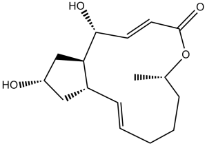
(1S,2E,7S,10E,12R,13R,15S)-12,15-Dihydroxy-7-methyl-8-oxabicyclo[11.3.0]hexadeca-2,10-dien-9-one
|
|
| 别名 |
|
|
| HS Tariff Code |
2934.99.9001
|
|
| 存储方式 |
Powder -20°C 3 years 4°C 2 years In solvent -80°C 6 months -20°C 1 month |
|
| 运输条件 |
Room temperature (This product is stable at ambient temperature for a few days during ordinary shipping and time spent in Customs)
|
| 溶解度 (体外实验) |
|
|||
|---|---|---|---|---|
| 溶解度 (体内实验) |
配方 1 中的溶解度: ≥ 2.5 mg/mL (8.92 mM) (饱和度未知) in 10% DMSO + 40% PEG300 + 5% Tween80 + 45% Saline (这些助溶剂从左到右依次添加,逐一添加), 澄清溶液。
例如,若需制备1 mL的工作液,可将100 μL 25.0 mg/mL澄清DMSO储备液加入到400 μL PEG300中,混匀;然后向上述溶液中加入50 μL Tween-80,混匀;加入450 μL生理盐水定容至1 mL。 *生理盐水的制备:将 0.9 g 氯化钠溶解在 100 mL ddH₂O中,得到澄清溶液。 配方 2 中的溶解度: ≥ 2.5 mg/mL (8.92 mM) (饱和度未知) in 10% DMSO + 90% (20% SBE-β-CD in Saline) (这些助溶剂从左到右依次添加,逐一添加), 澄清溶液。 例如,若需制备1 mL的工作液,可将 100 μL 25.0 mg/mL澄清DMSO储备液加入900 μL 20% SBE-β-CD生理盐水溶液中,混匀。 *20% SBE-β-CD 生理盐水溶液的制备(4°C,1 周):将 2 g SBE-β-CD 溶解于 10 mL 生理盐水中,得到澄清溶液。 View More
配方 3 中的溶解度: ≥ 2.5 mg/mL (8.92 mM) (饱和度未知) in 10% DMSO + 90% Corn Oil (这些助溶剂从左到右依次添加,逐一添加), 澄清溶液。 配方 4 中的溶解度: ≥ 2.5 mg/mL (8.92 mM) (饱和度未知) in 10% EtOH + 40% PEG300 + 5% Tween80 + 45% Saline (这些助溶剂从左到右依次添加,逐一添加), 澄清溶液。 例如,若需制备1 mL的工作液,可将100 μL 25.0 mg/mL 澄清 EtOH 储备液加入400 μL PEG300 中,混匀;再向上述溶液中加入50 μL Tween-80,混匀;然后加入450 μL 生理盐水定容至1 mL。 *生理盐水的制备:将 0.9 g 氯化钠溶解在 100 mL ddH₂O中,得到澄清溶液。 配方 5 中的溶解度: ≥ 2.5 mg/mL (8.92 mM) (饱和度未知) in 10% EtOH + 90% (20% SBE-β-CD in Saline) (这些助溶剂从左到右依次添加,逐一添加), 澄清溶液。 例如,若需制备1 mL的工作液,可将100μL 25.0mg/mL澄清EtOH储备液加入到900μL 20%SBE-β-CD生理盐水中,混匀。 *20% SBE-β-CD 生理盐水溶液的制备(4°C,1 周):将 2 g SBE-β-CD 溶解于 10 mL 生理盐水中,得到澄清溶液。 配方 6 中的溶解度: ≥ 2.5 mg/mL (8.92 mM) (饱和度未知) in 10% EtOH + 90% Corn Oil (这些助溶剂从左到右依次添加,逐一添加), 澄清溶液。 例如,若需制备1 mL的工作液,可将 100 μL 25.0 mg/mL 澄清乙醇储备液加入到 900 μL 玉米油中并混合均匀。 1、请先配制澄清的储备液(如:用DMSO配置50 或 100 mg/mL母液(储备液)); 2、取适量母液,按从左到右的顺序依次添加助溶剂,澄清后再加入下一助溶剂。以 下列配方为例说明 (注意此配方只用于说明,并不一定代表此产品 的实际溶解配方): 10% DMSO → 40% PEG300 → 5% Tween-80 → 45% ddH2O (或 saline); 假设最终工作液的体积为 1 mL, 浓度为5 mg/mL: 取 100 μL 50 mg/mL 的澄清 DMSO 储备液加到 400 μL PEG300 中,混合均匀/澄清;向上述体系中加入50 μL Tween-80,混合均匀/澄清;然后继续加入450 μL ddH2O (或 saline)定容至 1 mL; 3、溶剂前显示的百分比是指该溶剂在最终溶液/工作液中的体积所占比例; 4、 如产品在配制过程中出现沉淀/析出,可通过加热(≤50℃)或超声的方式助溶; 5、为保证最佳实验结果,工作液请现配现用! 6、如不确定怎么将母液配置成体内动物实验的工作液,请查看说明书或联系我们; 7、 以上所有助溶剂都可在 Invivochem.cn网站购买。 |
| 制备储备液 | 1 mg | 5 mg | 10 mg | |
| 1 mM | 3.5668 mL | 17.8342 mL | 35.6684 mL | |
| 5 mM | 0.7134 mL | 3.5668 mL | 7.1337 mL | |
| 10 mM | 0.3567 mL | 1.7834 mL | 3.5668 mL |
1、根据实验需要选择合适的溶剂配制储备液 (母液):对于大多数产品,InvivoChem推荐用DMSO配置母液 (比如:5、10、20mM或者10、20、50 mg/mL浓度),个别水溶性高的产品可直接溶于水。产品在DMSO 、水或其他溶剂中的具体溶解度详见上”溶解度 (体外)”部分;
2、如果您找不到您想要的溶解度信息,或者很难将产品溶解在溶液中,请联系我们;
3、建议使用下列计算器进行相关计算(摩尔浓度计算器、稀释计算器、分子量计算器、重组计算器等);
4、母液配好之后,将其分装到常规用量,并储存在-20°C或-80°C,尽量减少反复冻融循环。
计算结果:
工作液浓度: mg/mL;
DMSO母液配制方法: mg 药物溶于 μL DMSO溶液(母液浓度 mg/mL)。如该浓度超过该批次药物DMSO溶解度,请首先与我们联系。
体内配方配制方法:取 μL DMSO母液,加入 μL PEG300,混匀澄清后加入μL Tween 80,混匀澄清后加入 μL ddH2O,混匀澄清。
(1) 请确保溶液澄清之后,再加入下一种溶剂 (助溶剂) 。可利用涡旋、超声或水浴加热等方法助溶;
(2) 一定要按顺序加入溶剂 (助溶剂) 。
| NCT Number | Recruitment | interventions | Conditions | Sponsor/Collaborators | Start Date | Phases |
| NCT05969353 | Recruiting | Other: accupunture | Assessing the Effectiveness of BFA as a Non-pharmacologic Pain Management Intervention: A Randomised Sham Controlled Study |
Bnai Zion Medical Center | July 23, 2023 | Not Applicable |
| NCT04094246 | Recruiting | Procedure: Battlefield Acupuncture | Shoulder Injuries Pain,Postoperative |
Keller Army Community Hospital | September 25, 2019 | Not Applicable |
| NCT06333938 | Not yet recruiting NEW |
Device: Bridge Device: BFA |
Anesthesia Surgery |
Durham VA Medical Center | June 2024 | Phase 4 |
| NCT06128772 | Not yet recruiting | Other: Battlefield Acupuncture | Chronic Pain Substance Use Disorders |
Edith Nourse Rogers Memorial Veterans Hospital |
November 30, 2023 | Not Applicable |
|
|---|
Inhibition of intracellular protein trafficking by Brefeldin A |
Brefeldin A inhibits STING-induced IRF activity in THP1-Dual™ cells |
 Tepotinib hydrochloride monohydrate
 BKN-1
BKN-1
 NEO214
 MJO445
InvivoChem的所有产品仅用于作科学研究,不面向患者销售
Copyright 2020 InvivoChem LLC | All Rights Reserved 粤ICP备20063088号-1


 COA
COA

")
")
 463611831
