| 规格 | 价格 | 库存 | 数量 |
|---|---|---|---|
| 10 mM * 1 mL in DMSO |
|
||
| 5mg |
|
||
| 10mg |
|
||
| 50mg |
|
||
| 100mg |
|
||
| 250mg |
|
||
| 500mg |
|
||
| 5g |
|
||
| Other Sizes |
|

| 靶点 |
5-Lipoxygenase (5-LO); TRPV1; Caffeic acid targets histamine receptor H1 (H1R), transient receptor potential vanilloid 1 (TRPV1), Mas - related G - protein coupled receptor A3 (MrgprA3), transient receptor potential ankyrin 1 (TrpA1), Mas - related G - protein coupled receptor C11 (MrgprC11), and 5 - lipoxygenase (5 - LO).
|
|---|---|
| 体外研究 (In Vitro) |
咖啡酸调节组胺诱导的反应。当光浓度从 0.1 mM 增加到 1 mM 时,咖啡酸的调节作用逐渐增强,模拟正常的剂量调节反应。在用 1 mM 咖啡因处理的 HEK293T-TRPV1 细胞中,辣椒素诱导的反应显着降低。较低剂量的咖啡酸可抑制辣椒素引起的反应。实验表明,咖啡酸可以显着抑制组胺敏感的背根神经节(DRG)神经元。施用咖啡酸 (1 mM) 可使对组胺应用做出反应的 DRG 神经元百分比从 12.5% 降低至 2.1%。 1 mM 咖啡酸可显着抑制 TRPA1 表达细胞中异硫氰酸烯丙酯 (AITC) 诱导的细胞内钙升高。咖啡酸还可能抑制 AITC 诱导的 TRPA1 激活 [1]。
在表达H1R和TRPV1的HEK293T细胞中,咖啡酸可显著阻断组胺诱导的细胞内钙升高。在小鼠背根神经节(DRG)原代培养物中,其也能抑制组胺诱导的细胞内钙升高。在表达MrgprA3和TrpA1的DRG和HEK293T细胞中,咖啡酸可抑制氯喹诱导的反应。此外,它还可降低细胞中由Sligrl - NH2通过MrgprC11诱导的细胞内钙变化,但不能抑制β - 丙氨酸通过MrgprD诱导的反应。 |
| 体内研究 (In Vivo) |
在小鼠模型中,当使用咖啡酸(500 mg/kg)时,观察到组胺诱导的抓挠(30.50±10.87次/1小时,n=6)。此外,虽然似乎有下降趋势(49.40±12.35次/1小时,n=5),但较低剂量咖啡酸(100 mg/kg)在组胺诱导的抓伤中的抗抓伤作用尚未确定具有重要意义。 500 mg/kg 咖啡酸可显着减少氯缓冲液引起的划伤(161.6±31.42 次/1 h,n=5)[1]。在海马区域,咖啡因酸显着降低 5-LO mRNA(P<0.01)。 I/R-咖啡酸组的5-LO蛋白表达显着低于潜在再灌注(I/R)未治疗组(P<0.05或P<0.01)。在比较 I/R-咖啡酸组和 I/R 未治疗组时尤其如此。在低剂量和高剂量咖啡酸组中,整个过程中寻找平台的潜伏期都很显着,并且整个平台潜伏期都在I/R中。 R 组-咖啡酸(50 毫克/千克)。高剂量咖啡酸组海马神经元核固缩明显减少,固缩率为(13.3)±3.0)%,而低剂量组细胞损伤仍然明显(63.6±2.8)%[2] 。
在小鼠抓挠行为实验中,咖啡酸对组胺、氯喹和Sligrl - NH2诱导的抓挠行为有抑制作用。在大鼠全脑缺血再灌注模型中,咖啡酸(10、30、50 mg/kg)可缩短莫里斯水迷宫中的逃避潜伏期,减轻海马神经元损伤,增加神经元数量。同时降低NF - κBp65和5 - LO的表达,减少丙二醛(MDA)含量,增加超氧化物歧化酶(SOD)活性。 |
| 细胞实验 |
瘙痒是一种令人不快的感觉,会引起抓挠的欲望。尽管瘙痒通常被认为是一种微不足道的“令人担忧”的感觉,但它可能会使人衰弱和疲惫,导致生活质量下降。在目前的研究中,研究了咖啡酸是否可以用于缓解各种瘙痒剂引起的瘙痒感的问题,包括组胺、氯喹、SLIGRL-NH2和β-丙氨酸。事实证明,在表达H1R和TRPV1的HEK293T细胞中,组胺诱导的细胞内钙增加被咖啡酸显著阻断,这些分子是组胺诱导的瘙痒在感觉神经元中传播所需的分子。此外,在小鼠背根神经节(DRG)的原代培养物中,咖啡酸抑制组胺诱导的细胞内钙增加。当使用氯喹(一种已知可诱导组胺非依赖性瘙痒的抗疟疾药物)时,还发现咖啡酸抑制表达MRGPRA3和TRPA1的DRG和HEK293T细胞的诱导反应,这是氯喹介导的瘙痒的潜在分子实体。同样,通过PAR2和MRGPRC11的瘙痒诱导剂SLIGRL-NH2引起的细胞内钙变化也被咖啡酸降低。然而,研究发现,咖啡酸不能通过其特异性受体MRGPRD抑制β-丙氨酸诱导的反应[1]。
对于HEK293T细胞,先转染使其表达H1R和TRPV1,然后用不同浓度的咖啡酸处理,最后用组胺刺激,通过钙敏感荧光探针检测细胞内钙浓度。对于DRG原代培养物,在组胺刺激前加入咖啡酸,再以同样方式检测细胞内钙水平。对于表达MrgprA3和TrpA1或MrgprC11的细胞,操作类似,即先加入咖啡酸,再用相应的致痒剂刺激,最后检测细胞内钙变化。 |
| 动物实验 |
实验设计[2]
将大鼠分为五组:假手术组(n = 9)、缺血/再灌注未处理组(n = 9)、缺血/再灌注咖啡酸组(10 mg · kg−1)(n = 9)、缺血/再灌注咖啡酸组(30 mg · kg−1)(n = 9)和缺血/再灌注咖啡酸组(50 mg · kg−1)(n = 9)。在缺血/再灌注咖啡酸组中,大鼠在缺血前30分钟腹腔注射10、30和50 mg · kg−1的咖啡酸(用0.3%羧甲基纤维素钠配制)。假手术组和缺血/再灌注(I/R)组均注射等体积的0.3%羧甲基纤维素钠溶液。 全脑缺血/再灌注模型的建立[2] 大鼠腹腔注射水合氯醛(400 mg/kg)麻醉,并固定于仰卧位。全脑缺血的建立方法如前所述。在颈部做正中切口,然后向右侧延长1 cm。钝性分离后,仔细暴露双侧颈总动脉和右侧颈总静脉。灌注2 ml肝素化生理盐水(100 mL 0.9%生理盐水,含250 U肝素)后,结扎颈总静脉远端。从右侧颈总静脉抽取约占总血容量30%的血液,导致低血压。通过双侧颈总动脉夹闭联合低血压诱导全脑缺血。缺血20分钟后,解除动脉夹闭,并将抽取的血液回输。假手术组大鼠接受与上述相同的操作,但不包括双侧颈总动脉闭塞和右侧颈总静脉取血。在小鼠抓挠行为测试中,将咖啡酸溶解于合适的溶剂中,并通过灌胃法给予小鼠。一段时间后,皮下注射组胺、氯喹或Sligrl-NH2,并计数特定时间内小鼠的抓挠次数。在全脑缺血再灌注大鼠模型中,45只大鼠随机分为5组:假手术组、缺血再灌注未治疗组和3个咖啡酸治疗组(10、30、50 mg/kg)。咖啡酸溶于适宜溶剂,于双侧颈动脉闭塞20分钟前30分钟腹腔注射给药,随后进行再灌注。再灌注后进行Morris水迷宫实验,然后收集海马组织进行HE染色、SOD活性检测、MDA含量检测以及免疫组化检测NF-κBp65表达。 |
| 药代性质 (ADME/PK) |
代谢/代谢物
参与咖啡酸代谢的酶尚未被鉴定。以下实验中,咖啡酸 (CA)、绿原酸 (CGA) 和二氢咖啡酸 (DHCA) 与肝细胞孵育,结果表明它们可被细胞色素 P450、儿茶酚-O-甲基转移酶 (COMT) 和 β-氧化酶代谢。COMT 对 CA 或 DHCA 进行 O-甲基化后生成的阿魏酸 (FA) 或二氢阿魏酸 (DHFA) 也可被 CYP1A1/2 进行 O-去甲基化,但不能被 CYP2E1 进行 O-去甲基化。DHCA 或 DHFA 还会发生侧链脱氢反应,分别生成 CA 和 FA,而硫代乙醇酸(一种 β-氧化酶酰基辅酶 A 脱氢酶的抑制剂)可抑制该反应。 NADPH/微粒体(CYP2E1)催化的谷胱甘肽结合物形成速率按DHCA>CA>CGA递减的顺序排列,这与COMT对其O-甲基化的速率顺序相反。NADPH/P450生成的CA和DHCA-o-醌可能抑制COMT,但它们可以很容易地形成谷胱甘肽结合物。CA、DHCA和DHFA在分离的大鼠肝细胞中相互代谢,并可代谢为FA,而FA仅代谢为CA,不代谢为DHCA或DHFA。CA、DHCA、FA、DHFA和CGA均表现出剂量依赖性的肝细胞毒性,测定的LD50(2小时)按毒性递减的顺序排列为DHCA>CA>DHFA>CGA>FA。总之,已有证据表明,O-甲基化、GSH结合、氢化和脱氢反应均参与了CA和DHCA的肝脏代谢。CA和DHCA的O-甲基化途径是一种解毒途径,而P450催化的邻醌生成途径则是一种毒性途径。 在大鼠体内,绿原酸在胃和肠道中水解为咖啡酸和奎宁酸。已鉴定出多种代谢产物。间香豆酸和间羟基马尿酸的葡萄糖醛酸苷似乎是人体内的主要代谢产物。在人体志愿者口服咖啡酸后,O-甲基化衍生物(阿魏酸、二氢阿魏酸和香草酸)迅速从尿液中排出,而间羟基苯基衍生物则出现较晚。脱羟基反应归因于肠道细菌的作用。 咖啡酸已知的人体代谢产物包括(2S,3S,4S,5R)-6-[4-[(E)-2-羧基乙烯基]-2-羟基苯氧基]-3,4,5-三羟基氧杂环己烷-2-羧酸和(2S,3S,4S,5R)-6-[5-[(E)-2-羧基乙烯基]-2-羟基苯氧基]-3,4,5-三羟基氧杂环己烷-2-羧酸。 |
| 毒性/毒理 (Toxicokinetics/TK) |
相互作用
咖啡酸以浓度依赖的方式增强了C2C12细胞对放射性葡萄糖的摄取。苯肾上腺素对C2C12细胞摄取放射性葡萄糖也具有类似的作用。哌唑嗪减弱了咖啡酸的作用,其作用方式与阻断苯肾上腺素的作用类似。 9周龄雌性ICR/Ha小鼠饲喂含0.06 mmol/g(10 g/kg饲料)咖啡酸(纯度99%)的饲料。从实验第8天开始,小鼠每周两次灌胃给予1 mg苯并[a]芘,持续4周。最后一次苯并[a]芘处理3天后,停止饲喂含咖啡酸的饲料。小鼠在211日龄时处死。在17只有效小鼠中,咖啡酸显著降低了前胃肿瘤(≥1 mm)/只小鼠(组织学未明确)的数量(p < 0.05)(3.1个肿瘤/只,而单独使用苯并[a]芘治疗的38只小鼠为5.0个肿瘤/只)。 解毒剂和急救措施 基本治疗:保持呼吸道通畅。必要时进行吸痰。观察呼吸功能不全的迹象,必要时辅助呼吸。使用无创呼吸面罩以10至15升/分钟的流量给予氧气。监测肺水肿,必要时进行治疗……。监测休克,必要时进行治疗……。如果眼睛受到污染,立即用水冲洗眼睛。在运输过程中,持续用生理盐水冲洗每只眼睛……。不要使用催吐剂。如果误服,应漱口并给予5 ml/kg至200 ml的水稀释,前提是患者能够吞咽、有强烈的咽反射且不流涎。活性炭无效……。切勿尝试中和,因为会发生放热反应。皮肤烧伤经去污后,用干燥的无菌敷料覆盖……。/有机酸及相关化合物/Bronstein, AC, PL Currance;《危险物质暴露的急救》。第2版。圣路易斯,密苏里州。Mosby Lifeline出版社。1994年,第152-153页。 高级治疗:对于意识不清、严重肺水肿或呼吸骤停的患者,应考虑经口或经鼻气管插管以控制气道。一旦出现上呼吸道梗阻的迹象,可能需要尽早插管。使用球囊面罩进行正压通气可能有效。监测心律,必要时治疗心律失常……。建立静脉通路,使用5%葡萄糖溶液/SRP:“保持通路畅通”,最小流速/。如有低血容量体征,使用乳酸林格氏液。注意液体超负荷的体征。考虑药物治疗肺水肿……。对于伴有低血容量体征的低血压,谨慎输液。如果患者体液量正常但血压偏低,考虑使用血管加压药。注意液体超负荷的体征……。使用盐酸丙美卡因辅助眼部冲洗……。/有机酸及相关化合物/Bronstein, AC, PL Currance;《危险物质暴露的紧急护理》。第2版。圣路易斯,密苏里州。Mosby Lifeline出版社。1994年,第100页。 153 医学监测 致癌物预防措施:凡需进行医学监测者,尤其是在接触致癌物后,应就可能有用或强制性的细胞遗传学和/或其他检测做出临时决定。/化学致癌物/ 非人类毒性摘录 /实验动物:亚慢性或慢性前暴露/ 五只六周龄雄性Fischer 344大鼠,在基础饲料中添加20 g/kg的咖啡酸(纯度>98%),持续四周。所有处理组动物的前胃均出现上皮增生。五只未处理的对照组动物未检测到增生。国际癌症研究机构(IARC)。《人类化学物质致癌风险评估专著》。日内瓦:世界卫生组织,国际癌症研究机构,1972年至今。 (多卷本著作)。可访问: /实验动物:亚慢性或亚慢性暴露/ 一组15只7周龄雄性叙利亚金仓鼠,饲喂含1%(10 g/kg饲料)咖啡酸(纯度>98%)的饲料,持续20周。选择1%的剂量水平是作为大鼠LD50的四分之一。对胃和膀胱进行组织病理学和放射自显影检查。在15只处理组动物中,14只出现轻度前胃上皮增生(其中1只为重度),而在15只未处理组动物中,7只出现轻度前胃上皮增生(p < 0.001)。对 (3)H-胸苷掺入的评估显示,与未处理的大鼠相比,腺胃前胃和幽门区域的标记细胞数量有所增加,但差异无统计学意义。国际癌症研究机构 (IARC)。《人类致癌化学品风险评估专著》。日内瓦:世界卫生组织国际癌症研究机构,1972年至今。(多卷本)。可访问: /实验动物:慢性暴露或致癌性/ 本研究探讨了咖啡酸在F344大鼠(雌雄均有)和C57BL/6N x C3H/HeN F1小鼠中的致癌潜力。将30只动物分别喂食含0%和2.0%咖啡酸的饲料,大鼠喂食104周,小鼠喂食96周后,详细的组织病理学检查显示,大鼠前胃鳞状细胞乳头状瘤或癌的发生率较高(雄性77%,雌性80%),而小鼠的发生率较低(雄性13%,雌性3%)。在3只大鼠和2只小鼠中观察到这些鳞状细胞癌侵入腹腔。此外,在接受治疗的大鼠中,肾小管细胞增生和腺瘤的发生率较高(雄性分别为73%和13%),而雌性分别为20%和0%,这些病变与毒性损伤明显相关。在小鼠中,接受治疗的雌性小鼠也出现了肾小管细胞增生和肿瘤(分别为97%和28%),而雄性小鼠的发生率较低(分别为27%和3%)。小鼠未出现明显的肾毒性损伤。经处理的雄性小鼠中也出现了肺泡II型细胞肿瘤(27%),具有统计学意义。因此,本研究表明,咖啡酸对F344大鼠和C57BL/6N x C3H/HeN F1小鼠(雌雄均有)的前胃鳞状细胞上皮、雄性大鼠和雌性小鼠的肾小管细胞以及雄性小鼠的肺泡II型细胞均具有致癌活性。PMID:1913684 /实验动物:慢性暴露或致癌性/ 两组各20只50日龄的雌性Sprague-Dawley大鼠,通过灌胃法给予0.5 ml芝麻油中溶于25 mg/kg体重的7,12-二甲基苯并[a]蒽。一周后,动物饲喂含0.5%(5 g/kg饲料)咖啡酸(纯度>99%)的饲料,持续51周。检查了乳腺、耳管、胃、肝脏和肾脏。单独使用7,12-二甲基苯并蒽处理的动物,其前胃乳头状瘤的发生率显著升高(6/19 vs 0/19;p < 0.01)。未发现其他肿瘤发生率的显著升高。国际癌症研究机构(IARC)。《人类致癌化学品风险评估专著》。日内瓦:世界卫生组织国际癌症研究机构,1972年至今。(多卷本)。可访问:https://monographs.iarc.fr/ENG/Classification/index.php,第V56卷,第124页(1993年)。 |
| 参考文献 | |
| 其他信息 |
根据世界卫生组织国际癌症研究机构(IARC)的说法,咖啡酸可能致癌。
3,4-二羟基肉桂酸在氯仿或石油醚溶液中呈黄色棱柱状或片状,或呈淡黄色颗粒状。碱性溶液中颜色由黄色变为橙色。(NTP, 1992) 咖啡酸是一种羟基肉桂酸,是肉桂酸苯环上3位和4位被羟基取代后形成的。它以顺式和反式两种形式存在,其中反式更为常见。咖啡酸是一种植物代谢产物,同时也是EC 1.13.11.33(花生四烯酸15-脂氧合酶)抑制剂、EC 2.5.1.18(谷胱甘肽转移酶)抑制剂、EC 1.13.11.34(花生四烯酸5-脂氧合酶)抑制剂、抗氧化剂和EC 3.5.1.98(组蛋白去乙酰化酶)抑制剂。它是一种羟基肉桂酸,属于儿茶酚类化合物。 据报道,丹参、白花丹参以及其他有相关数据的生物体中均含有咖啡酸。 咖啡酸是一种口服生物利用度高的羟基肉桂酸衍生物和多酚,具有潜在的抗氧化、抗炎和抗肿瘤活性。服用后,咖啡酸可作为抗氧化剂,防止氧化应激,从而预防自由基引起的DNA损伤。咖啡酸靶向并抑制鳞状细胞癌扩增的组蛋白去甲基化酶(HDM)癌蛋白基因1(GASC1;JMJD2C;KDM4C),从而抑制癌细胞增殖。GASC1是含Jumonji(Jmj)结构域蛋白KDM4亚组的成员,它能使组蛋白H3上的赖氨酸9和赖氨酸36(H3K9和H3K36)去甲基化,并在肿瘤细胞发育中发挥关键作用。 咖啡酸是酿酒酵母(Saccharomyces cerevisiae)中发现或产生的代谢产物。 另见:黑升麻(部分);紫草根(部分)。牛蒡根(部分)……查看更多…… 作用机制 咖啡酸苯乙酯 (CAPE) 由咖啡酸和苯乙醇(比例 1:5)在室温下以二环己基碳二亚胺 (DCC) 为缩合剂合成,产率约为 38%。CAPE 可抑制人白血病 HL-60 细胞的生长。它还能抑制 HL-60 细胞中 DNA、RNA 和蛋白质的合成,IC50 值分别为 1.0 M、5.0 M 和 1.5 M。 为了解咖啡酸的降血糖作用,本研究采用成肌细胞 C2C12 来研究其对葡萄糖的摄取。咖啡酸以浓度依赖的方式增强了 C2C12 细胞对放射性葡萄糖的摄取。在C2C12细胞中也观察到了苯肾上腺素对放射性葡萄糖摄取的类似作用。哌唑嗪减弱咖啡酸的作用方式与阻断苯肾上腺素的作用方式相似。咖啡酸对α1-肾上腺素能受体的作用通过[3H]哌唑嗪在C2C12细胞中的结合置换得到进一步证实。此外,苯肾上腺素在C2C12细胞中增加葡萄糖摄取的作用可被α1A-肾上腺素能受体拮抗剂坦索罗辛和WB 4101抑制,但不受α1B-肾上腺素能受体拮抗剂氯乙基可乐定(CEC)的影响。因此,可以认为C2C12细胞中存在α1A-肾上腺素能受体。在与这些拮抗剂共同孵育的C2C12细胞中也观察到了咖啡酸作用的类似抑制。 α1A-肾上腺素能受体的激活似乎是咖啡酸在C2C12细胞中发挥作用的原因。在磷脂酶C特异性抑制剂U73312存在的情况下,咖啡酸刺激的放射性葡萄糖进入C2C12细胞的摄取呈浓度依赖性降低,而U73343(U73312的阴性对照)则无此作用。此外,chelerythrine 和 GF 109203X 能减弱咖啡酸在足以抑制蛋白激酶 C 的浓度下的作用。因此,所得数据表明,咖啡酸激活 C2C12 细胞中的 α1A-肾上腺素能受体可能通过磷脂酶 C-蛋白激酶 C 通路增加葡萄糖摄取。 研究表明,膳食中 2% 的咖啡酸(CA,3,4-二羟基肉桂酸)可导致 F344 大鼠和 B6C3F1 小鼠的前胃和肾脏发生癌变。鉴于咖啡酸存在于咖啡和多种食物中,并采用 0% 至 2% 剂量范围内的线性插值法推算癌症发生率,人类患癌风险相当高。在两个靶器官中,肿瘤形成均先于增生,这可能是其致癌作用的主要机制。本研究在雄性F344大鼠中,以不同膳食浓度(0、0.05%、0.14%、0.40%和1.64%)的CA喂养4周后,探讨了该效应的剂量反应关系。腹腔注射2小时后,通过免疫组织化学分析掺入的5-溴-2'-脱氧尿苷(BrdU)来观察处于DNA复制S期的细胞。在前胃中,0.40%和1.64%浓度下,每毫米切片长度的上皮细胞总数和BrdU阳性细胞的单位长度标记指数(ULLI)均增加约2.5倍。最低浓度(0.05%)未观察到任何效应。0.14%浓度下,这两个指标均下降了约三分之一。在肾脏中,近端肾小管细胞的标记指数也显示出J形(或U形)剂量反应,在1.64%时增加了1.8倍。在非靶器官——腺胃和肝脏中,未观察到剂量相关效应。数据表明,癌症诱导的器官特异性与细胞分裂刺激之间存在良好的相关性。就剂量反应关系以及将动物肿瘤数据外推至人类癌症风险而言,线性外推似乎并不合适。 咖啡酸是一种广泛分布于药用植物中的酚类化合物。它通过抑制多种瘙痒传导通路发挥止痒作用,并且对大鼠全脑缺血再灌注损伤具有保护作用,这可能与抑制5-脂氧合酶(5-LO)和调节氧化应激相关指标有关。 |
| 分子式 |
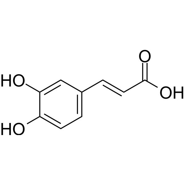
C9H8O4
|
|---|---|
| 分子量 |
180.1574
|
| 精确质量 |
180.042
|
| CAS号 |
331-39-5
|
| 相关CAS号 |
trans-Caffeic acid;501-16-6;Caffeic acid phenethyl ester;104594-70-9;Caffeic acid-13C3;1185245-82-2
|
| PubChem CID |
689043
|
| 外观&性状 |
Off-white to light yellow solid
|
| 密度 |
1.5±0.1 g/cm3
|
| 沸点 |
416.8±35.0 °C at 760 mmHg
|
| 熔点 |
433 to 437 °F (decomposes) (NTP, 1992)
; Decomposes at 223-225 °C (Softens at 194 °C).
; 225 °C
|
| 闪点 |
220.0±22.4 °C
|
| 蒸汽压 |
0.0±1.0 mmHg at 25°C
|
| 折射率 |
1.707
|
| LogP |
1.42
|
| tPSA |
77.76
|
| 氢键供体(HBD)数目 |
3
|
| 氢键受体(HBA)数目 |
4
|
| 可旋转键数目(RBC) |
2
|
| 重原子数目 |
13
|
| 分子复杂度/Complexity |
212
|
| 定义原子立体中心数目 |
0
|
| SMILES |
O=C(O)/C=C/C1=CC=C(O)C(O)=C1
|
| InChi Key |
QAIPRVGONGVQAS-DUXPYHPUSA-N
|
| InChi Code |
InChI=1S/C9H8O4/c10-7-3-1-6(5-8(7)11)2-4-9(12)13/h1-5,10-11H,(H,12,13)/b4-2+
|
| 化学名 |
(E)-3-(3,4-dihydroxyphenyl)prop-2-enoic acid
|
| 别名 |
caffeic acid; 3,4-Dihydroxycinnamic acid; 331-39-5; 3,4-Dihydroxybenzeneacrylic acid; (E)-3-(3,4-dihydroxyphenyl)prop-2-enoic acid; Cinnamic acid, 3,4-dihydroxy-; 3-(3,4-Dihydroxyphenyl)-2-propenoic acid; 2-Propenoic acid, 3-(3,4-dihydroxyphenyl)-;
|
| HS Tariff Code |
2934.99.9001
|
| 存储方式 |
Powder -20°C 3 years 4°C 2 years In solvent -80°C 6 months -20°C 1 month |
| 运输条件 |
Room temperature (This product is stable at ambient temperature for a few days during ordinary shipping and time spent in Customs)
|
| 溶解度 (体外实验) |
DMSO : ~100 mg/mL (~555.06 mM)
H2O : < 0.1 mg/mL |
|---|---|
| 溶解度 (体内实验) |
配方 1 中的溶解度: ≥ 2.5 mg/mL (13.88 mM) (饱和度未知) in 10% DMSO + 90% (20% SBE-β-CD in Saline) (这些助溶剂从左到右依次添加,逐一添加), 澄清溶液。
例如,若需制备1 mL的工作液,可将100 μL 25.0 mg/mL澄清DMSO储备液加入900 μL 20% SBE-β-CD生理盐水溶液中,混匀。 *20% SBE-β-CD 生理盐水溶液的制备(4°C,1 周):将 2 g SBE-β-CD 溶解于 10 mL 生理盐水中,得到澄清溶液。 配方 2 中的溶解度: ≥ 2.5 mg/mL (13.88 mM) (饱和度未知) in 10% DMSO + 90% Corn Oil (这些助溶剂从左到右依次添加,逐一添加), 澄清溶液。 例如,若需制备1 mL的工作液,可将 100 μL 25.0 mg/mL 澄清 DMSO 储备液加入到 900 μL 玉米油中并混合均匀。 View More
配方 3 中的溶解度: ≥ 2.08 mg/mL (11.55 mM) (饱和度未知) in 10% DMSO + 40% PEG300 + 5% Tween80 + 45% Saline (这些助溶剂从左到右依次添加,逐一添加), 澄清溶液。 1、请先配制澄清的储备液(如:用DMSO配置50 或 100 mg/mL母液(储备液)); 2、取适量母液,按从左到右的顺序依次添加助溶剂,澄清后再加入下一助溶剂。以 下列配方为例说明 (注意此配方只用于说明,并不一定代表此产品 的实际溶解配方): 10% DMSO → 40% PEG300 → 5% Tween-80 → 45% ddH2O (或 saline); 假设最终工作液的体积为 1 mL, 浓度为5 mg/mL: 取 100 μL 50 mg/mL 的澄清 DMSO 储备液加到 400 μL PEG300 中,混合均匀/澄清;向上述体系中加入50 μL Tween-80,混合均匀/澄清;然后继续加入450 μL ddH2O (或 saline)定容至 1 mL; 3、溶剂前显示的百分比是指该溶剂在最终溶液/工作液中的体积所占比例; 4、 如产品在配制过程中出现沉淀/析出,可通过加热(≤50℃)或超声的方式助溶; 5、为保证最佳实验结果,工作液请现配现用! 6、如不确定怎么将母液配置成体内动物实验的工作液,请查看说明书或联系我们; 7、 以上所有助溶剂都可在 Invivochem.cn网站购买。 |
| 制备储备液 | 1 mg | 5 mg | 10 mg | |
| 1 mM | 5.5506 mL | 27.7531 mL | 55.5062 mL | |
| 5 mM | 1.1101 mL | 5.5506 mL | 11.1012 mL | |
| 10 mM | 0.5551 mL | 2.7753 mL | 5.5506 mL |
1、根据实验需要选择合适的溶剂配制储备液 (母液):对于大多数产品,InvivoChem推荐用DMSO配置母液 (比如:5、10、20mM或者10、20、50 mg/mL浓度),个别水溶性高的产品可直接溶于水。产品在DMSO 、水或其他溶剂中的具体溶解度详见上”溶解度 (体外)”部分;
2、如果您找不到您想要的溶解度信息,或者很难将产品溶解在溶液中,请联系我们;
3、建议使用下列计算器进行相关计算(摩尔浓度计算器、稀释计算器、分子量计算器、重组计算器等);
4、母液配好之后,将其分装到常规用量,并储存在-20°C或-80°C,尽量减少反复冻融循环。
计算结果:
工作液浓度: mg/mL;
DMSO母液配制方法: mg 药物溶于 μL DMSO溶液(母液浓度 mg/mL)。如该浓度超过该批次药物DMSO溶解度,请首先与我们联系。
体内配方配制方法:取 μL DMSO母液,加入 μL PEG300,混匀澄清后加入μL Tween 80,混匀澄清后加入 μL ddH2O,混匀澄清。
(1) 请确保溶液澄清之后,再加入下一种溶剂 (助溶剂) 。可利用涡旋、超声或水浴加热等方法助溶;
(2) 一定要按顺序加入溶剂 (助溶剂) 。
Link: https://clinicaltrials.gov/ct2/show/NCT04648917
Conditions:Esophagus Cancer, Stage IIILink: https://clinicaltrials.gov/ct2/show/NCT02556814
Conditions:Immune ThrombocytopeniaLink: https://clinicaltrials.gov/ct2/show/NCT02351622
Conditions:Immune Thrombocytopenia 匹伐加宾
匹伐加宾
 磷酸氢化可的松
 BAY-784
 Gly-PEG3-endo-BCN
InvivoChem的所有产品仅用于作科学研究,不面向患者销售
Copyright 2020 InvivoChem LLC | All Rights Reserved 粤ICP备20063088号-1


 COA
COA
 463611831
