| 规格 | 价格 | 库存 | 数量 |
|---|---|---|---|
| 100mg |
|
||
| Other Sizes |
|

| 靶点 |
Microbial Metabolite
|
|---|---|
| 体外研究 (In Vitro) |
反式咖啡酸对燕麦胚芽鞘 2 毫米切段具有促生长活性。稀溶液的反式咖啡酸可显著促进豌豆上胚轴的直立生长 [1]
|
| 药代性质 (ADME/PK) |
代谢/代谢物
参与咖啡酸代谢的酶尚未被鉴定。以下实验中,咖啡酸 (CA)、绿原酸 (CGA) 和二氢咖啡酸 (DHCA) 与肝细胞孵育,结果表明它们可被细胞色素 P450、儿茶酚-O-甲基转移酶 (COMT) 和 β-氧化酶代谢。COMT 对 CA 或 DHCA 进行 O-甲基化后生成的阿魏酸 (FA) 或二氢阿魏酸 (DHFA) 也可被 CYP1A1/2 进行 O-去甲基化,但不能被 CYP2E1 进行 O-去甲基化。DHCA 或 DHFA 还会发生侧链脱氢反应,分别生成 CA 和 FA,而硫代乙醇酸(一种 β-氧化酶酰基辅酶 A 脱氢酶的抑制剂)可抑制该反应。 NADPH/微粒体(CYP2E1)催化的谷胱甘肽结合物形成速率按DHCA>CA>CGA递减的顺序排列,这与COMT对其O-甲基化的速率顺序相反。NADPH/P450生成的CA和DHCA-o-醌可能抑制COMT,但它们可以很容易地形成谷胱甘肽结合物。CA、DHCA和DHFA在分离的大鼠肝细胞中相互代谢,并可代谢为FA,而FA仅代谢为CA,不代谢为DHCA或DHFA。CA、DHCA、FA、DHFA和CGA均表现出剂量依赖性的肝细胞毒性,测定的LD50(2小时)按毒性递减的顺序排列为DHCA>CA>DHFA>CGA>FA。总之,已有证据表明,O-甲基化、GSH结合、氢化和脱氢反应均参与了CA和DHCA的肝脏代谢。CA和DHCA的O-甲基化途径是一种解毒途径,而P450催化的邻醌生成途径则是一种毒性途径。 在大鼠体内,绿原酸在胃和肠道中水解为咖啡酸和奎宁酸。已鉴定出多种代谢产物。间香豆酸和间羟基马尿酸的葡萄糖醛酸苷似乎是人体内的主要代谢产物。在人体志愿者口服咖啡酸后,O-甲基化衍生物(阿魏酸、二氢阿魏酸和香草酸)迅速从尿液中排出,而间羟基苯基衍生物则出现较晚。脱羟基反应归因于肠道细菌的作用。 咖啡酸已知的代谢产物包括(2S,3S,4S,5R)-6-[5-[(E)-2-羧基乙烯基]-2-羟基苯氧基]-3,4,5-三羟基氧杂环己烷-2-羧酸和(2S,3S,4S,5R)-6-[4-[(E)-2-羧基乙烯基]-2-羟基苯氧基]-3,4,5-三羟基氧杂环己烷-2-羧酸。 |
| 毒性/毒理 (Toxicokinetics/TK) |
相互作用
咖啡酸以浓度依赖的方式增强了C2C12细胞对放射性葡萄糖的摄取。苯肾上腺素对C2C12细胞摄取放射性葡萄糖也具有类似的作用。哌唑嗪减弱了咖啡酸的作用,其作用方式与阻断苯肾上腺素的作用类似。 9周龄雌性ICR/Ha小鼠饲喂含0.06 mmol/g(10 g/kg饲料)咖啡酸(纯度99%)的饲料。从实验第8天开始,小鼠每周两次灌胃给予1 mg苯并[a]芘,持续4周。最后一次苯并[a]芘处理3天后,停止饲喂含咖啡酸的饲料。小鼠在211日龄时处死。在 17 只有效小鼠中,咖啡酸显著降低了前胃肿瘤(≥ 1 mm)/只小鼠(组织学未具体说明)的数量(p < 0.05)(3.1 个肿瘤/只小鼠,而 38 只单独用苯并(a)芘治疗的小鼠为 5.0 个肿瘤/只小鼠)。 |
| 参考文献 | |
| 其他信息 |
根据世界卫生组织国际癌症研究机构(IARC)的说法,咖啡酸可能致癌。
3,4-二羟基肉桂酸在氯仿或石油醚溶液中呈黄色棱柱状或片状,或呈淡黄色颗粒状。碱性溶液中颜色由黄色变为橙色。(NTP, 1992) 咖啡酸是一种羟基肉桂酸,是肉桂酸苯环上3位和4位被羟基取代后形成的。它以顺式和反式两种形式存在,其中反式更为常见。咖啡酸是一种植物代谢产物,同时也是EC 1.13.11.33(花生四烯酸15-脂氧合酶)抑制剂、EC 2.5.1.18(谷胱甘肽转移酶)抑制剂、EC 1.13.11.34(花生四烯酸5-脂氧合酶)抑制剂、抗氧化剂和EC 3.5.1.98(组蛋白去乙酰化酶)抑制剂。它是一种羟基肉桂酸,属于儿茶酚类化合物。 据报道,丹参、白花丹参以及其他有相关数据的生物体中均含有咖啡酸。 咖啡酸是一种口服生物利用度高的羟基肉桂酸衍生物和多酚,具有潜在的抗氧化、抗炎和抗肿瘤活性。服用后,咖啡酸可作为抗氧化剂,防止氧化应激,从而预防自由基引起的DNA损伤。咖啡酸靶向并抑制鳞状细胞癌扩增的组蛋白去甲基化酶(HDM)癌蛋白基因1(GASC1;JMJD2C;KDM4C),从而抑制癌细胞增殖。GASC1是含Jumonji(Jmj)结构域蛋白KDM4亚组的成员,它能使组蛋白H3上的赖氨酸9和赖氨酸36(H3K9和H3K36)去甲基化,并在肿瘤细胞发育中发挥关键作用。 咖啡酸是酿酒酵母(Saccharomyces cerevisiae)中发现或产生的代谢产物。 另见:黑升麻(部分);紫草根(部分)。牛蒡根(部分)……查看更多…… 作用机制 咖啡酸苯乙酯 (CAPE) 由咖啡酸和苯乙醇(比例 1:5)在室温下以二环己基碳二亚胺 (DCC) 为缩合剂合成,产率约为 38%。CAPE 可抑制人白血病 HL-60 细胞的生长。它还能抑制 HL-60 细胞中 DNA、RNA 和蛋白质的合成,IC50 值分别为 1.0 M、5.0 M 和 1.5 M。 为了解咖啡酸的降血糖作用,本研究采用成肌细胞 C2C12 来研究其对葡萄糖的摄取。咖啡酸以浓度依赖的方式增强了 C2C12 细胞对放射性葡萄糖的摄取。在C2C12细胞中也观察到了苯肾上腺素对放射性葡萄糖摄取的类似作用。哌唑嗪减弱咖啡酸的作用方式与阻断苯肾上腺素的作用方式相似。咖啡酸对α1-肾上腺素能受体的作用通过[3H]哌唑嗪在C2C12细胞中的结合置换得到进一步证实。此外,苯肾上腺素在C2C12细胞中增加葡萄糖摄取的作用可被α1A-肾上腺素能受体拮抗剂坦索罗辛和WB 4101抑制,但不受α1B-肾上腺素能受体拮抗剂氯乙基可乐定(CEC)的影响。因此,可以认为C2C12细胞中存在α1A-肾上腺素能受体。在与这些拮抗剂共同孵育的C2C12细胞中也观察到了咖啡酸作用的类似抑制。 α1A-肾上腺素能受体的激活似乎是咖啡酸在C2C12细胞中发挥作用的原因。在磷脂酶C特异性抑制剂U73312存在的情况下,咖啡酸刺激的放射性葡萄糖进入C2C12细胞的摄取呈浓度依赖性降低,而U73343(U73312的阴性对照)则无此作用。此外,chelerythrine 和 GF 109203X 能减弱咖啡酸在足以抑制蛋白激酶 C 的浓度下的作用。因此,所得数据表明,咖啡酸激活 C2C12 细胞中的 α1A-肾上腺素能受体可能通过磷脂酶 C-蛋白激酶 C 通路增加葡萄糖摄取。 研究表明,膳食中 2% 的咖啡酸(CA,3,4-二羟基肉桂酸)可导致 F344 大鼠和 B6C3F1 小鼠的前胃和肾脏发生癌变。鉴于咖啡酸存在于咖啡和多种食物中,并采用 0% 至 2% 剂量范围内的线性插值法推算癌症发生率,人类患癌风险相当高。在两个靶器官中,肿瘤形成均先于增生,这可能是其致癌作用的主要机制。本研究在雄性F344大鼠中,以不同膳食浓度(0、0.05%、0.14%、0.40%和1.64%)的CA喂养4周后,探讨了该效应的剂量反应关系。腹腔注射2小时后,通过免疫组织化学分析掺入的5-溴-2'-脱氧尿苷(BrdU)来观察处于DNA复制S期的细胞。在前胃中,0.40%和1.64%浓度下,每毫米切片长度的上皮细胞总数和BrdU阳性细胞的单位长度标记指数(ULLI)均增加约2.5倍。最低浓度(0.05%)未观察到任何效应。0.14%浓度下,这两个指标均下降了约三分之一。在肾脏中,近端肾小管细胞的标记指数也显示出 J 形(或 U 形)剂量反应,在 1.64% 时增加了 1.8 倍。在非靶器官——腺胃和肝脏中,未观察到剂量相关效应。数据表明,癌症诱导的器官特异性与细胞分裂刺激之间存在良好的相关性。就剂量反应关系以及将动物肿瘤数据外推至人类癌症风险而言,线性外推似乎并不合适。 反式咖啡酸可能是一种非常重要的天然生长调节剂,其促生长作用与吲哚-3-乙酸类似。它是彩叶草叶片乙醚提取物中的活性生长物质之一[1]。 |
| 分子式 |
C9H8O4
|
|---|---|
| 分子量 |
180.16
|
| 精确质量 |
180.042
|
| 元素分析 |
C, 60.00; H, 4.48; O, 35.52
|
| CAS号 |
501-16-6
|
| 相关CAS号 |
Caffeic acid; 331-39-5
|
| PubChem CID |
689043
|
| 外观&性状 |
White to off-white solid powder
|
| 密度 |
1.5±0.1 g/cm3
|
| 沸点 |
416.8±35.0 °C at 760 mmHg
|
| 熔点 |
211-213ºC (dec.)(lit.)
|
| 闪点 |
220.0±22.4 °C
|
| 蒸汽压 |
0.0±1.0 mmHg at 25°C
|
| 折射率 |
1.707
|
| LogP |
1.42
|
| tPSA |
77.76
|
| 氢键供体(HBD)数目 |
3
|
| 氢键受体(HBA)数目 |
4
|
| 可旋转键数目(RBC) |
2
|
| 重原子数目 |
13
|
| 分子复杂度/Complexity |
212
|
| 定义原子立体中心数目 |
0
|
| SMILES |
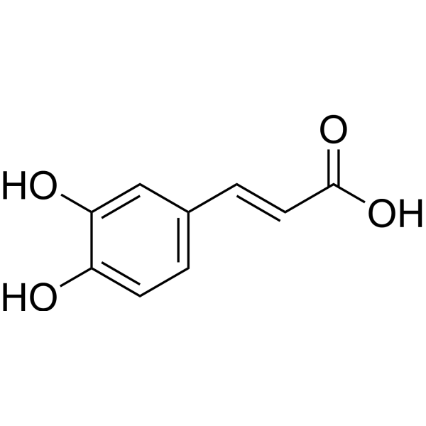
C1=CC(=C(C=C1/C=C/C(=O)O)O)O
|
| InChi Key |
QAIPRVGONGVQAS-DUXPYHPUSA-N
|
| InChi Code |
InChI=1S/C9H8O4/c10-7-3-1-6(5-8(7)11)2-4-9(12)13/h1-5,10-11H,(H,12,13)/b4-2+
|
| 化学名 |
(E)-3-(3,4-dihydroxyphenyl)prop-2-enoic acid
|
| 别名 |
Caffeic acid; AI3-63211; caffeic acid; 3,4-Dihydroxycinnamic acid; 331-39-5; 3,4-Dihydroxybenzeneacrylic acid; (E)-3-(3,4-dihydroxyphenyl)prop-2-enoic acid; Cinnamic acid, 3,4-dihydroxy-; 3-(3,4-Dihydroxyphenyl)-2-propenoic acid; ...; 501-16-6;
|
| HS Tariff Code |
2934.99.9001
|
| 存储方式 |
Powder -20°C 3 years 4°C 2 years In solvent -80°C 6 months -20°C 1 month |
| 运输条件 |
Room temperature (This product is stable at ambient temperature for a few days during ordinary shipping and time spent in Customs)
|
| 溶解度 (体外实验) |
May dissolve in DMSO (in most cases), if not, try other solvents such as H2O, Ethanol, or DMF with a minute amount of products to avoid loss of samples
|
|---|---|
| 溶解度 (体内实验) |
注意: 如下所列的是一些常用的体内动物实验溶解配方,主要用于溶解难溶或不溶于水的产品(水溶度<1 mg/mL)。 建议您先取少量样品进行尝试,如该配方可行,再根据实验需求增加样品量。
注射用配方
注射用配方1: DMSO : Tween 80: Saline = 10 : 5 : 85 (如: 100 μL DMSO → 50 μL Tween 80 → 850 μL Saline)(IP/IV/IM/SC等) *生理盐水/Saline的制备:将0.9g氯化钠/NaCl溶解在100 mL ddH ₂ O中,得到澄清溶液。 注射用配方 2: DMSO : PEG300 :Tween 80 : Saline = 10 : 40 : 5 : 45 (如: 100 μL DMSO → 400 μL PEG300 → 50 μL Tween 80 → 450 μL Saline) 注射用配方 3: DMSO : Corn oil = 10 : 90 (如: 100 μL DMSO → 900 μL Corn oil) 示例: 以注射用配方 3 (DMSO : Corn oil = 10 : 90) 为例说明, 如果要配制 1 mL 2.5 mg/mL的工作液, 您可以取 100 μL 25 mg/mL 澄清的 DMSO 储备液,加到 900 μL Corn oil/玉米油中, 混合均匀。 View More
注射用配方 4: DMSO : 20% SBE-β-CD in Saline = 10 : 90 [如:100 μL DMSO → 900 μL (20% SBE-β-CD in Saline)] 口服配方
口服配方 1: 悬浮于0.5% CMC Na (羧甲基纤维素钠) 口服配方 2: 悬浮于0.5% Carboxymethyl cellulose (羧甲基纤维素) 示例: 以口服配方 1 (悬浮于 0.5% CMC Na)为例说明, 如果要配制 100 mL 2.5 mg/mL 的工作液, 您可以先取0.5g CMC Na并将其溶解于100mL ddH2O中,得到0.5%CMC-Na澄清溶液;然后将250 mg待测化合物加到100 mL前述 0.5%CMC Na溶液中,得到悬浮液。 View More
口服配方 3: 溶解于 PEG400 (聚乙二醇400) 请根据您的实验动物和给药方式选择适当的溶解配方/方案: 1、请先配制澄清的储备液(如:用DMSO配置50 或 100 mg/mL母液(储备液)); 2、取适量母液,按从左到右的顺序依次添加助溶剂,澄清后再加入下一助溶剂。以 下列配方为例说明 (注意此配方只用于说明,并不一定代表此产品 的实际溶解配方): 10% DMSO → 40% PEG300 → 5% Tween-80 → 45% ddH2O (或 saline); 假设最终工作液的体积为 1 mL, 浓度为5 mg/mL: 取 100 μL 50 mg/mL 的澄清 DMSO 储备液加到 400 μL PEG300 中,混合均匀/澄清;向上述体系中加入50 μL Tween-80,混合均匀/澄清;然后继续加入450 μL ddH2O (或 saline)定容至 1 mL; 3、溶剂前显示的百分比是指该溶剂在最终溶液/工作液中的体积所占比例; 4、 如产品在配制过程中出现沉淀/析出,可通过加热(≤50℃)或超声的方式助溶; 5、为保证最佳实验结果,工作液请现配现用! 6、如不确定怎么将母液配置成体内动物实验的工作液,请查看说明书或联系我们; 7、 以上所有助溶剂都可在 Invivochem.cn网站购买。 |
| 制备储备液 | 1 mg | 5 mg | 10 mg | |
| 1 mM | 5.5506 mL | 27.7531 mL | 55.5062 mL | |
| 5 mM | 1.1101 mL | 5.5506 mL | 11.1012 mL | |
| 10 mM | 0.5551 mL | 2.7753 mL | 5.5506 mL |
1、根据实验需要选择合适的溶剂配制储备液 (母液):对于大多数产品,InvivoChem推荐用DMSO配置母液 (比如:5、10、20mM或者10、20、50 mg/mL浓度),个别水溶性高的产品可直接溶于水。产品在DMSO 、水或其他溶剂中的具体溶解度详见上”溶解度 (体外)”部分;
2、如果您找不到您想要的溶解度信息,或者很难将产品溶解在溶液中,请联系我们;
3、建议使用下列计算器进行相关计算(摩尔浓度计算器、稀释计算器、分子量计算器、重组计算器等);
4、母液配好之后,将其分装到常规用量,并储存在-20°C或-80°C,尽量减少反复冻融循环。
计算结果:
工作液浓度: mg/mL;
DMSO母液配制方法: mg 药物溶于 μL DMSO溶液(母液浓度 mg/mL)。如该浓度超过该批次药物DMSO溶解度,请首先与我们联系。
体内配方配制方法:取 μL DMSO母液,加入 μL PEG300,混匀澄清后加入μL Tween 80,混匀澄清后加入 μL ddH2O,混匀澄清。
(1) 请确保溶液澄清之后,再加入下一种溶剂 (助溶剂) 。可利用涡旋、超声或水浴加热等方法助溶;
(2) 一定要按顺序加入溶剂 (助溶剂) 。
 Prinsepiol
 Cyanidin 5-O-glucoside chloride
 (-)-Mitorubrinol
 Procyanidin B3-3-O-gallate
InvivoChem的所有产品仅用于作科学研究,不面向患者销售
Copyright 2020 InvivoChem LLC | All Rights Reserved 粤ICP备20063088号-1


 463611831
