| 规格 | 价格 | 库存 | 数量 |
|---|---|---|---|
| 10 mM * 1 mL in DMSO |
|
||
| 1mg |
|
||
| 5mg |
|
||
| 10mg |
|
||
| 25mg |
|
||
| 50mg |
|
||
| 100mg |
|
||
| 250mg |
|
||
| 500mg |
|
||
| Other Sizes |
|
| 靶点 |
Akt2 (IC50 = 6 nM); p70S6K (IC50 = 120 nM); PKA (IC50 = 168 nM); Autophagy; Apoptosis
|
|---|---|
| 体外研究 (In Vitro) |
CCT128930 对于人胶质母细胞瘤细胞 (U87MG) 的 GI50 值为 6.3 M,对于人前列腺癌细胞 (LNCaP) 的 GI50 值为 0.35 M,对于人前列腺癌细胞 (PC3) 的 GI50 值为 1.9 M,所有这些都是 PTEN 缺陷的人肿瘤细胞系。 1]。当暴露于化合物 CCT128930(0.1-60 M;1 小时)时,人胶质母细胞瘤细胞系 U87MG 在丝氨酸 473 处表现出 AKT 磷酸化的初始诱导,最高可达 20 M,随后在较高浓度[1] 时磷酸化降低。由于相应总蛋白和 GAPDH 水平相对恒定,CCT128930 在 10 M 时抑制下游靶标 pSer235/236 S6RP,在 5 M 时抑制 AKT 的直接底物(Ser9 GSK3、pThr246 PRAS40 和 pT24 FOXO1/p32 FOXO3a) [1]。
CCT128930是一种新型的ATP竞争性AKT抑制剂,是通过基于片段和结构的方法发现的。它是一种强效、先进的吡咯并嘧啶先导化合物,通过靶向单个氨基酸差异,对AKT的选择性高于PKACCT128930在体外多个肿瘤细胞系中表现出明显的抗增殖活性,并抑制了一系列AKT底物的磷酸化,这与AKT抑制一致CCT128930在PTEN缺失的U87MG人胶质母细胞瘤细胞中引起G(1)阻滞,与AKT通路阻断一致。[1] PI3K/Akt/mTOR通路在肿瘤进展和抗癌药物耐药性中起着重要作用。本研究的目的是确定新型Akt小分子抑制剂CCT128930对HepG2肝癌癌症细胞的抗肿瘤作用。我们的结果表明,在低浓度下,CCT128930增加但不抑制HepG2和A549细胞中Akt的磷酸化。CCT128930通过下调cyclinD1和Cdc25A,上调p21、p27和p53,诱导细胞周期阻滞在G1期,从而抑制细胞增殖。更高剂量(20μM)的CCT128930通过激活caspase-3、caspase-9和PARP引发细胞凋亡。CCT128930处理增加了HepG2细胞中ERK和JNK的磷酸化。CCT128930激活了HepG2细胞的DNA损伤反应,其特征是H2AX、ATM(共济失调毛细血管扩张症突变)、Chk1和Chk2的磷酸化。当暴露于更高浓度的CCT128930时,HepG2细胞表现出自噬,同时LC3-II和Beclin-1水平升高。使用氯喹阻断自噬会放大CCT128930诱导的凋亡细胞死亡和H2AX的磷酸化。本研究的结果推进了我们目前对CCT128930在癌症细胞中抗癌机制的理解[2]。 |
| 体内研究 (In Vivo) |
腹膜内注射 25 mg/kg 的 CCT128930 在已建立的 PTEN 缺失 U87MG 人胶质母细胞瘤异种移植物中显示出显着的抗肿瘤作用,第 12 天处理:对照 (T/C) 比率为 48%。40 mg/kg 的 CCT128930 也发挥有效的抗肿瘤作用在 HER2 阳性、PIK3CA 突变型 BT474 人乳腺癌异种移植物中发挥作用,第 22 天完全生长停滞,T/C 比为 29%。静脉注射时,CCT128930 达到 6.4 M 的血浆峰值浓度,然后被快速清除分布容积高、半衰期短、曲线下面积 (AUC0-) 为 4.6 M h。 CCT128930 的腹膜内给药导致血浆药物浓度峰值为 1.3 M,相关 AUC0- 为 1.3 Mh。 CCT128930口服时,血浆峰浓度仅为0.43 M,AUC0-低至0.4 Mh。[1]
CCT128930的体内药效学活性[1] 在体外证明了CCT128930对多种AKT生物标志物的浓度依赖性和时间依赖性抑制作用,并在体内证明了肿瘤暴露的有希望水平,然后在携带U87MG人胶质母细胞瘤肿瘤的同一小鼠中评估了该化合物的药效学作用,用于药代动力学研究(图4B)。图4C和D总结了CCT128930治疗(50 mg/kg i.p.×4天)对最后一次给药后2小时和6小时收获的U87MG异种移植物中几种AKT生物标志物的影响(见图4B)CCT128930在2小时和6小时的时间点均导致AKT上Ser473磷酸化显著增加(分别为P<0.001和P<0.01),与体外生物标志物结果一致(比较图3和图2)。此外,在2小时和6小时的时间点,Ser9 GSK3β(分别为P<0.001和P<0.01)、Ser235/236 S6RP(分别为P<0.05和P<0.01)和Thr246 PRAS40(分别为<0.05和P<0.05)的磷酸化明显降低,蛋白质的总形式保持相对恒定(图4C和D)。这些观察结果与体内U87MG肿瘤中CCT128930对AKT活性的抑制一致。 CCT128930的抗肿瘤活性[1] 接下来,在两种分子相关的人肿瘤异种移植物模型中评估了CCT128930的抗肿瘤活性。图5A显示,在已建立的PTEN缺失的U87MG人胶质母细胞瘤异种移植物中,25 mg/kg腹腔注射(7天×5次)的CCT128930具有明显的抗肿瘤作用,在第12天的治疗:对照(T/C)比率为48%。这种疗法没有减肥的效果。用CCT128930(40mg/kg bid×7天5次)治疗HER2-阳性、PIK3CA-突变BT474人癌症异种移植物也具有显著的抗肿瘤作用,在第22天完全停止生长,T/C比率为29%。该方案与最小的体重减轻有关,在治疗的第15天,最低体重仅为初始体重的94.8%。这些结果清楚地表明,在与PI3K通路激活分子相关的两种人类肿瘤异种移植物模型中,CCT128930作为单一药物具有抗肿瘤活性。 |
| 酶活实验 |
使用 10 μM CCT128930,ATP 浓度相当于每种酶的 Km,对 50 种不同的人类激酶进行分析。
|
| 细胞实验 |
将细胞接种到 96 孔板中并贴壁 36 小时,以确保处理前呈指数生长。使用 96 小时 SRB 测定法测定体外抗增殖活性。 TCA 固定的细胞用溶解在 1% 乙酸中的 0.4%(重量/体积)SRB 染色 30 分钟。染色期结束时,除去 SRB,并用 1% 乙酸快速冲洗培养物四次以除去未结合的染料。将乙酸从烧杯中直接倒入培养孔中。该程序允许快速进行冲洗,从而不会发生蛋白质结合染料的解吸。通过在水槽上猛烈地弹动板来去除残留的洗涤溶液,这确保了漂洗溶液的完全去除。由于 96 孔板中的毛细管作用很强,当板简单倒置时,仅靠重力排水通常无法去除冲洗溶液。冲洗后,将培养物风干直至看不到残留水分。使用 10 mM 无缓冲 Tris 碱 (pH 10.5) 在旋转摇床上溶解结合染料 5 分钟。 OD 在 UVmax 微量滴定板读数器或 Beckman DU-70 分光光度计中读取。为了获得最大灵敏度,OD 在 564 nm 处测量。然而,由于读数与染料浓度仅低于 1.8 OD 单位呈线性关系,因此通常使用次优波长,以便实验中的所有样品都保持在线性 OD 范围内。对于大多数细胞系,大约 490-530 nm 的波长非常适合此目的。
细胞活力测定和细胞集落形成测定[2] 如其他地方报道的那样,MTT法用于检测细胞存活率。简而言之,将细胞铺在96孔板上24小时。然后取出培养基,用不同浓度的CCT128930处理细胞。加入20μL MTT(5mg/mL)4小时。去除上清液后,加入150μL DMSO以溶解甲酰胺晶体,在490 nm处检测吸光度。对于细胞集落形成试验,以每孔1500个细胞(6孔细胞培养板)接种HepG2细胞,并用不同浓度的CCT128930处理2周。将细胞固定在1%戊二醛中,并用0.5%结晶紫染色。在倒置显微镜下计数>30个细胞的菌落。 细胞周期分析[2] 细胞用CCT128930处理24小时。处理结束时,收集细胞,用冰冷的磷酸盐缓冲盐水(PBS)洗涤,并在4°C的70%冷乙醇中固定过夜。用PBS洗涤后,用RNase A消化细胞并用PI染色。使用带有CellQuest软件的FACSCalibur流式细胞术分析样品的DNA含量。 |
| 动物实验 |
6-8 周龄雌性 CrTacNCr-Fox1nu 小鼠 [1]
25 mg/kg(U87MG 人胶质母细胞瘤异种移植)或 40 mg/kg(BT474 人乳腺癌异种移植) 每日腹腔注射,连续 5 天(U87MG 人胶质母细胞瘤异种移植);每日两次腹腔注射,连续 5 天(BT474 人乳腺癌异种移植) 体内人肿瘤异种移植研究 [1] 将 PTEN 缺失的 U87MG 人胶质母细胞瘤细胞 (2×10⁶) 皮下注射 (sc) 到 6-8 周龄雌性 CrTacNCr-Fox1nu 小鼠的右侧腹部。对于HER2阳性、PIK3CA突变的BT474人乳腺癌异种移植瘤,将细胞(5 × 10⁶)悬浮于添加Matrigel(1:1)的培养基中,皮下注射到预先皮下植入雌二醇缓释片(0.025 mg,90天缓释剂 #NE-121)3天的雌性小鼠乳腺脂肪垫中。将动物随机分组,当肿瘤平均体积达到约100 mm³时,开始给予载体或CCT128930治疗。对照组小鼠仅接受载体(10% DMSO、5% Tween 20、85% 生理盐水)注射,治疗组小鼠分别接受 50 mg/kg CCT128930 腹腔注射(ip),每日一次,连续 5 天(U87MG 人胶质母细胞瘤异种移植模型)或 40 mg/kg CCT128930 腹腔注射,每日两次,连续 5 天(BT474 人乳腺癌异种移植模型)。每周监测三次肿瘤大小和体重。肿瘤大小通过游标卡尺测量两个正交直径进行评估,体积根据公式 V = 4/3π[(d1+d2)/4]3 计算。研究结束时,切除肿瘤并称重。 为了评估CCT128930的药代动力学和药效学特征,向携带U87MG人胶质母细胞瘤异种移植瘤的小鼠单次腹腔注射该化合物(50 mg/kg)。分别于给药后2小时和6小时采集血浆和肿瘤样本。通过心脏穿刺采集小鼠血液,收集血浆样本并冷冻于-20°C直至分析。解剖肿瘤,将其分成大致相等的两块,并立即置于液氮中速冻直至分析。在药效学研究中,使用含有 50 mmol/L Tris (pH 7.4)、1 mmol/L NaCl、1 mmol/L EDTA、1% Triton X-100、1 mmol/L NaF、1 mmol/L NaVO4、5 μmol/L 苯戊酸酯、5 μmol/L Vbphen、10 mg/mL TLCK、每 10 mL 缓冲液中加入 1 片 Complete 抑制剂片剂、蛋白酶抑制剂混合物以及磷酸酶抑制剂 1 和 2 的缓冲液匀浆肿瘤组织。使用 Bradford 试剂测定蛋白质含量,并如上所述使用免疫印迹法分析样品。 药代动力学分析 [1] 使用 LC-MS 测定生物样品中 CCT128930 的浓度。药物采用甲醇提取,色谱柱为Synergi-Polar RP柱(5.0 cm × 4.6 mm内径,4 µm粒径),使用Waters 600MS泵和717型自动进样器,流动相为0.1%甲酸/甲醇,流速0.6 mL/min,梯度洗脱12分钟。检测采用液相色谱-质谱联用仪(LC-MS),使用TSQ700三重四极杆质谱仪,通过电喷雾电离正离子模式对分析物进行电离,监测[M+H]+离子跃迁342.8→146.6.4。喷雾电压优化为5.5 kV,毛细管温度优化为260℃。该检测方法在 10 – 10,000 nM CCT128930 范围内呈线性。 PRAS40 免疫荧光研究 [1] 将 U87MG 人胶质母细胞瘤细胞以 5 × 10⁴ 个细胞/孔的密度接种于 24 孔板的盖玻片上,培养 36 小时后,用递增浓度的 CCT128930 处理 24 小时。细胞用 3.8% 甲醛固定,并用 0.01% Triton X-100 透化。BALB/c 小鼠每天腹腔注射 50 mg/kg CCT128930,连续 4 天,然后拔下胡须,立即将胡须根部浸入 10% 福尔马林溶液中固定 30 分钟,最后保存在 4% 生理盐水中,4°C 保存。从健康志愿者身上采集毛囊,并按上述方法进行处理。胡须和毛囊在分析前用柠檬酸缓冲液进行抗原修复。细胞、胡须和毛囊分别用抗磷酸化Thr246 PRAS40抗体和抗PRAS40抗体(细胞1:200;胡须和毛囊1:50)染色,然后用1:1000 AlexaFluor® 488标记的山羊抗兔IgG抗体进行显色。细胞核用1:10000 TOPRO-3进行复染。细胞或毛囊用Vectashield封片剂封片,并使用Leica SP1共聚焦激光扫描荧光显微镜进行观察和图像采集。所有图像均使用与相应载体处理的对照样品相同的检测器设置拍摄。光学放大倍数为250倍,软件(数字)放大倍数为2倍,总放大倍数为500倍。对于卵泡,沿z轴从球体中心采集图像,并包含球体的整个横截面。使用INCell Investigator Developer Toolbox v1.6软件对单个细胞的荧光强度进行定量。使用TOPRO-3识别图像中的所有细胞核。然后扩展细胞核边界周围的区域以识别细胞质区域。将细胞核和细胞质分割结果连接起来,以便仅对含有细胞核的细胞内的细胞质区域进行定量。然后分别报告pThr246 PRAS40图像和总PRAS40图像中每个细胞的平均荧光强度。 |
| 药代性质 (ADME/PK) |
CCT128930 的药代动力学 [1]
测定 CCT128930 的药代动力学是为了确定体内是否能达到治疗活性药物浓度。图 4A 展示了单次静脉注射 25 mg/kg CCT128930 后的药代动力学曲线,补充表 1 对此进行了总结。静脉注射后,CCT128930 在血浆中的峰浓度为 6.4 µM,其消除半衰期相对较短,分布容积较大,清除速度快,AUC0-∞ 为 4.6 µMh。腹腔注射后,血浆药物峰浓度降低了4倍,血浆清除率与静脉注射相似。相应的AUC0-∞为1.3 µM·h,腹腔注射生物利用度为29%。 口服CCT128930的药代动力学特征与其他给药途径相似,但血浆峰浓度仅为0.43 µM,血浆清除率与静脉注射相似,表明不存在首过代谢(补充表1)。这导致AUC相应降低,口服生物利用度仅为8.5%。更重要的是,腹腔注射后,肿瘤中CCT128930的峰浓度是相应血浆值的6倍,达到8 µM,并且有药物滞留的证据,表现为半衰期延长2倍,表观清除率降低6倍。这导致肿瘤药物暴露量远高于血浆暴露量,AUC0-∞为25.8 µM·h。肿瘤与血浆药物浓度比未达到稳态,而是在30分钟时为4:1,6小时时为163:1,证实了药物在组织中的滞留。假设药物动力学呈线性,这些数据支持使用更高剂量和重复给药,以在体内达到潜在的治疗性肿瘤药物浓度。 图4B显示,在连续4天以50 mg/kg的剂量腹腔注射CCT128930后,U87MG人胶质母细胞瘤肿瘤中的药物浓度始终远高于相应的血浆浓度,末次给药后2小时和6小时的肿瘤与血浆药物浓度比分别为27:1和42:1。此外,末次给药后至少6小时内,U87MG肿瘤中的药物浓度远高于CCT128930的GI50,并且始终比体外生物标志物调节所需的浓度高5倍(图2)。代谢研究显示,24小时后,仅有0.23%的给药剂量(25 mg/kg,腹腔注射)以未改变的形式从尿液中排出(数据未显示)。上述结果表明,在耐受性良好的剂量下,CCT128930在肿瘤组织中达到了药理活性浓度。 |
| 参考文献 |
|
| 其他信息 |
AKT在癌症中经常发生异常调控,使其成为极具吸引力的抗癌药物靶点。CCT128930是一种新型ATP竞争性AKT抑制剂,采用基于片段和结构的策略发现。它是一种高效的先进先导吡咯并嘧啶化合物,通过靶向单个氨基酸差异,对AKT的选择性优于PKA。体外实验表明,CCT128930具有显著的抗增殖活性,并能抑制多种肿瘤细胞系中一系列AKT底物的磷酸化,这与AKT抑制作用相符。CCT128930可导致PTEN缺失的U87MG人胶质母细胞瘤细胞发生G1期阻滞,这与AKT通路阻断相符。药代动力学研究表明,在人肿瘤异种移植模型中可以达到潜在的活性浓度。此外,CCT128930还能阻断U87MG肿瘤异种移植模型中多个下游AKT生物标志物的磷酸化,表明其在体内抑制AKT活性。在U87MG和HER2阳性、PIK3CA突变型BT474人乳腺癌异种移植模型中均观察到CCT128930的抗肿瘤活性,这与其药代动力学和药效学特性相符。本文介绍了一种定量免疫荧光分析方法,用于检测毛囊中AKT底物PRAS40的磷酸化水平和总蛋白表达。在体内小鼠胡须毛囊和体外人毛囊中,CCT128930处理均导致pThr246 PRAS40显著降低,而总PRAS40水平变化甚微。总之,CCT128930 是一种新型、选择性强且高效的 AKT 抑制剂,可在体外和体内阻断 AKT 活性,并诱导显著的抗肿瘤反应。我们还开发了一种用于检测人毛囊中 AKT 抑制的新型生物标志物检测方法,目前正在进行临床试验。[1]
总之,我们评估了这种新型 AKT 抑制剂 CCT128930 的药理学和治疗特性。它是吡咯并嘧啶类化合物的典型代表,我们通过靶向单个氨基酸差异,实现了对 AKT 相对于 PKA 的选择性,这是我们首次发现的。我们已证实,CCT128930 可在体外和体内抑制 AKT 下游标志物的磷酸化,并在具有合适药代动力学和药效学特性的分子相关人类癌症模型中表现出良好的单药抗肿瘤活性。我们还利用这种AKT抑制剂开发了一种新型检测方法,用于量化PI3K-AKT通路阻断后正常组织中药效学生物标志物的变化,该方法可能在其他相关通路抑制剂的临床试验中具有更广泛的应用前景。[1]近期报道表明,尽管细胞凋亡作为细胞对DNA损伤的反应已被广泛研究,但自噬在决定细胞命运方面发挥着重要作用。自噬是真核细胞中普遍存在且高度保守的通路,可响应多种条件,例如营养匮乏、生长因子撤离和氧化应激。LC3从胞质形式(LC3-I)转化为蛋白水解和脂化形式(LC3-II)是自噬的特征性标志。我们发现CCT128930可以诱导HepG2细胞发生自噬,表现为LC3-II水平升高。研究自噬抑制剂是否能增强CCT128930的抗癌活性也很有意义。我们注意到,CCT128930与氯喹(CQ)联用可诱导HepG2细胞凋亡,达到预期效果。靶向自噬通路可能是一种有前景的治疗策略,可通过诱导细胞凋亡来增强CCT128930对肝细胞癌(HCC)的杀伤作用。总之,我们的结果表明CCT128930能够抑制癌细胞生长。CCT128930可以剂量依赖性地诱导细胞周期阻滞、DNA损伤和自噬。CCT128930处理细胞后,ERK1/2和JNK1/2的磷酸化水平显著升高。使用溶酶体蛋白酶抑制剂氯喹抑制自噬可增强CCT128930在癌细胞中的凋亡诱导活性和抗癌活性。[2] |
| 分子式 |
C18H20CLN5
|
|---|---|
| 分子量 |
341.84
|
| 精确质量 |
341.14
|
| 元素分析 |
C, 63.24; H, 5.90; Cl, 10.37; N, 20.49
|
| CAS号 |
885499-61-6
|
| 相关CAS号 |
CCT128930 hydrochloride;2453324-32-6
|
| PubChem CID |
17751819
|
| 外观&性状 |
Off-white to yellow solid powder
|
| 密度 |
1.3±0.1 g/cm3
|
| 沸点 |
547.9±50.0 °C at 760 mmHg
|
| 闪点 |
285.2±30.1 °C
|
| 蒸汽压 |
0.0±1.5 mmHg at 25°C
|
| 折射率 |
1.680
|
| LogP |
2.93
|
| tPSA |
70.83
|
| 氢键供体(HBD)数目 |
2
|
| 氢键受体(HBA)数目 |
4
|
| 可旋转键数目(RBC) |
3
|
| 重原子数目 |
24
|
| 分子复杂度/Complexity |
418
|
| 定义原子立体中心数目 |
0
|
| SMILES |
ClC1C=CC(CC2(CCN(C3C4=C(NC=C4)N=CN=3)CC2)N)=CC=1
|
| InChi Key |
RZIDZIGAXXNODG-UHFFFAOYSA-N
|
| InChi Code |
InChI=1S/C18H20ClN5/c19-14-3-1-13(2-4-14)11-18(20)6-9-24(10-7-18)17-15-5-8-21-16(15)22-12-23-17/h1-5,8,12H,6-7,9-11,20H2,(H,21,22,23)
|
| 化学名 |
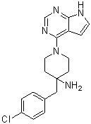
4-(4-chlorobenzyl)-1-(7H-pyrrolo[2,3-d]pyrimidin-4-yl)piperidin-4-amine
|
| 别名 |
CCT128930; CCT-128930; CCT128930; 885499-61-6; 4-(4-chlorobenzyl)-1-(7H-pyrrolo[2,3-d]pyrimidin-4-yl)piperidin-4-amine; CCT-128,930; 4-[(4-chlorophenyl)methyl]-1-(7H-pyrrolo[2,3-d]pyrimidin-4-yl)piperidin-4-amine; CHEMBL263664; 4-[(4-chlorophenyl)methyl]-1-{7H-pyrrolo[2,3-d]pyrimidin-4-yl}piperidin-4-amine; CCT 128,930; CCT 128930
|
| HS Tariff Code |
2934.99.9001
|
| 存储方式 |
Powder -20°C 3 years 4°C 2 years In solvent -80°C 6 months -20°C 1 month |
| 运输条件 |
Room temperature (This product is stable at ambient temperature for a few days during ordinary shipping and time spent in Customs)
|
| 溶解度 (体外实验) |
|
|||
|---|---|---|---|---|
| 溶解度 (体内实验) |
配方 1 中的溶解度: ≥ 2.08 mg/mL (6.08 mM) (饱和度未知) in 10% DMSO + 40% PEG300 + 5% Tween80 + 45% Saline (这些助溶剂从左到右依次添加,逐一添加), 澄清溶液。
例如,若需制备1 mL的工作液,可将100 μL 20.8 mg/mL澄清DMSO储备液加入400 μL PEG300中,混匀;然后向上述溶液中加入50 μL Tween-80,混匀;加入450 μL生理盐水定容至1 mL。 *生理盐水的制备:将 0.9 g 氯化钠溶解在 100 mL ddH₂O中,得到澄清溶液。 配方 2 中的溶解度: ≥ 2.08 mg/mL (6.08 mM) (饱和度未知) in 10% DMSO + 90% (20% SBE-β-CD in Saline) (这些助溶剂从左到右依次添加,逐一添加), 澄清溶液。 例如,若需制备1 mL的工作液,可将 100 μL 20.8 mg/mL澄清DMSO储备液加入900 μL 20% SBE-β-CD生理盐水溶液中,混匀。 *20% SBE-β-CD 生理盐水溶液的制备(4°C,1 周):将 2 g SBE-β-CD 溶解于 10 mL 生理盐水中,得到澄清溶液。 View More
配方 3 中的溶解度: ≥ 2.08 mg/mL (6.08 mM) (饱和度未知) in 10% DMSO + 90% Corn Oil (这些助溶剂从左到右依次添加,逐一添加), 澄清溶液。 配方 4 中的溶解度: 1% DMSO+30% polyethylene glycol+1% Tween 80: 11mg/mL 1、请先配制澄清的储备液(如:用DMSO配置50 或 100 mg/mL母液(储备液)); 2、取适量母液,按从左到右的顺序依次添加助溶剂,澄清后再加入下一助溶剂。以 下列配方为例说明 (注意此配方只用于说明,并不一定代表此产品 的实际溶解配方): 10% DMSO → 40% PEG300 → 5% Tween-80 → 45% ddH2O (或 saline); 假设最终工作液的体积为 1 mL, 浓度为5 mg/mL: 取 100 μL 50 mg/mL 的澄清 DMSO 储备液加到 400 μL PEG300 中,混合均匀/澄清;向上述体系中加入50 μL Tween-80,混合均匀/澄清;然后继续加入450 μL ddH2O (或 saline)定容至 1 mL; 3、溶剂前显示的百分比是指该溶剂在最终溶液/工作液中的体积所占比例; 4、 如产品在配制过程中出现沉淀/析出,可通过加热(≤50℃)或超声的方式助溶; 5、为保证最佳实验结果,工作液请现配现用! 6、如不确定怎么将母液配置成体内动物实验的工作液,请查看说明书或联系我们; 7、 以上所有助溶剂都可在 Invivochem.cn网站购买。 |
| 制备储备液 | 1 mg | 5 mg | 10 mg | |
| 1 mM | 2.9253 mL | 14.6267 mL | 29.2535 mL | |
| 5 mM | 0.5851 mL | 2.9253 mL | 5.8507 mL | |
| 10 mM | 0.2925 mL | 1.4627 mL | 2.9253 mL |
1、根据实验需要选择合适的溶剂配制储备液 (母液):对于大多数产品,InvivoChem推荐用DMSO配置母液 (比如:5、10、20mM或者10、20、50 mg/mL浓度),个别水溶性高的产品可直接溶于水。产品在DMSO 、水或其他溶剂中的具体溶解度详见上”溶解度 (体外)”部分;
2、如果您找不到您想要的溶解度信息,或者很难将产品溶解在溶液中,请联系我们;
3、建议使用下列计算器进行相关计算(摩尔浓度计算器、稀释计算器、分子量计算器、重组计算器等);
4、母液配好之后,将其分装到常规用量,并储存在-20°C或-80°C,尽量减少反复冻融循环。
计算结果:
工作液浓度: mg/mL;
DMSO母液配制方法: mg 药物溶于 μL DMSO溶液(母液浓度 mg/mL)。如该浓度超过该批次药物DMSO溶解度,请首先与我们联系。
体内配方配制方法:取 μL DMSO母液,加入 μL PEG300,混匀澄清后加入μL Tween 80,混匀澄清后加入 μL ddH2O,混匀澄清。
(1) 请确保溶液澄清之后,再加入下一种溶剂 (助溶剂) 。可利用涡旋、超声或水浴加热等方法助溶;
(2) 一定要按顺序加入溶剂 (助溶剂) 。
Effect of CCT128930 exposure on expression of AKT biomarkers and cell cycle proteins in a panel of human tumor cell lines. Mol Cancer Ther. 2011, 10(2), 360-371. |
Pharmacokinetic behavior and pharmacodynamic effects of CCT128930 in vivo. |
Antitumor activity of CCT128930 in human tumor xenografts. |
InvivoChem的所有产品仅用于作科学研究,不面向患者销售
Copyright 2020 InvivoChem LLC | All Rights Reserved 粤ICP备20063088号-1


 COA
COA


 463611831
