| 规格 | 价格 | 库存 | 数量 |
|---|---|---|---|
| 10 mM * 1 mL in DMSO |
|
||
| 1mg |
|
||
| 5mg |
|
||
| 25mg |
|
||
| 50mg |
|
||
| 100mg |
|
||
| 250mg |
|
||
| 500mg |
|
||
| 1g |
|
||
| Other Sizes |
|
| 靶点 |
GSK-3β(IC50 = 0.58 nM); GSK-3α(IC50 = 0.65 nM); cdc2(IC50 = 3700 nM)
Sirtuin 1 (SIRT1), a NAD⁺-dependent deacetylase. For Selisistat (SEN0014196; EX 527), the Ki value for SIRT1 was 13 nM (using a fluorogenic peptide substrate). It exhibited high selectivity over other sirtuins: IC50 > 10 μM (SIRT2), IC50 > 10 μM (SIRT3), IC50 > 10 μM (SIRT4), confirming SIRT1-specific inhibition [2] - SIRT1 (no additional Ki/IC50 values; focus on SIRT1 inhibition-mediated reduction of mutant huntingtin (mHtt) aggregation in Huntington’s disease models) [1] - SIRT1 (no new potency data; inhibition of SIRT1 upregulated SIRT4 to alleviate hepatic steatosis) [3] - SIRT1 (no new potency data; focus on clinical pharmacokinetics and safety in healthy volunteers) [4] |
|---|---|
| 体外研究 (In Vitro) |
Selisistat (1-10 μM) 可降低转染细胞中人 SirT1 和果蝇 Sir2 的脱乙酰化活性[1]。
Selisistat在哺乳动物细胞中的特异性研究[1] 为了确定selisistat对sirtuins的特异性和活性,我们用GCN5(一种组蛋白乙酰转移酶)和核因子κB (NFκB) p65亚基(一种特征的SirT1底物)转染HEK293细胞。通过在转染细胞中乙酰化的p65与总p65蛋白的比例可以看出,GCN5积极地使p65乙酰化。当人类SirT1也与GCN5和p65共转染时,p65乙酰化水平降低了约80%。当果蝇Sir2共转染到细胞中时,p65的GCN5乙酰化降低了约70%。在这些细胞中添加selisistat可抑制SirT1去乙酰化,在10 μm处恢复50%的p65乙酰化。同样,selisistat阻断果蝇Sir2对p65去乙酰化的能力,被抑制的乙酰化活性恢复60%。这些数据表明,selisistat抑制果蝇sirr2和人类SirT1的去乙酰化活性。 Selisistat对培养的HD动物细胞模型具有保护作用[1] 考虑到基因降低Sir2对果蝇HD病理的积极作用,我们试图确定selisistat是否在哺乳动物HD模型中表现出积极作用。表达mHtt外显子1片段的大鼠嗜铬细胞瘤细胞(PC-12)被广泛用于研究mHtt的毒性和聚集性。PC-12细胞诱导表达人类Htt外显子1片段,扩增聚谷氨酰胺重复序列,在转基因表达后出现聚集、转录变化和细胞毒性。在该模型中,诱导mHtt表达导致毒性(以乳酸脱氢酶[LDH]释放来测量)的显著增加,而用浓度为1 μm和10 μm的selisistat处理可显著降低毒性。 在STHdhQ111/Q111细胞(亨廷顿病细胞模型)中,Selisistat(1 μM、5 μM)处理48小时可剂量依赖性减少mHtt聚集(1 μM时减少40%,5 μM时减少65%),通过免疫荧光检测。蛋白质印迹(Western blot)显示,5 μM时SIRT1底物PGC-1α的乙酰化水平升高,凋亡标志物切割型caspase-3减少50% [1] - 在荧光法SIRT1活性实验中,Selisistat抑制SIRT1的Ki值为13 nM。在10 μM浓度下,对I/II类组蛋白去乙酰化酶(HDAC1、HDAC2、HDAC6)及其他Sirtuins(SIRT2–SIRT7)的抑制率<10%,展现出广泛的靶点选择性 [2] - 在原代大鼠肝细胞和肝星状细胞(HSCs)中,Selisistat(1 μM、10 μM)处理72小时可通过qRT-PCR检测到SIRT4 mRNA上调(10 μM时增加2.5倍),减少肝细胞脂质积累(10 μM时减少45%),并通过Western blot检测到HSC活化受抑(10 μM时α-SMA蛋白减少50%) [3] - 在人肝微粒体中,Selisistat主要通过CYP3A4(占总代谢的70%)和CYP2D6(20%)代谢,代谢稳定性半衰期为60分钟。在浓度高达10 μM时,不抑制CYP1A2、CYP2C9或CYP2C19 [4] |
| 体内研究 (In Vivo) |
在亨廷顿病 (HD) 的 R6/2 小鼠模型中,硒尼司他(每日口服 5 和 20 mg/kg;转基因 R6/2 小鼠从 4.5 周龄开始直至死亡)具有保护作用[1]。
在本研究中,Selisistat (SEN0014196;ex527)(5µg/kg),每周给药两次,连续10周,降低血清甘油三酯(TG)、总胆固醇、丙氨酸转氨酶(ALT)和天冬氨酸转氨酶(AST)水平,减轻肝纤维化,马松三色和肝脂肪油红- o染色证实。EX-527上调HFD喂养大鼠肝脏中SIRT2、SIRT3和SIRT4的表达,下调转化生长因子-β1 (TGF-β1)和α-平滑肌肌动蛋白(α-SMA)的表达。它降低了血清中促炎细胞因子的产生和羟脯氨酸水平以及SMAD4的表达,恢复了凋亡蛋白(Bcl-2、Bax和cleaved caspase-3)的表达。这些数据表明SIRT4/SMAD4轴在肝纤维化中起关键作用。SIRT4上调有可能对抗hfd诱导的脂质积累、炎症和纤维形成。我们证明EX-527在抑制hfd诱导的肝纤维化进展方面是一个有希望的候选药物。[3] 在R6/2小鼠(亨廷顿病模型)中,口服Selisistat(10 mg/kg、30 mg/kg,每日一次,连续6周)可剂量依赖性改善运动功能(30 mg/kg时转棒实验表现提升30%),并通过免疫组化检测到纹状体中mHtt包涵体减少55%(30 mg/kg)。与溶媒组相比,30 mg/kg剂量组生存期延长15% [1] - 在亨廷顿病转基因果蝇模型中,喂食含Selisistat(10 μM)的食物7天,可减少mHtt诱导的神经退行性变(眼部退化评分从4降至1.5),并改善运动能力(攀爬能力提升40%) [1] - 在高脂饮食喂养的Zucker肥胖大鼠(肝脂肪变模型)中,口服Selisistat(5 mg/kg、15 mg/kg,每日一次,连续8周)可剂量依赖性减少肝脏甘油三酯含量(15 mg/kg时减少35%)和胶原沉积(纤维化标志物,15 mg/kg时减少50%)。血清ALT/AST水平降低40%(15 mg/kg),Western blot显示肝脏SIRT4蛋白上调2倍 [3] - 在健康人类志愿者(n=24)中,单次口服Selisistat(10 mg、30 mg、60 mg)呈现剂量依赖性药代动力学:Cmax分别为0.2 μM(10 mg)、0.6 μM(30 mg)、1.1 μM(60 mg);Tmax均为1.5小时;终端半衰期均为8.2小时。未观察到QT间期或肝肾功能标志物的显著变化 [4] |
| 酶活实验 |
I类和II类HDAC荧光测定。[2]
使用含有I类和II类HDAC的HeLa细胞提取物和H4- k16 (Ac)底物(代表赖氨酸16上乙酰化的组蛋白H4残基12−16),在上述荧光法中测量了I类和II类HDAC去乙酰化酶的活性。 烟酰胺释放试验。[2] SIRT1的活性是用非荧光法测定的,使用的是代表赖氨酸382乙酰化残基368 - 386的p53肽底物。如前所述,该实验测量了[羰基-14C]-NAD中[14C]烟酰胺的释放。 在未标记烟酰胺浓度为52 μM的情况下,使用上述方法测量烟酰胺交换。添加的烟酰胺通过酶催化交换促进[14C]烟酰胺从标记的NAD释放。[14C]烟酰胺从NAD释放后,未标记的烟酰胺与酶结合并转化为未标记的NAD。 NAD糖水解酶(NADase)酶活性在烟酰胺释放试验中测量,如上所述。采用阴离子交换色谱法纯化猪脑NADase粗酶。每孔含有0.5 μg纯化酶和NAD,浓度为18.55 μM (KM的70%)。 微粒体的稳定性。[2] 利用大鼠肝微粒体评估体外代谢稳定性。将浓度为10 μM的化合物与大鼠肝微粒体(1 mg蛋白质/mL)在37°C下孵育,并在0、5、15、30和60 min后用HPLC/MS进行定量。对照组不含微粒体。 细胞色素P450抑制试验。[2] 细胞色素P450检测在384孔微孔板上进行,使用重组人同工酶3A4、2D6、1A2、2C9和2C19与荧光底物孵育,如前所述。 荧光法SIRT1抑制实验:将重组人SIRT1蛋白与荧光乙酰化肽底物(Ac-Arg-His-Lys-Lys(Ac)-AMC)及NAD⁺(500 μM)共同孵育于实验缓冲液(25 mM Tris-HCl pH 8.0、137 mM NaCl、2.7 mM KCl、1 mM MgCl₂)中。加入系列稀释的Selisistat(0.1 nM–10 μM),37°C孵育60分钟。加入显色液(含去乙酰化特异性蛋白酶),检测荧光强度(激发光360 nm,发射光460 nm),采用竞争性抑制模型计算Ki值 [2] - Sirtuins选择性实验:采用相同荧光法检测Selisistat对SIRT2、SIRT3、SIRT4、SIRT5、SIRT6、SIRT7的抑制作用。使用重组Sirtuin蛋白及其对应荧光底物,在浓度高达10 μM时测定IC50值 [2] |
| 细胞实验 |
hERG试验。[2]
用hERG钾通道稳定转染中国仓鼠卵巢(CHO)细胞。hERG通道的阻断引起膜电位的变化,这是用电位测定染料测量的。染料负载细胞用10 μM化合物和2 mM氯化钾孵育。使用Tecan Safire荧光仪测量384孔微孔板格式的荧光变化。测量了化合物对缺乏hERG通道的对照CHO细胞的影响,并用于纠正非特异性猝灭和毒性。 表达PC-12外显子1的突变/野生型Htt细胞株[1] 从Rubinsztein教授的实验室获得了稳定表达人类HD基因gfp标记的外显子1片段的PC-12 7210(外显子1 mutQ74)细胞(PC12-Q74)(24)。四环素(Tet-on)诱导的mHTT构建物包括人类Htt序列(NM_00211)的核苷酸1-297,并包括74 CAG重复扩增,一旦表达,对细胞具有毒性。将细胞接种于96孔聚d-赖氨酸(MW 70-150 kDa)预涂板上,密度为45K细胞/100 μl培养基/孔,培养液中含有2% HS、1% FBS、100 mU/ml青霉素/链霉素和1%谷氨酰胺,实验前在37℃、90%相对湿度、10% CO2气氛的培养箱中培养24 h。实验当天,将不含血清的相同培养基添加到孔中,以使先前的血清浓度最终稀释为1:3。在无血清培养基中加入强力霉素(终浓度为1 μg/ml)进行转基因诱导。加入selisistat(从DMSO 10 mm原液中加入)以获得结果中描述的最终浓度,在仅接受DMSO的对照组中省略其添加。所有处理和对照的DMSO终浓度均为0.1%。72h时,用LDH- mix细胞毒性测试试剂盒测定培养基中细胞释放的LDH水平,用分光光度计测定490 nm(读数)和720 nm(空白)处的吸光度 慢病毒感染培养纹状体神经元[1] 采用慢病毒载体实现HD体外模型(27)。这些模型涉及慢病毒介导的纹状体神经元培养中野生型Htt(18个谷氨酰胺重复序列,18Q)或mHtt(82个谷氨酰胺重复序列,82Q) n端171个氨基酸片段的过表达。对于慢病毒介导的蛋白表达,在播种后24小时感染培养物。第4天,将一半培养基替换为添加2x浓度selisistat的新鲜培养基。此后每周进行1次复方处理,加入1倍浓度的复方新鲜培养基。强启动子结构(高表达,5-10倍内源性)在体外2-4周内导致polyq依赖性细胞死亡,通过降低neun阳性和neun阳性细胞数量来评估。在体外1-2周(高表达)或2-4周(中等表达)时,暴露于Htt- n171 - 82q而非Htt- n171 - 18q的细胞也会出现细胞内Htt包涵体。 HEK293细胞转染及治疗[1] HEK 293-T细胞在含有10% FBS, 1% Penstrep, 1% G-Max的DMEM中生长,37℃,10% CO2。将8 × 10⁵个细胞接种在MW6板上,24 h后,按照制造商的说明,用Lipofectamine 2000转染2.5µg总质粒DNA。从OriGene Technologies购买表达GCN5 (NM_021078.1)、p65 (NM_021975.3)、human_SirT1 (NM_012238.4)的质粒,从DGRC订购表达果蝇基因Sir2 cDNA (LD07439)的质粒,克隆到pcDNA载体中。转染4小时后,取出Opti-MEM培养基,将selisistat在培养基中稀释至0.1、1和10µm (DMSO 0.1%, v/v,作为对照),加入细胞中。转染24 h后,收集细胞,用RIPA缓冲液(150 mm NaCl, 1.0% NP-40, 0.5%脱氧胆酸钠,0.1% SDS, 50 mm Tris, pH 8.0)和蛋白酶和磷酸酶抑制剂(Complete EDTA-free蛋白酶抑制剂鸡尾酒,Roche和PhosSTOP抑制剂鸡尾酒,Roche)裂解。总裂解物在3000g下离心5分钟澄清,并根据制造商的说明用BCA定量蛋白质量。 亨廷顿病细胞实验:将STHdhQ111/Q111细胞(含扩展CAG重复序列的小鼠纹状体细胞)接种于24孔板,用Selisistat(1 μM、5 μM)或溶媒处理48小时。检测mHtt聚集时,固定并透化细胞,用抗mHtt抗体(EM48)及荧光二抗染色,通过图像分析定量聚集物;检测凋亡时,裂解细胞并通过Western blot检测切割型caspase-3 [1] - 肝脂肪变细胞实验:将原代大鼠肝细胞接种于96孔板,用油酸(0.2 mM)联合Selisistat(1 μM、10 μM)处理72小时。用油红O染色脂质,在510 nm处检测吸光度;将原代大鼠HSCs用Selisistat(1 μM、10 μM)处理72小时,通过Western blot检测α-SMA(HSC活化标志物) [3] - 代谢酶实验:将人肝微粒体与Selisistat(1 μM)及选择性CYP底物(如CYP3A4用咪达唑仑,CYP2D6用右美沙芬)共同孵育30分钟。通过LC-MS/MS定量代谢产物,确定各CYP酶对Selisistat代谢的贡献 [4] |
| 动物实验 |
溶于DMSO;每只大鼠约5 μg;脑室内注射。雄性Sprague-Dawley大鼠果蝇杂交[1]
为了比较正常背景下和Sir2改变背景下表达Htt的动物的表型,将elav-Gal4[C155]; Sir2[17]/CyO果蝇与UAS-Httex1p Q93纯合子(p463品系)杂交。为了在HD背景下产生Sir2纯合缺失,将elav-Gal4[C155]; Sir2[17]/CyO果蝇与含有Sir2[17]和第二个染色体标记的UAS-Httex1p Q93果蝇杂交。杂交在22.5°C下进行。羽化后,成虫在25°C下饲养于标准玉米粉糖蜜培养基上(用于遗传学研究)或含有0.1% DMSO或指定浓度selisistat (0.1–10 µm)的培养基上(用于药理学研究)。每日提供新鲜食物。7日龄时按所述方法进行伪瞳孔分析。在寿命实验中,将新孵化的处女蝇以25–30只为一组进行饲养。通过计数存活的蝇的数量来计算存活率,并每隔2–3天更换一次蝇。将7日龄的蝇置于高9.5厘米、直径2.4厘米的聚苯乙烯泡沫塑料小瓶中进行爬虫实验。小瓶放置在前后开口的饲养箱中。在小瓶后方放置一个灯箱以提高对蝇的观察效果。使用 Exilim EX-FH20 摄像机以 40 fps 的帧率对果蝇进行视频录制。计算摇晃后果蝇爬过小瓶中点的百分比随时间的变化。每个小瓶大约使用 10-15 只果蝇进行爬升实验。 药物处理[1] 在小鼠 3.5-4 周龄进行测试以确定所有动物的基线行为表现后,于 4.5 周龄开始进行药物处理。将 18 只小鼠(雌雄各 9 只)分配到每个 R6/2 组。在开始测试之前,根据体重、CAG 重复序列、出生日期和窝仔数对各实验组的小鼠进行平衡。在 3.5-4 周龄时,对小鼠进行旷场、转棒和握力测试,并在开始药物处理之前重新平衡各处理组的分组,以确保各处理组之间的初始行为表现相似。用于重新平衡各组的附加数据包括转棒跌倒时间、开放场中的总运动距离和总站立次数以及握力。小鼠每日(QD)经口灌胃(PO;10 ml/kg)给予塞利司他(5 和 20 mg/kg)或其赋形剂(0.5% 羟丙基甲基纤维素 Methocel K4M Premium,溶于无菌水中;0.5% HPMC)。每周配制混悬液,并分装至棕色小瓶(光敏)中,用于每日给药;粉末状药物储存于 4°C 的干燥器中。赋形剂每两个月配制一次,并储存于 4°C。每个小瓶在给药前均进行涡旋振荡,瓶内放置一个小型搅拌子,并在给药过程中置于搅拌器上。另设一组用于药代动力学评估的动物,从 3 周龄至 10 周龄开始给予塞利司他。末次给药后,处死动物,并在给药后0.25、0.5、1、6和24小时,从每组三只小鼠中采集躯干血样。血样置于肝素涂层试管中,置于冰水浴中,直至在4℃下以2700 rpm离心。去除上清液,将血浆储存于-80℃,直至使用LC-MS/MS方法进行分析,该方法的定量下限为5 ng/ml。药代动力学参数估计采用WinNonlin 5.01.1版软件进行。体内药代动力学分析。 [2] C57bl/6J小鼠分别通过静脉注射(iv)或灌胃给予10 mg/kg的化合物1(selisistat)或化合物35,溶于含4% DMSO和10%环糊精的磷酸盐缓冲液中。分别于给药后5、15、30、60和90分钟以及2、4、6、8和24小时采集血浆。样品由Absorption Systems公司采用液相色谱-质谱联用仪(LCMS)进行分析。血浆样品采用固相萃取法在96孔板中制备。取50 μL血浆与300 μL含内标物(华法林,浓度为50 ng/mL)的1%磷酸溶液混合。将血浆样品转移至 Waters Oasis HLB 30 mg 萃取板,用 5% 甲醇/水溶液洗涤,并用乙腈洗脱。洗脱液在 37 °C 下用氮气吹干,并重新溶解于 20% 乙腈水溶液中。[2] 将样品 (25 μL) 注入 Keystone Hypersil BDS C18 色谱柱(30 × 2.1 mm,3 μm),流速为 0.3 mL/min。流动相为 2.5 mM NH4OH-甲酸(pH 3.5)至 2.5 mM NH4OH-甲酸(90% 乙腈),梯度洗脱时间为 3 分钟。使用配备电喷雾接口的 PE Sciex API4000 质谱仪采集质谱图。定量分析采用标准曲线法,将化合物 1 或 35 添加到空白肝素化雄性 C57BL/6J 小鼠血浆中(终浓度分别为 0.3、1、3、10、30、100、300 和 1000 ng/mL),并以此为标准曲线进行分析。口服生物利用度百分比通过口服和静脉给药后直至最后一个可定量时间点的曲线下面积比值计算得出。末端消除半衰期根据静脉给药后获得的数据计算得出。雄性 ZDF 大鼠(体重 300 ± 25 g)购自 Central Lab Animal Inc.。大鼠饲养于正常温度(24 ± 0.5 °C)、相对湿度(54–58%)和 12 小时光照/12 小时黑暗循环的条件下,并严格控制特定病原体。实验开始前,大鼠在实验室条件下适应 10 天。高脂饮食(HFD)由碳水化合物、脂肪(60%)、蛋白质、矿物质、纤维和维生素组成,购自Research Diets公司,并喂饲大鼠11周以诱导糖尿病。对喂饲高脂饮食的大鼠腹腔注射EX-527(selisistat)(5 μg/kg,每周两次),持续10周。正常饮食组大鼠饲喂不含脂肪的饮食。使用血糖仪测定血糖水平。血糖水平超过300 mg/dL的大鼠被判定为糖尿病,并用于后续研究。所有实验用ZDF大鼠随机分为三组(n = 6)。治疗21周后,对大鼠进行麻醉。通过腹腔静脉采集血液,并转移至肝素化试管中。将血液以 2000×g 离心 10 分钟后获得血清,并立即转移至 −80 °C 保存,直至进行后续分析。收集主要器官(肝脏),用生理盐水灌注后,保存于 −80 °C 以备后续分析,如图 1 所示。[3]亨廷顿病小鼠模型(R6/2 小鼠):将 4 周龄雄性 R6/2 小鼠随机分为 3 组(每组 n=12):赋形剂组(0.5% 羟丙基甲基纤维素 + 0.1% Tween 80)、Selisistat 10 mg/kg 组和 30 mg/kg 组。药物溶于赋形剂中,每日灌胃一次,持续 6 周。每周通过转棒试验评估小鼠的运动功能。研究结束时,采集脑组织进行免疫组织化学(mHtt包涵体)和蛋白质印迹(PGC-1α)分析[1] - 果蝇亨廷顿病模型:表达mHtt(120Q)的转基因果蝇喂食含Selisistat(10 μM)或载体的食物,持续7天。眼部退化程度按0-5分评分,并通过攀爬实验测量运动活性[1] - Zucker肥胖大鼠肝脂肪变性模型:雄性Zucker肥胖大鼠(6周龄)喂食高脂饮食,并随机分为3组(每组n=8):载体组(0.5%甲基纤维素)、Selisistat 5 mg/kg组和Selisistat 15 mg/kg组。药物每日口服一次,持续8周。采用比色法试剂盒测定血清ALT/AST水平,并采集肝组织进行甘油三酯定量(酶法试剂盒)和胶原染色(Masson三色染色法)[3] - 健康志愿者临床研究:24名健康成年人(12名男性,12名女性)被随机分配接受单次口服Selisistat(10 mg、30 mg、60 mg)或安慰剂。分别于给药后0、0.5、1、2、4、8、12、24和48小时采集血样,用于血浆Selisistat定量(LC-MS/MS)。在基线和给药后24小时测量心电图(QT间期)和临床化学指标(ALT、AST、肌酐)[4] |
| 药代性质 (ADME/PK) |
药代动力学结果[4]
男性受试者在空腹状态下单次口服5至600 mg塞利司他后,药物吸收迅速,但吸收速率似乎呈剂量依赖性,塞利司他的中位达峰时间(tmax)从5 mg剂量组的给药后1小时增加到600 mg剂量组的给药后4小时(图1)。塞利司他的消除呈双相性,表观末端血浆半衰期似乎随剂量增加而延长(平均值范围从5 mg剂量组的1.6小时到600 mg剂量组的6.1小时)。塞利司他的AUC(0,∞)在5至300 mg剂量范围内呈剂量比例增加,在300 mg至600 mg剂量水平之间出现显著的超比例增加,表明一种或多种清除机制在高剂量下接近饱和(图2A和表2)。在所有男性受试者中,尿液中原形药物的排泄比例相对于剂量而言均较低,各剂量组给药后24小时内排泄量均低于0.02%。多次给药后,尿液中排泄的药物比例仍然较低,但随时间推移而增加,这与观察到的血浆蓄积一致。食物对男性受试者单次服用selisistat的药代动力学影响甚微。高脂早餐后,药物吸收速率有所延迟,但吸收程度基本不变。selisistat多次口服给药的药代动力学研究表明,其达峰时间(tmax)和表观末端半衰期均无剂量或时间依赖性。在每个剂量水平下,受试者晨起谷浓度的塞利司他血浆浓度显示,通常在第 4 天达到稳态。与单剂量研究结果一致,在每日一次 100 mg 至 300 mg 的剂量范围内,稳态 AUC(0,τ) 呈超比例增加(图 2B),而稳态 Cmax 则呈剂量比例增加。此外,每日两次 100 mg 剂量组的稳态 AUC(0,τ) 约为每日一次 100 mg 剂量组的两倍(表 3)。 在单剂量阶段,AUC(0,∞) 和 Cmax 的受试者间变异性 (%CV) 分别为 35%–71% 和 23%–46%。在所有剂量水平下,AUC(0,∞) 和 Cmax 的受试者间变异性汇总分别为 56% 和 33%。在多剂量阶段,男性受试者间变异性(%CV)为17%~59%,女性为28%~68%。单次和多次给药后,女性的全身暴露量均高于男性。女性的AUC(0,∞)、AUC(0,τ)和Cmax值分别比男性高1.1倍、2.2~2.3倍和1.7~1.9倍。白种人和非白种人受试者的全身暴露量或药代动力学参数估计值无差异。在健康志愿者中,口服Selisistat显示出剂量比例药代动力学特征:10 mg、30 mg和60 mg剂量组的Cmax分别为0.2 ± 0.05 μM、0.6 ± 0.1 μM和1.1 ± 0.2 μM。 Tmax 为 1.5 ± 0.3 小时(所有剂量);AUC₀-∞ 分别为 1.8 ± 0.4 μM·h、5.5 ± 0.8 μM·h 和 10.2 ± 1.5 μM·h;末端半衰期 (t₁/₂) 为 8.2 ± 1.0 小时(所有剂量)[4] - 在 Sprague-Dawley 大鼠中,Selisistat 的口服生物利用度为 42%(10 mg/kg 剂量),Cmax = 0.8 μM,Tmax = 1.2 小时,t₁/₂ = 7.5 小时[4] - 在人肝微粒体中,Selisistat 由 CYP3A4 (70%) 和 CYP2D6 (20%) 代谢;未检测到 CYP1A2、CYP2C9 或 CYP2C19 的代谢。在人体中,尿液中原形药物的排泄量小于给药剂量的5%[4] |
| 毒性/毒理 (Toxicokinetics/TK) |
安全性[4]
研究期间未报告严重不良事件,也无受试者因不良事件退出研究。健康男性受试者单次口服塞利司他剂量高达 600 mg 时,以及女性受试者单次口服塞利司他剂量为 300 mg 时,均被认为安全且耐受性良好(表 5)。健康男性受试者每日一次口服塞利司他剂量高达 300 mg,持续 7 天,以及健康女性受试者每日两次口服塞利司他剂量为 100 mg,持续 7 天,也被认为安全且耐受性良好。男性受试者药物相关不良事件发生率较低,且随着塞利司他剂量的增加,不良事件发生率并未增加。不良事件发生率未超过安慰剂组。与单次给药相比,多次给药后未观察到不良事件报告数量的增加(表 6)。男性和女性受试者报告的大多数不良事件均为轻度,无需治疗即可缓解。研究期间仅发生一例严重不良事件。一名18岁男性受试者在服用150 mg药物后1小时18分钟出现体位性晕厥。研究者认为该事件可能与研究药物相关。饮食状况对不良事件无影响。单次口服selisistat后,最常见的药物相关不良事件是头痛,男性受试者发生率为12%,女性受试者为83%。多次口服selisistat后,男性受试者不良事件发生率较低。女性受试者中,6名受试者中有3名报告至少发生过一次胃肠道不适。总体而言,服用药物和安慰剂的女性不良事件发生率均高于男性(表5和表6)。临床实验室评估结果(包括肝功能检查、血液学参数、生命体征或心脏功能)未发现与剂量或治疗相关的趋势。具体而言,12导联安全心电图记录的各项参数均未发现与治疗或剂量相关的趋势,且在任何剂量水平的selisistat治疗中,心电图形态均未发现具有临床意义的异常。12导联安全心电图评估结果显示,所有受试者的QTc间期均未超过480 ms,且较基线值增加超过60 ms。体格检查、姿势控制和神经系统检查均未发现具有临床意义的异常,摇摆平台测试表现也未见变化。 心电图参数的浓度-效应模型[4] 以受试者间ΔQTcF的标准差衡量的QTc数据变异性较低,单次递增剂量(SAD)和多次递增剂量(MAD)部分分别为5.3 ms和6.8 ms15。表7显示了第1部分(血浆浓度最高)中各剂量组QTcF相对于基线的变化。各时间点和剂量组的模式表明,selisistat对QTc间期没有剂量依赖性效应。在观察到的血浆浓度范围内,单次给予5 mg至600 mg selisistat后,未观察到ΔΔQTcF的显著浓度依赖性效应。具有截距的线性模型能够很好地拟合数据,估计的总体截距和斜率分别为 0.9 ms(90% CI −0.2, 2.0)和 −0.00026 ms/ng ml−1(90% CI −0.00063, 0.00010)(图 3A)。MAD 部分的数据分析也得到了类似的结果,截距为 2.8 ms(90% CI −0.16, 5.71),估计斜率为 −0.00011 ms/ng ml−1(90% CI −0.00087, 0.00066;图 3B)。使用该模型预测,单次服用 600 mg 后,在观察到的几何 Cmax 为 26.6 μm 时,ΔΔQTcF 效应为 −0.9 ms(90% CI −3.3, 1.4)。每日一次服用300 mg,连续7天后,观察到的Cmax水平为22.5 μM,可预测ΔΔQTcF效应约为2.8 ms(90% CI -0.1, 5.6)。对于血浆浓度超过平均Cmax水平(例如30 μM)的情况,使用同一模型可预测QTcF效应为3.7 ms(90% CI -0.1, 7.5)。在研究的SAD和MAD部分中,所有观察到的血浆浓度下,预测的ΔΔQTcF效应的90% CI上限均低于10 ms(图3A、B)。 在用Selisistat(10 mg/kg、30 mg/kg)治疗6周的R6/2小鼠中,未观察到体重、食物摄入量或毒性临床症状(嗜睡、共济失调)的显著变化。肝肾组织学检查未见异常[1] - 在接受Selisistat(15 mg/kg)治疗8周的Zucker肥胖大鼠中,血清肌酐和尿素氮(肾功能指标)与对照组相比无变化,且心脏、脾脏或胰腺未观察到组织病理学损伤[3] - 在健康志愿者中,Selisistat(最高剂量60 mg)耐受性良好;不良反应轻微(头痛、恶心),发生率<10%。未观察到血清ALT、AST、肌酐或QT间期(心电图)的显著变化[4] - 通过平衡透析法测定,Selisistat在人血浆中的血浆蛋白结合率为95%[4] |
| 参考文献 |
|
| 其他信息 |
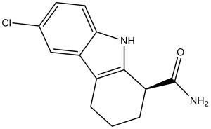
6-氯-2,3,4,9-四氢-1H-咔唑-1-甲酰胺是咔唑类化合物的一种,其结构为2,3,4,9-四氢-1H-咔唑,在1位被氨基咔唑基取代,6位被氯取代。它属于咔唑类化合物、单羧酸酰胺和有机氯化合物。
它是SIRT1的选择性抑制剂,不抑制组蛋白去乙酰化酶(HDAC)或其他sirtuin去乙酰化酶家族成员(SIRT1、SIRT2、SIRT3、HDAC和NADase的IC50值分别为98、19600、48700、>100000和>100000 nM)。增强p53乙酰化以响应DNA损伤剂。 针对人源sirtuin SIRT1的高通量筛选发现了一系列吲哚类化合物,它们作为强效抑制剂,对SIRT1具有选择性,优于其他脱乙酰酶和NAD加工酶。本文所述的最有效化合物对SIRT1的IC50值为60-100 nM,比先前报道的SIRT抑制剂提高了500倍。制备对映体纯的吲哚衍生物使其能够在体外和体内进行表征。动力学分析表明,这些抑制剂在烟酰胺从酶中释放后结合,并阻止脱乙酰肽和O-乙酰-ADP-核糖(酶催化脱乙酰化的产物)的释放。这些SIRT1抑制剂分子量小、可透过细胞膜、口服生物利用度高且代谢稳定。这些化合物为研究SIRT1的生物学特性以及探索SIRT1抑制剂的治疗用途提供了化学工具。[2] 已知Sirtuin (SIRT)能够预防非酒精性脂肪性肝病(NAFLD);然而,SIRT4在肝纤维化进展中的作用尚不清楚。我们假设选择性SIRT1抑制剂EX-527能够抑制高脂饮食(HFD)诱导的肝纤维化进展。我们发现NAFLD患者肝脏中SIRT4的表达显著低于正常受试者。在本研究中,每周两次、持续十周给予HFD大鼠EX-527 (5 µg/kg),结果显示血清甘油三酯(TG)、总胆固醇、丙氨酸氨基转移酶(ALT)和天冬氨酸氨基转移酶(AST)水平降低,Masson三色染色和油红O染色证实肝纤维化程度减轻。 EX-527 上调了高脂饮食喂养大鼠肝脏中 SIRT2、SIRT3 和 SIRT4 的表达,但下调了转化生长因子-β1 (TGF-β1) 和 α-平滑肌肌动蛋白 (α-SMA) 的表达。它降低了血清中促炎细胞因子的产生和羟脯氨酸水平,并降低了 SMAD4 的表达,同时恢复了凋亡蛋白(Bcl-2、Bax 和 cleaved caspase-3)的表达。这些数据表明 SIRT4/SMAD4 轴在肝纤维化发生中起着关键作用。SIRT4 的上调有可能对抗高脂饮食诱导的脂质积累、炎症和纤维化。我们证实 EX-527 是一种有前景的候选药物,可抑制高脂饮食 (HFD) 诱导的肝纤维化进展。[3]目的:Selisistat (SEN0014196) 是一种首创的 SirT1 抑制剂,正在开发用于治疗亨廷顿病。这项首次人体研究旨在探讨健康男性和女性受试者单次和多次服用 selisistat 的安全性、药代动力学和药物基因组学。 方法:在这项双盲、随机、安慰剂对照研究中,7 组受试者(每组 8 人)分别接受单次 5、25、75、150、300 和 600 mg 剂量的 selisistat,另有 4 组受试者(每组 8 人)每日一次分别服用 100、200 和 300 mg 的 selisistat,持续 7 天。整个研究过程中均进行了血液采样和安全性评估。 结果:Selisistat 吸收迅速,在 5-300 mg 剂量范围内,全身暴露量与剂量成正比增加。重复给药 4 天内即可达到稳态血浆浓度。药物相关不良事件的发生率与剂量水平或给药次数无关,且与安慰剂组相当。未报告严重不良事件,也无受试者因不良事件退出研究。临床实验室参数或生命体征未见异常变化趋势。心率或心电图参数(包括 QTc 间期和 T 波形态)也未见异常变化趋势。体格检查、神经系统检查和姿势控制检查均未发现异常。在外周血中观察到转录改变。 结论:健康男性和女性受试者单次服用高达 600 mg 的 Selisistat 以及多次服用高达 300 mg/天的 Selisistat 均安全且耐受性良好。[4] Selisistat (SEN0014196; EX 527) 是一种强效、选择性的 SIRT1 抑制剂,最初开发用于治疗神经退行性疾病(例如亨廷顿病),其作用机制是通过减少突变亨廷顿蛋白 (mHtt) 的聚集和神经炎症。[1][2] - 在肝脂肪变性和纤维化中,Selisistat 通过抑制 SIRT1 上调 SIRT4 发挥治疗作用,从而减少肝脏脂质积累并抑制肝星状细胞活化。[3] - Selisistat 在健康人体内显示出良好的药代动力学特征(剂量比例暴露、半衰期长),且未观察到明显的毒性。剂量最高可达 60 毫克,支持其临床开发潜力[4] - Selisistat 对 SIRT1 相对于其他 sirtuins 和 HDAC 的高选择性可最大限度地减少脱靶效应,这是其用于治疗慢性疾病(例如神经退行性疾病、代谢紊乱)的关键优势[2] |
| 分子式 |
C13H13CLN2O
|
|---|---|
| 分子量 |
248.7081
|
| 精确质量 |
248.071
|
| 元素分析 |
C, 62.78; H, 5.27; Cl, 14.25; N, 11.26; O, 6.43
|
| CAS号 |
49843-98-3
|
| 相关CAS号 |
(S)-Selisistat;848193-68-0;(R)-Selisistat;848193-69-1
|
| PubChem CID |
5113032
|
| 外观&性状 |
Off-white to light yellow solid powder
|
| 密度 |
1.4±0.1 g/cm3
|
| 沸点 |
531.7±38.0 °C at 760 mmHg
|
| 熔点 |
179.0 to 183.0 °C
|
| 闪点 |
275.4±26.8 °C
|
| 蒸汽压 |
0.0±1.4 mmHg at 25°C
|
| 折射率 |
1.688
|
| LogP |
2.5
|
| tPSA |
58.9
|
| 氢键供体(HBD)数目 |
2
|
| 氢键受体(HBA)数目 |
1
|
| 可旋转键数目(RBC) |
1
|
| 重原子数目 |
17
|
| 分子复杂度/Complexity |
323
|
| 定义原子立体中心数目 |
0
|
| SMILES |
ClC1C([H])=C([H])C2=C(C=1[H])C1C([H])([H])C([H])([H])C([H])([H])C([H])(C(N([H])[H])=O)C=1N2[H]
|
| InChi Key |
FUZYTVDVLBBXDL-UHFFFAOYSA-N
|
| InChi Code |
InChI=1S/C13H13ClN2O/c14-7-4-5-11-10(6-7)8-2-1-3-9(13(15)17)12(8)16-11/h4-6,9,16H,1-3H2,(H2,15,17)
|
| 化学名 |
6-Chloro-2,3,4,9-tetrahydro-1H-carbazole-1-carboxamide
|
| 别名 |
Selisistat; EX 527; SEN 0014196; 6-chloro-2,3,4,9-tetrahydro-1H-carbazole-1-carboxamide; SEN0014196; SIRT1 Inhibitor III; EX527; SEN-0014196; SEN0014196; EX-527
|
| HS Tariff Code |
2934.99.9001
|
| 存储方式 |
Powder -20°C 3 years 4°C 2 years In solvent -80°C 6 months -20°C 1 month |
| 运输条件 |
Room temperature (This product is stable at ambient temperature for a few days during ordinary shipping and time spent in Customs)
|
| 溶解度 (体外实验) |
|
|||
|---|---|---|---|---|
| 溶解度 (体内实验) |
配方 1 中的溶解度: ≥ 2.5 mg/mL (10.05 mM) (饱和度未知) in 10% DMSO + 40% PEG300 + 5% Tween80 + 45% Saline (这些助溶剂从左到右依次添加,逐一添加), 澄清溶液。
例如,若需制备1 mL的工作液,可将100 μL 25.0 mg/mL澄清DMSO储备液加入到400 μL PEG300中,混匀;然后向上述溶液中加入50 μL Tween-80,混匀;加入450 μL生理盐水定容至1 mL。 *生理盐水的制备:将 0.9 g 氯化钠溶解在 100 mL ddH₂O中,得到澄清溶液。 配方 2 中的溶解度: ≥ 2.5 mg/mL (10.05 mM) (饱和度未知) in 10% DMSO + 90% (20% SBE-β-CD in Saline) (这些助溶剂从左到右依次添加,逐一添加), 澄清溶液。 例如,若需制备1 mL的工作液,可将 100 μL 25.0 mg/mL澄清DMSO储备液加入900 μL 20% SBE-β-CD生理盐水溶液中,混匀。 *20% SBE-β-CD 生理盐水溶液的制备(4°C,1 周):将 2 g SBE-β-CD 溶解于 10 mL 生理盐水中,得到澄清溶液。 View More
配方 3 中的溶解度: ≥ 2.5 mg/mL (10.05 mM) (饱和度未知) in 10% DMSO + 90% Corn Oil (这些助溶剂从左到右依次添加,逐一添加), 澄清溶液。 配方 4 中的溶解度: 1% DMSO+30% polyethylene glycol+1% Tween 80:14mg/mL 1、请先配制澄清的储备液(如:用DMSO配置50 或 100 mg/mL母液(储备液)); 2、取适量母液,按从左到右的顺序依次添加助溶剂,澄清后再加入下一助溶剂。以 下列配方为例说明 (注意此配方只用于说明,并不一定代表此产品 的实际溶解配方): 10% DMSO → 40% PEG300 → 5% Tween-80 → 45% ddH2O (或 saline); 假设最终工作液的体积为 1 mL, 浓度为5 mg/mL: 取 100 μL 50 mg/mL 的澄清 DMSO 储备液加到 400 μL PEG300 中,混合均匀/澄清;向上述体系中加入50 μL Tween-80,混合均匀/澄清;然后继续加入450 μL ddH2O (或 saline)定容至 1 mL; 3、溶剂前显示的百分比是指该溶剂在最终溶液/工作液中的体积所占比例; 4、 如产品在配制过程中出现沉淀/析出,可通过加热(≤50℃)或超声的方式助溶; 5、为保证最佳实验结果,工作液请现配现用! 6、如不确定怎么将母液配置成体内动物实验的工作液,请查看说明书或联系我们; 7、 以上所有助溶剂都可在 Invivochem.cn网站购买。 |
| 制备储备液 | 1 mg | 5 mg | 10 mg | |
| 1 mM | 4.0207 mL | 20.1037 mL | 40.2075 mL | |
| 5 mM | 0.8041 mL | 4.0207 mL | 8.0415 mL | |
| 10 mM | 0.4021 mL | 2.0104 mL | 4.0207 mL |
1、根据实验需要选择合适的溶剂配制储备液 (母液):对于大多数产品,InvivoChem推荐用DMSO配置母液 (比如:5、10、20mM或者10、20、50 mg/mL浓度),个别水溶性高的产品可直接溶于水。产品在DMSO 、水或其他溶剂中的具体溶解度详见上”溶解度 (体外)”部分;
2、如果您找不到您想要的溶解度信息,或者很难将产品溶解在溶液中,请联系我们;
3、建议使用下列计算器进行相关计算(摩尔浓度计算器、稀释计算器、分子量计算器、重组计算器等);
4、母液配好之后,将其分装到常规用量,并储存在-20°C或-80°C,尽量减少反复冻融循环。
计算结果:
工作液浓度: mg/mL;
DMSO母液配制方法: mg 药物溶于 μL DMSO溶液(母液浓度 mg/mL)。如该浓度超过该批次药物DMSO溶解度,请首先与我们联系。
体内配方配制方法:取 μL DMSO母液,加入 μL PEG300,混匀澄清后加入μL Tween 80,混匀澄清后加入 μL ddH2O,混匀澄清。
(1) 请确保溶液澄清之后,再加入下一种溶剂 (助溶剂) 。可利用涡旋、超声或水浴加热等方法助溶;
(2) 一定要按顺序加入溶剂 (助溶剂) 。
| NCT Number | Recruitment | interventions | Conditions | Sponsor/Collaborators | Start Date | Phases |
| NCT04184323 | Withdrawn | Drug: EX-527 (Selisistat) Drug: Placebo |
Endometriosis Uterine Diseases |
Wake Forest University Health Sciences | January 2022 | Phase 2 |
Pharmacologic blockade of SIRT1 blunts the orexigenic action of ghrelin.Diabetes.2011 Apr;60(4):1177-85. |
Mice lacking p53 do not respond to ghrelin injection.Diabetes.2011 Apr;60(4):1177-85. |
Pharmacologic blockade of SIRT1 does not modify the ghrelin-induced GH secretion.Diabetes.2011 Apr;60(4):1177-85. |
 SIRT1 activator 1
 SIRT5 inhibitor 8
 SIRT5 inhibitor 9
 CypE-IN-1
InvivoChem的所有产品仅用于作科学研究,不面向患者销售
Copyright 2020 InvivoChem LLC | All Rights Reserved 粤ICP备20063088号-1


 COA
COA
")
")
")
 463611831
