| 规格 | 价格 | 库存 | 数量 |
|---|---|---|---|
| 25mg |
|
||
| 50mg |
|
||
| 100mg |
|
||
| 250mg |
|
||
| 500mg |
|
||
| Other Sizes |
|

| 靶点 |
GABA receptor
|
|---|---|
| 体外研究 (In Vitro) |
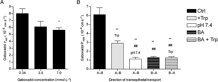
盐酸加波沙朵/Gaboxadol(0.34、3.5和7.0 μM)以剂量依赖性方式降低Caco-2单层细胞的通透性,平均Papp值为8.1 × 10⁻⁶ cm·s-1、6.1 × 10 -1 cm·s-1(0.34、3.5 和 7 μM 加波沙朵),5.6 × 10-6 cm·s-1(0.34、3.5 和 7 μM 加波沙朵)[3]。
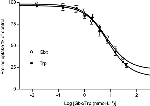
Gaboxadol在体外通过hPAT1转运的特性 [3] 在Gaboxadol/加博沙多浓度增加的情况下,通过测量hPAT1底物脯氨酸进入cco -2细胞单层的顶端摄取,研究了加博沙多和hPAT1之间的相互作用(图1)。加博沙多降低cco -2细胞单层的顶端脯氨酸摄取,估计抑制剂亲和力(Ki值)为6.6 mmol·L−1。同样,已知的PAT1抑制剂色氨酸也降低了脯氨酸的顶端吸收,Ki值为7.7 mmol·L−1。研究了三种浓度(0.34、3.5和7.0 mmol·L−1)下加博沙多在caco2细胞单层上的经上皮转运(A-B)通量。对于0.34、3.5和7 mmol·L−1的加博沙多,加博沙多转运的平均Papp值分别为8.1 × 10⁻⁶ cm·s−1、6.1 × 10⁻⁶ cm·s−1和5.6 × 10⁻⁶ cm·s−1(图2)。因此,随着加博沙多浓度的增加,加博沙多通过Caco-2细胞单层的通透性降低(P < 0.05)。在35 mmol·L−1色氨酸存在的情况下,研究了加博沙多在caco2细胞单层中使用3.5 mmol·L−1加博沙多的顶端浓度,有或没有pH梯度,双向运输(图2)。加博沙多在A-B方向的转运量约为B-A方向的5倍(P < 0.005)。色氨酸的存在使加博沙多的通透性降低了53% (Papp为2.9 × 10⁻⁶ cm·s−1,P < 0.005)。在没有质子梯度的情况下,加博沙多的渗透率降低了82%,为1.1 × 10⁻⁶ cm·s−1 (P < 0.005)。在色氨酸存在、质子梯度不存在的情况下,加博沙多在B-A方向的渗透性与[3H]-甘露醇的渗透性相似(Papp为1.6±0.36 × 10⁻⁶ cm·s−1)。此外,在转运实验中,加博沙多和色氨酸的存在并没有改变美托洛尔或甘露醇在Caco-2细胞单层间转运的通透性。甘露醇和美托洛尔的渗透率分别为1.6±0.36 × 10⁻⁶ cm·s−1和6.9±0.99 × 10⁻⁶ cm·s−1。综上所述,加博沙多通过Caco-2细胞单层的上皮转运是ph依赖的,可以被色氨酸抑制,并在A-B方向极化。综上所述,这些观察结果表明,hPAT1介导了加博沙多在人肠上皮细胞腔膜上的转运,这一转运步骤在很大程度上决定了加博沙多的上皮转运。 加博沙多/Gaboxadol转运通过hPAT1体外的表征[3] 在加博沙多浓度增加的情况下,通过测量hPAT1底物脯氨酸进入cco -2细胞单层的顶端摄取,研究了加博沙多和hPAT1之间的相互作用(图1)。加博沙多降低cco -2细胞单层的顶端脯氨酸摄取,估计抑制剂亲和力(Ki值)为6.6 mmol·L−1。同样,已知的PAT1抑制剂色氨酸也降低了脯氨酸的顶端吸收,Ki值为7.7 mmol·L−1。研究了三种浓度(0.34、3.5和7.0 mmol·L−1)下加博沙多在caco2细胞单层上的经上皮转运(A-B)通量。对于0.34、3.5和7 mmol·L−1的加博沙多,加博沙多转运的平均Papp值分别为8.1 × 10⁻⁶ cm·s−1、6.1 × 10⁻⁶ cm·s−1和5.6 × 10⁻⁶ cm·s−1(图2)。因此,随着加博沙多浓度的增加,加博沙多通过Caco-2细胞单层的通透性降低(P < 0.05)。在35 mmol·L−1色氨酸存在的情况下,研究了加博沙多在caco2细胞单层中使用3.5 mmol·L−1加博沙多的顶端浓度,有或没有pH梯度,双向运输(图2)。加博沙多在A-B方向的转运量约为B-A方向的5倍(P < 0.005)。色氨酸的存在使加博沙多的通透性降低了53% (Papp为2.9 × 10⁻⁶ cm·s−1,P < 0.005)。在没有质子梯度的情况下,加博沙多的渗透率降低了82%,为1.1 × 10⁻⁶ cm·s−1 (P < 0.005)。在色氨酸存在、质子梯度不存在的情况下,加博沙多在B-A方向的渗透性与[3H]-甘露醇的渗透性相似(Papp为1.6±0.36 × 10⁻⁶ cm·s−1)。此外,在转运实验中,加博沙多和色氨酸的存在并没有改变美托洛尔或甘露醇在Caco-2细胞单层间转运的通透性。甘露醇和美托洛尔的渗透率分别为1.6±0.36 × 10⁻⁶ cm·s−1和6.9±0.99 × 10⁻⁶ cm·s−1。综上所述,加博沙多通过Caco-2细胞单层的上皮转运是ph依赖的,可以被色氨酸抑制,并在A-B方向极化。综上所述,这些观察结果表明,hPAT1介导了加博沙多在人肠上皮细胞腔膜上的转运,这一转运步骤在很大程度上决定了加博沙多的上皮转运。 Gaboxadol是Caco-2细胞单层中质子偶联氨基酸转运体hPAT1的底物[3] 加博沙多抑制cco -2细胞对hPAT1底物脯氨酸的顶端吸收,Ki值为6.6 mmol·L−1。这种亲和力与最近观察到的其他hPAT1底物如GABA (3.1 mmol·L−1)和GABA类似物muscimol (1.7 mmol·L−1)和THPO (11.3 mmol·L−1)的亲和力相当(Larsen et al., 2008)。色氨酸的亲和力与加博沙多相当,为7.7 mmol·L−1。Metzner等人(2005)先前将色氨酸描述为PAT1的抑制剂,并报道了cco -2细胞中通过hPAT1摄取脯氨酸的Ki值为4.7 mmol·L−1。考虑到两个实验室对hPAT1脯氨酸亲和力的微小差异;Metzner等人报告的Kt值为1.4 mmol·L−1,而Larsen等人报告的Km值分别为3.6 mmol·L−1,两项研究中色氨酸对hPAT1的亲和力相当(Metzner等人,2005;Larsen et al., 2008)。根据其他小组发表的结果(Thwaites et al., 1993;Metzner et al., 2005),我们发现ccao -2细胞的大部分顶端脯氨酸运输是由hPAT1介导的,没有观察到脯氨酸的其他钠依赖性或钠依赖性转运体的证据(Larsen et al., 2008)。其他氨基酸转运蛋白,如顶钠依赖性氨基酸转运蛋白B0 (B0AT1), B0,+ (ATB0,+)和系统(ASC) (ASCT2)不太可能参与加博沙多的转运。它们都存在于Caco-2细胞中,但它们的底物移位不是质子偶联的。此外,与PAT1相比,这些转运蛋白对底物的亲和力值更高,例如ASC (ASCT2),约为100 μ mol·L−1 (Uchiyama et al., 2005);B0 (B0AT1), 500-700µmol·L−1 (Broer et al., 2004)和B0,+ (ATB0,+),约150µmol·L−1 (Hatanaka et al., 2002)。 Gaboxadol在Caco-2细胞单层间的上皮运输呈尖向基底侧极化。色氨酸可以抑制加博沙多的转运,并依赖于顶端供体溶液的pH。此外,随着加博沙多浓度的增加,加博沙多在根尖-基底侧方向的Papp降低。这与加博沙多通过hPAT1转运一致,该途径约占总上皮转运的80%。在小鼠小肠和Caco-2细胞中发现了氨基酸转运系统b0,+,分别占丙氨酸和精氨酸总转运量的15-85% (Wenzel et al., 2001;Dave et al., 2004)。然而,色氨酸与系统b0 +的结合尚未明确显示(Su et al., 1992;Tate et al., 1992),此外,阳离子氨基酸、两性离子氨基酸和胱氨酸对体系b0,+具有µmol·L−1的亲和性(Palacin, 1994)。因此,如果加博沙多通过系统b0,+运输到任何显著程度,它在Caco-2细胞中应该是明显的。在体外,经上皮的加博沙多转运还具有ph依赖性,而PAT1是目前已知的肠道中唯一的质子偶联氨基酸转运蛋白。加巴喷丁(也是两性离子γ-氨基类似物)在大鼠小肠中的渗透性被证明是质子独立的(Nguyen et al., 2007),因此加巴喷丁和加博沙多存在不同的根尖转运机制。结果表明,加博沙多是Caco-2细胞单层中hPAT1的底物,hPAT1介导加博沙多通过肠道肠细胞的管腔膜运输,这似乎对由此产生的上皮运输很重要。加博沙多跨基底外膜外排的机制尚不清楚。 加博沙多/Gaboxadol的体内吸收 在狗[3] 口服给药后,加博沙多在犬体内吸收迅速,Tmax约为0.46 h,生物利用度高达85%。这些观察结果与先前关于人体口服吸收的研究一致,表明加博沙多的Tmax约为0.5 h,生物利用度约为90% (Schultz等人,1981;Lund et al., 2006)。一旦被吸收,加博沙多主要以加博沙多的形式从尿液中排出,而一小部分以葡萄糖醛酸偶联物的形式排出,在大鼠和小鼠中占2-7%,在两个人类受试者中占30-35% (Schultz et al., 1981;Lund et al., 2006;Shadle et al., 2006)。总的来说,这表明在狗体内,加博沙多被迅速和完全吸收,可能在小肠近端,吸收后代谢最小。 |
| 体内研究 (In Vivo) |
盐酸加波沙朵/Gaboxadol(腹膜内注射;0.5、1、1.5、2、3、4 或 5 mg/kg;每天三次;间隔三天)使 Fmr1 KO2 小鼠的步行距离正常化至 0.5 mg/kg WT 活动水平,此外,该化学物质对 Fmr1 KO2 小鼠的运动活性没有影响 [2]。
在这里,我们试图评估Gaboxadol(也称为OV101和THIP),一种选择性和有效的含有δ亚基的突触外GABAA受体(dSEGA)的激动剂,通过评估其在相对未表征的FXS小鼠模型(Fmr1 KO2小鼠)中的异常行为正常化能力,作为FXS的治疗剂。四个行为领域(多动、焦虑、攻击和重复行为)通过一系列行为分析进行了探讨。结果显示,与野生型(WT)相比,Fmr1 KO2小鼠过度活跃,具有异常焦虑样行为,更易怒和更具攻击性,重复行为频率增加,这些都是FXS个体的行为缺陷。加博沙多/Gaboxadol治疗使Fmr1 KO2小鼠中观察到的所有异常行为恢复到WT水平,为其治疗FXS的潜在益处提供了证据。我们发现,仅加博沙多就能增强突触外GABA受体,足以使FXS模型中的许多行为缺陷正常化,这些缺陷的终点可直接转化为FXS的临床表现。综上所述,这些数据支持未来对FXS患者加博沙多的评估,特别是关于多动症、焦虑、易怒、攻击和重复行为的症状。[2] 加博沙多使Fmr1 KO2小鼠的过度活动正常化[2] 多动是人类FXS的一个显著特征(Bailey et al., 2008;Wheeler et al., 2014;Hagerman et al., 2017),并已在先前表征的荷兰-比利时Fmr1 KO小鼠中可靠地复制(Olmos-Serrano et al., 2010;Kazdoba et al., 2014)。为了测试Fmr1 KO2小鼠是否表现出运动亢进,以及加博沙多是否能使这种异常行为正常化,我们给Fmr1 KO2小鼠注射了载药或加博沙多(0.5-5 mg/kg, i.p),并在OFT测试前30分钟给WT窝仔注射载药。记录30 min在OFT内行走的总距离(cm)。结果显示,Fmr1 KO2小鼠的行走距离较WT窝中对照组显著增加(图1,F(8,81) = 21.27, p < 0.0001),与其他FXS模型的结果一致。加博沙多(0.5 mg/kg)使Fmr1 KO2小鼠的行走距离正常化到WT活性水平(图1)。高剂量的加博沙多(1 - 5 mg/kg, ig)对Fmr1 KO2小鼠的运动活性没有影响(图1)。这些结果不能归因于加博沙多的镇静作用,因为在WT C57Bl/6或BALB/c小鼠中,加博沙多高达2.0 mg/kg的剂量在60分钟的OFT中对运动活动没有影响(数据未显示),这与先前的研究一致,表明加博沙多对WT小鼠(Olmos-Serrano等人,2011)或大鼠(Silverman等人,2016)的运动没有影响。 Fmr1 KO2小鼠焦虑样行为经加博沙多归一化 [2] 为了评估加博沙多对Fmr1 KO2小鼠焦虑样行为的影响,采用了三种不同的行为测试:OFT、LDT和SAT的中心移动距离。中心移动距离的增加被解释为焦虑的减少,并利用了小鼠在进入新环境时保持在周边的固有偏好。Fmr1 KO2小鼠注射加博沙多(0.5-5 mg/kg, i.p), WT窝仔在放置于OFT中30分钟前注射载药。与WT对照组相比,Fmr1 KO2小鼠在中心行走的总距离显著增加(图2A, F(8,81) = 21.32, p < 0.0001)。加博沙多治疗(0.5 mg/kg, i.p.p)使Fmr1 KO2对中心距离的影响正常化,达到与WT对照组相当的水平(图2A)。在本实验中,高剂量的加博沙多(1-5 mg/kg)对Fmr1 KO2小鼠没有影响(图2A)。 加博沙多使Fmr1 KO2小鼠的易怒和攻击行为正常化 [2] 与其他形式的综合征自闭症一样,很大一部分FXS患者表现出易怒、社交焦虑和攻击性。这些异常行为可以在啮齿类动物中建模,通过表征测试小鼠和新笼伴侣之间的si。为了验证Fmr1 KO2突变体易怒和攻击性增加的假设,我们量化了摇尾、咬人行为、攀爬行为和攻击延迟的实例。小鼠入笼前30 min分别注射载药或加博沙多(0.5 ~ 5mg /kg, i.p)。 尾巴嘎嘎作响,或尾巴的快速振动,反映了攻击性和战斗倾向。与WT对照组相比,Fmr1 KO2小鼠的摇尾频率显著增加(图3A, F(8,81) = 16.03, p < 0.0001)。加博沙多(0.5、1.5和5.0 mg/kg)使Fmr1 KO2小鼠的效果正常化,达到与WT对照组相当的水平(图3A)。 Gaboxadol使Fmr1 KO2小鼠的重复行为正常化[2] 坚持和重复行为在FXS患者中很常见,并且具有高度破坏性(Arron et al., 2011;Leekam et al., 2011;Hall et al., 2016)。为了验证这些特征可能在Fmr1 KO2动物中观察到的假设,我们量化了WT和Fmr1 KO2突变小鼠的打圈、自我梳理和刻板印象。小鼠分别注射载药或加博沙多(0.5 ~ 5mg /kg, i.p)后,在实验室内测量逆时针转数(CCW)。与WT对照组相比,Fmr1 KO2小鼠在5分钟测试期间CCW转数显著增加(图4A, F(8,81) = 25.46, p < 0.0001)。向Fmr1 KO2小鼠注射加博沙多(0.5、1.0 mg/kg)后,CCW转数恢复到WT水平(图4A)。基因型对顺时针旋转无影响(p = 0.386,数据未显示)。 加博沙多的体内吸收 在狗[3] 口服给药后,加博沙多在犬体内吸收迅速,Tmax约为0.46 h,生物利用度高达85%。这些观察结果与先前关于人体口服吸收的研究一致,表明加博沙多的Tmax约为0.5 h,生物利用度约为90% (Schultz等人,1981;Lund et al., 2006)。一旦被吸收,加博沙多主要以加博沙多的形式从尿液中排出,而一小部分以葡萄糖醛酸偶联物的形式排出,在大鼠和小鼠中占2-7%,在两个人类受试者中占30-35% (Schultz et al., 1981;Lund et al., 2006;Shadle et al., 2006)。总的来说,这表明在狗体内,加博沙多被迅速和完全吸收,可能在小肠近端,吸收后代谢最小。 联合给药色氨酸后,Gaboxadol/加博沙多的体内吸收[3] 同时给药hPAT1抑制剂色氨酸对加博沙多的吸收谱有剂量依赖性,导致Cmax降低和Tmax增加。加博沙多吸收率的降低可由胃排空率的改变引起。在人类中,胃排空的速度随着一餐所摄入的卡路里数量的增加而降低(Calbet和MacLean, 1997;Sunesen et al., 2005),在狗的实验中,膳食成分也被证明可以延长胃排空(Mizuta et al., 1990)。为了排除观察到的对加博沙多吸收的影响是胃排空改变的结果,研究了常被用作胃排空标志的扑热息痛的累积吸收曲线(Calbet and MacLean, 1997;Sunesen et al., 2005),在色氨酸存在的情况下进行了研究。高剂量色氨酸对扑热息痛的胃排空无显著影响。然而,色氨酸对加博沙多的ka有显著影响。由于共给色氨酸改变了加博沙多Tmax, Cmax和ka,而Fa, ke和AUC不变,色氨酸的作用可能是色氨酸和加博沙多在吸收部位相互作用的结果,而不是由于胃排空的改变。其他研究表明,加博沙多与血浆蛋白结合程度较低,不被细胞色素p -450代谢(Lund et al., 2006)。因此,根据体外实验结果表明,hPAT1介导了Caco-2单层中大部分加博沙多的腔内转运,似乎体内观察也可以解释为pat1介导的狗对加博沙多的吸收,而这种吸收被色氨酸的共同给药所减少。从色氨酸对加博沙多Cmax的影响或对肠道吸收速率常数ka的影响来估计色氨酸抑制加博沙多肠道运输的体内亲和值。IC50值分别为10.1和12.6 mmol·L−1。如前所述,在Caco-2细胞中,hPAT1对色氨酸的体外亲和力(通过hPAT1抑制脯氨酸运输)为7.7 mmol·L−1。考虑到色氨酸的抑制作用是针对两种不同的化合物(脯氨酸和加博沙多)测量的,并且体内转运不仅包括腔内转运,还包括体循环中的外观,其中遇到了加博沙多跨几个膜的转移,IC50值彼此惊人地接近。hPAT1底物的特点是在毫摩尔范围内具有亲和力,并且在整个肠道中表达的转运蛋白具有高容量(Chen et al., 2003)。加博沙多和色氨酸的摩尔给药比高达1:41,由于它们对hPAT1的亲和力相当,Cmax和ka的降低可能是由于加博沙多和色氨酸在吸收部位,即小肠肠细胞管腔膜上的PAT1蛋白处的竞争性相互作用。因此,最大血浆浓度随着Tmax的延长而出现,但由于容量过大和肠道中PAT1的表达,随着吸收沿着肠道的长度进行,吸收分数保持不变。因此,加博沙多的峰值血浆浓度可以通过修改吸收过程来降低,如这里所示,或者通过更经典的缓释制剂方法,如前面所建议的(Kjaer和Nielsen, 1983)。 |
| 细胞实验 |
细胞培养和体外实验方案如前所述(Larsen等人,2008年)。将第20 ~ 29代Caco-2细胞接种到Transwell™插入物(1.12 cm2, 0.4µm孔径)上,于接种后第25 ~ 28天进行实验。在汉克斯平衡盐溶液(HBSS)缓冲液中,测定了Gaboxadol/加博沙多在Caco-2细胞单分子层从根尖向基底侧方向(A-B)和基底侧向根尖方向(B-A)的顶端摄取和上皮转运。在所有实验中,基底外侧的缓冲液pH为7.4。除非另有说明,在加入Gaboxadol/加博沙多盐酸或35 mmol·L−1色氨酸后,将应用于顶室的缓冲液调至pH 6.0。研究了0.34、3.5或7.0 mmol·L−1Gaboxadol的转运情况。这些浓度的选择是基于人单次睡前口服15mg加博沙多,提供约0.34 mmol·L−1的通径浓度,以及所获得的加博沙多对hPAT1的亲和力。在根尖室中加入含有12.5 nmol·L−1(0.5µCi) L-(3H)脯氨酸和0-30 mmol·L−1加博沙多或0-35 mmol·L−1色氨酸的新鲜根尖HBSS培养基,进行根尖摄取实验。5 min后终止根尖摄取实验,对样品进行闪烁计数分析。[3]
|
| 动物实验 |
动物/疾病模型: Fmr1 KO2 小鼠(Fmr1 启动子和第一个外显子缺失,导致小鼠 mRNA 和蛋白质缺失)[2]
剂量: 0.5、1、1.5、2、3、4 或 5 mg/kg 给药途径:腹腔注射 (ip) 实验结果: Fmr1 KO2 小鼠的活动过度症状得到改善。 同一笼内的小鼠注射相同剂量的Gaboxadol或载体,突变体和对照组小鼠分开饲养。所有小鼠均以 5 只/笼的方式饲养于塑料笼(35 × 30 × 12 cm)中,并在实验前适应动物房环境至少一周。室温(21 ± 2°C)、相对湿度(55 ± 5%)、12 小时光照/黑暗循环(上午 7 点至晚上 7 点开灯)和空气交换(每小时 16 次)均由自动控制。所有小鼠均可自由摄取食物和水。所有测试均在光照期进行,由一位对基因型和药物处理不知情的实验人员完成。[2] Gaboxadol 处理和实验时间线:Fmr1 KO2 小鼠在每次测试日行为测试前 30 分钟注射载体(0.9% 无菌生理盐水)或 Gaboxadol(0.5、1、1.5、2、3、4 或 5 mg/kg,腹腔注射),每次测试间隔三天,以避免药物的累积效应。所有实验中均包含在同一时间点注射载体的野生型小鼠。对小鼠(每组 n = 10)进行行为学筛选,测试顺序如下,每次测试间隔 2-3 天:旷场测试(OFT;第 1 天)、连续通道测试(第 4 天)、明暗箱测试(第 7 天)、社交测试和攻击性测试(第 10 天)以及自我梳理和刻板行为测试(第 12 天)。[2] 犬对Gaboxadol的吸收[3] 所有动物护理和实验研究均已获得丹麦司法部指定的动物福利委员会的批准,并符合欧盟指令 86/609/EEC、丹麦动物实验法规以及美国国立卫生研究院《实验动物护理和使用指南》。选取六只成年雄性比格犬(体重15.9–21.7 kg),采用罗马象限设计随机分组,分别接受六种不同配方的加博沙多盐酸盐,试验持续六周。试验开始前,所有犬只禁食20–24小时,给药10小时后恢复喂食。加博沙多的给药途径有两种:静脉注射(1.0 mL·kg−1)或灌胃(5.0 mL·kg−1),后者通过软管直接灌胃给药。所有犬只均接受2.5 mg·kg−1的加博沙多。除加博沙多外,口服制剂中还分别含有0、2.5、10、50或150 mg·kg−1的色氨酸,以确保两种化合物同时发挥作用。所有溶液的pH值均调整至5.2,并使用Vapro蒸汽压渗透压计(型号552O,Wescor公司,美国犹他州洛根市)检测渗透压。静脉输液用葡萄糖调节至等渗。通过单次静脉穿刺从头静脉采集2 mL血样,并收集到含有200 IU肝素作为抗凝剂的Eppendorf管中。分别在给予加博沙多之前以及给药后5、15、30、60、90分钟、2、3、4、6、8和10小时采集血样。立即将血样在4–8°C下以2200 g离心15分钟,收集血浆,并储存于−80°C直至进一步分析。动物在两次治疗之间有6天的洗脱期。 犬胃排空的研究[3] 采用与之前描述的以对乙酰氨基酚为标记物的方案类似的方案,评估色氨酸对犬胃排空率的影响。选取6只犬(体重16.1–21.5 kg),随机分配接受三种不同配方的对乙酰氨基酚,进行交叉研究。犬分别接受50 mg·kg−1的对乙酰氨基酚,以静脉注射(1 mL·kg−1)或口服溶液(5 mL·kg−1)的形式给药,该溶液含有2.5 mg·kg−1的Gaboxadol和0或150 mg·kg−1的色氨酸。犬只禁食、给药、采血和洗脱均按先前所述进行。 分析方法[3] 血浆和缓冲液中Gaboxadol的定量:采用液液萃取法从血浆和缓冲液样品中提取Gaboxadol。将100 µL HBSS或血浆样品与25 µL内标物(d4-gaboxadol)和25 µL纯水混合。加入400 µL冷乙腈进行蛋白质沉淀。10000 g离心15分钟后,取425 µL上清液转移至玻璃管中,并在45℃下用氮气吹干。将样品重新溶解于80 µL甲醇/乙腈(30:70)混合液中,涡旋混合10分钟,并在3300×g下离心3分钟。随后采用亲水相互作用色谱-串联质谱(MS/MS)法对加博沙多进行定量分析,该方法参考了Kall等人(2007)的方法并进行了改进。液相色谱(LC)系统由安捷伦1100系列泵和脱气机组成。色谱柱为Phenomenex公司(美国加利福尼亚州托兰斯市)的Asahipak氨基柱(NH2P-50,150 × 2 mm),流动相为20.0 mmol·L⁻¹乙酸铵(pH 4)-乙腈(30:70),流速为0.2 mL·min⁻¹。进样量为20 μL,色谱柱保持在室温下。总运行时间为10 min,前5 min的洗脱液弃去。加博沙多在色谱柱上的洗脱时间约为8 min。所用的MS/MS系统由一台配备Turbo Ion Spray和Turbo V离子源的Sciex API 4000 MS/MS检测器(Applied Biosystems,美国加利福尼亚州福斯特城)组成。信号在0.5至2500 ng·mL⁻¹范围内呈线性关系,该方法的定量限为0.5 ng·mL⁻¹。所用软件为Analyst™(Applied Biosystems,版本4.0)。 |
| 药代性质 (ADME/PK) |
犬口服加博沙多吸收的药代动力学分析[3]
在比格犬中,口服或静脉注射2.5 mg·kg−1加博沙多后,监测了10小时内的血浆浓度曲线(图3)。犬口服加博沙多后的生物利用度Fa较高(超过80%)(表1)。口服联合使用2.5–150 mg·kg−1色氨酸并未显著改变加博沙多的AUC,且各制剂的平均相对生物利用度在75%(10 mg·kg−1色氨酸)至86.1%(2.5 mg·kg−1色氨酸)之间。此外,联合使用色氨酸也未改变加博沙多的消除速率常数(ke)和清除率(CL)。然而,同时给予150 mg·kg⁻¹色氨酸后,加博沙多尔血浆最大浓度(Cmax)从2502 ng·mL⁻¹降至1419 ng·mL⁻¹,即降低了57%。此外,达到最大血浆浓度所需时间(Tmax)从0.46 h延长至1.5 h(P < 0.01)。随后,将五个剂量组的Cmax值拟合到剂量反应曲线(图4),结果表明加博沙多尔的吸收与色氨酸浓度之间存在直接相互作用。色氨酸对加博沙多尔Cmax的体内IC50值估计为12.6 mg·kg⁻¹,相当于12.3 mmol·L⁻¹的色氨酸浓度(未校正胃肠液稀释的影响)。 加博沙多和对乙酰氨基酚的吸收速率常数[3] 如图5A所示的反卷积曲线所示,随着色氨酸剂量的增加,加博沙多的平均累积吸收分数逐渐发生变化。与单独服用加博沙多相比,在150 mg·kg−1色氨酸存在的情况下,加博沙多在0.5–1.25小时时间点的吸收显著降低。口服对乙酰氨基酚60分钟后吸收率为91.5 ± 3.3%(图5B),表明胃排空主要发生在给药后的第一个小时内。同时服用150.0 mg·kg−1色氨酸并未显著改变胃排空速率,因为在所测试的时间点,无论是否存在色氨酸,对乙酰氨基酚的吸收分数均无显著差异。血浆中对乙酰氨基酚的药代动力学参数Tmax、AUC和CL与对乙酰氨基酚和色氨酸联合给药后获得的参数无显著差异(结果未显示)。基于图5A所示的曲线,计算了加博沙多尔的吸收速率常数ka,并将其作为色氨酸剂量对数的函数绘制在图6A中。与色氨酸联合给药可降低加博沙多尔的ka值,其体内IC50值为10.3 mg·kg−1,相当于浓度为10.1 mmol·L−1的色氨酸口服溶液。图6B显示,150 mg·kg−1的PAT1抑制剂色氨酸显著降低了加博沙多尔的吸收速率常数(P < 0.01),而对乙酰氨基酚的吸收速率常数无显著影响。 |
| 参考文献 |
|
| 其他信息 |
GABA(C)受体在神经系统功能的诸多方面发挥着重要作用,包括记忆、近视、疼痛和睡眠,目前正受到广泛研究。已有证据表明,视网膜、海马、脊髓、上丘、垂体和肠道等多种组织中存在功能性GABA(C)受体。本文综述了多种可用于区分GABA(C)受体与其他主要抑制性神经递质GABA受体的神经化学物质。文中介绍了一些选择性激动剂(包括(+)-cAMP和5-甲基-IAA)、竞争性拮抗剂(如TPMPA、(±)-顺式-3-ACPBPA和aza-THIP)、正向调节剂(如别孕烯醇酮)和负向调节剂(如表孕烯醇酮和洛瑞克唑)。本文还介绍了可能有助于区分同源ρ1和ρ2 GABA(C)受体的神经化学物质(2-甲基-TACA和环噻嗪)。由于GABA(C)受体与GABA(A)和GABA(B)受体相比分布范围较小、丰度较低且结构相对简单,因此它们是极具吸引力的药物靶点。[1]
Gaboxadol在0.5 mg/kg剂量下可使Fmr1 KO2小鼠所有测试的行为缺陷恢复正常。虽然更高剂量也能使易激惹和攻击性行为恢复正常,但在其他评估的行为领域未观察到这种效果。此处观察到的疗效窗口略窄的一个解释可能是,先前的研究表明,张力性抑制不足或过度都会导致信息处理受损,而Gaboxadol正是增强这种生理过程。根据该模型,高剂量药物的行为获益会被药物引入的与脆性X综合征无关的缺陷所抵消(Duguid等人,2012)。我们的研究结果为加博沙多在逆转自闭症谱系障碍相关行为、攻击性和社交能力方面的潜在益处提供了强有力的证据。综上所述,这些结果支持以下假设:加博沙多增强突触外GABAA受体可能对脆性X综合征患者有益。总之,这些数据支持未来对加博沙多在脆性X综合征患者中的应用进行评估,尤其是在多动、焦虑、自闭症谱系障碍相关刻板行为、社交能力、易怒、攻击性和认知等症状方面。[2] 背景和目的:加博沙多一直被开发用于治疗慢性疼痛和失眠。加博沙多的临床应用表明,不良反应似乎与血清峰值浓度相关。本研究旨在体外和体内探讨加博沙多尔的肠道吸收机制。 实验方法:体外转运研究采用Caco-2细胞单层模型。体内药代动力学研究采用比格犬模型。加博沙多尔剂量为2.5 mg·kg(-1),分别以静脉注射(1.0 mL·kg(-1))或口服溶液(5.0 mL·kg(-1))的方式给药。 主要结果:加博沙多尔可能是人质子偶联氨基酸转运蛋白hPAT1的底物,并且能够抑制Caco-2细胞单层模型中hPAT1介导的L-[(3)H]脯氨酸的摄取,其抑制常数K(i)为6.6 mmol·L(-1)。加博沙多的跨上皮转运呈顶端至基底外侧方向的极化,且依赖于加博沙多的浓度和顶端缓冲溶液的pH值。在比格犬中,加博沙多的吸收几乎完全(绝对生物利用度F(a)为85.3%),达峰时间(T(max))为0.46小时。口服联合使用2.5-150 mg·kg⁻¹的PAT1抑制剂L-色氨酸,可显著降低加博沙多的吸收速率常数k(a)和C(max),并延长其T(max),而曲线下面积和清除率保持不变。结论和意义:加博沙多通过小肠肠细胞腔膜的吸收可能由PAT1介导。这项研究有助于降低加博沙多尔的吸收率,从而降低血浆峰浓度。[3] 总之,本研究首次表明,加博沙多尔在Caco-2细胞单层上的高渗透性很可能是由于PAT1介导的跨腔膜转运,从而导致高跨上皮转运。体外加博沙多尔转运动力学和在犬体内观察到的药代动力学结果支持以下结论:PAT1介导加博沙多尔在体外和体内均能跨黏膜转运。此外,本研究表明,可以利用转运蛋白的活性来调节或控制药物的肠道吸收。该制剂设计为降低加博沙多尔的血浆峰浓度提供了一种简便的方法,同时保持了较高的生物利用度。这可能有助于减少与高血浆峰浓度相关的副作用。[3] |
| 分子式 |
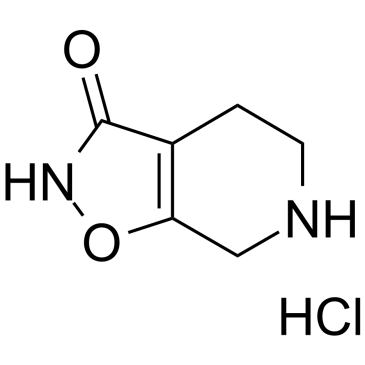
C6H9CLN2O2
|
|---|---|
| 分子量 |
176.6009
|
| 精确质量 |
176.035
|
| 元素分析 |
C, 40.81; H, 5.14; Cl, 20.07; N, 15.86; O, 18.12
|
| CAS号 |
85118-33-8
|
| 相关CAS号 |
THIP;64603-91-4
|
| PubChem CID |
5702253
|
| 外观&性状 |
White to off-white solid powder
|
| 沸点 |
295.7ºC at 760 mmHg
|
| 熔点 |
236 °C
|
| 闪点 |
132.6ºC
|
| LogP |
0.744
|
| tPSA |
58.03
|
| 氢键供体(HBD)数目 |
3
|
| 氢键受体(HBA)数目 |
3
|
| 可旋转键数目(RBC) |
0
|
| 重原子数目 |
11
|
| 分子复杂度/Complexity |
210
|
| 定义原子立体中心数目 |
0
|
| SMILES |
O=C1NOC2=C1CCNC2.[H]Cl
|
| InChi Key |
ZDZDSZQYRBZPNN-UHFFFAOYSA-N
|
| InChi Code |
InChI=1S/C6H8N2O2.ClH/c9-6-4-1-2-7-3-5(4)10-8-6/h7H,1-3H2,(H,8,9)1H
|
| 化学名 |
4,5,6,7-Tetrahydroisoxazolo(5,4-c)pyridin-3(2H)-one monohydrochloride
|
| 别名 |
OV-101; OV101; Lu-02-030; MK-0928; GABOXADOL HYDROCHLORIDE; 85118-33-8; 478RVH3TVD; EINECS 285-687-7; 4,5,6,7-Tetrahydroisoxazolo[5,4-c]pyridin-3-ol hydrochloride; DTXSID90234251; GABOXADOL HYDROCHLORIDE [MI]; ISOXAZOLO(5,4-C)PYRIDIN-3(2H)-ONE, 4,5,6,7-TETRAHYDRO-, HYDROCHLORIDE (1:1); MK 0928; Lu-02030; OV 101; Lu02-030; MK0928; Lu02030; THIP
|
| HS Tariff Code |
2934.99.9001
|
| 存储方式 |
Powder -20°C 3 years 4°C 2 years In solvent -80°C 6 months -20°C 1 month 注意: 请将本产品存放在密封且受保护的环境中(例如氮气保护),避免吸湿/受潮和光照。 |
| 运输条件 |
Room temperature (This product is stable at ambient temperature for a few days during ordinary shipping and time spent in Customs)
|
| 溶解度 (体外实验) |
H2O : ≥ 100 mg/mL (~566.25 mM)
DMSO : ~75 mg/mL (~424.69 mM) |
|---|---|
| 溶解度 (体内实验) |
配方 1 中的溶解度: ≥ 1.47 mg/mL (8.32 mM) (饱和度未知) in 10% DMSO + 40% PEG300 + 5% Tween80 + 45% Saline (这些助溶剂从左到右依次添加,逐一添加), 澄清溶液。
例如,若需制备1 mL的工作液,可将100 μL 14.7 mg/mL澄清DMSO储备液加入400 μL PEG300中,混匀;然后向上述溶液中加入50 μL Tween-80,混匀;加入450 μL生理盐水定容至1 mL。 *生理盐水的制备:将 0.9 g 氯化钠溶解在 100 mL ddH₂O中,得到澄清溶液。 配方 2 中的溶解度: ≥ 1.47 mg/mL (8.32 mM) (饱和度未知) in 10% DMSO + 90% (20% SBE-β-CD in Saline) (这些助溶剂从左到右依次添加,逐一添加), 澄清溶液。 例如,若需制备1 mL的工作液,可将 100 μL 14.7mg/mL澄清的DMSO储备液加入到900μL 20%SBE-β-CD生理盐水中,混匀。 *20% SBE-β-CD 生理盐水溶液的制备(4°C,1 周):将 2 g SBE-β-CD 溶解于 10 mL 生理盐水中,得到澄清溶液。 View More
配方 3 中的溶解度: ≥ 1.47 mg/mL (8.32 mM) (饱和度未知) in 10% DMSO + 90% Corn Oil (这些助溶剂从左到右依次添加,逐一添加), 澄清溶液。 配方 4 中的溶解度: 100 mg/mL (566.25 mM) in PBS (这些助溶剂从左到右依次添加,逐一添加), 澄清溶液; 超声助溶. 1、请先配制澄清的储备液(如:用DMSO配置50 或 100 mg/mL母液(储备液)); 2、取适量母液,按从左到右的顺序依次添加助溶剂,澄清后再加入下一助溶剂。以 下列配方为例说明 (注意此配方只用于说明,并不一定代表此产品 的实际溶解配方): 10% DMSO → 40% PEG300 → 5% Tween-80 → 45% ddH2O (或 saline); 假设最终工作液的体积为 1 mL, 浓度为5 mg/mL: 取 100 μL 50 mg/mL 的澄清 DMSO 储备液加到 400 μL PEG300 中,混合均匀/澄清;向上述体系中加入50 μL Tween-80,混合均匀/澄清;然后继续加入450 μL ddH2O (或 saline)定容至 1 mL; 3、溶剂前显示的百分比是指该溶剂在最终溶液/工作液中的体积所占比例; 4、 如产品在配制过程中出现沉淀/析出,可通过加热(≤50℃)或超声的方式助溶; 5、为保证最佳实验结果,工作液请现配现用! 6、如不确定怎么将母液配置成体内动物实验的工作液,请查看说明书或联系我们; 7、 以上所有助溶剂都可在 Invivochem.cn网站购买。 |
| 制备储备液 | 1 mg | 5 mg | 10 mg | |
| 1 mM | 5.6625 mL | 28.3126 mL | 56.6251 mL | |
| 5 mM | 1.1325 mL | 5.6625 mL | 11.3250 mL | |
| 10 mM | 0.5663 mL | 2.8313 mL | 5.6625 mL |
1、根据实验需要选择合适的溶剂配制储备液 (母液):对于大多数产品,InvivoChem推荐用DMSO配置母液 (比如:5、10、20mM或者10、20、50 mg/mL浓度),个别水溶性高的产品可直接溶于水。产品在DMSO 、水或其他溶剂中的具体溶解度详见上”溶解度 (体外)”部分;
2、如果您找不到您想要的溶解度信息,或者很难将产品溶解在溶液中,请联系我们;
3、建议使用下列计算器进行相关计算(摩尔浓度计算器、稀释计算器、分子量计算器、重组计算器等);
4、母液配好之后,将其分装到常规用量,并储存在-20°C或-80°C,尽量减少反复冻融循环。
计算结果:
工作液浓度: mg/mL;
DMSO母液配制方法: mg 药物溶于 μL DMSO溶液(母液浓度 mg/mL)。如该浓度超过该批次药物DMSO溶解度,请首先与我们联系。
体内配方配制方法:取 μL DMSO母液,加入 μL PEG300,混匀澄清后加入μL Tween 80,混匀澄清后加入 μL ddH2O,混匀澄清。
(1) 请确保溶液澄清之后,再加入下一种溶剂 (助溶剂) 。可利用涡旋、超声或水浴加热等方法助溶;
(2) 一定要按顺序加入溶剂 (助溶剂) 。
| NCT Number | Recruitment | interventions | Conditions | Sponsor/Collaborators | Start Date | Phases |
| NCT00209963 | Completed | Drug: Gaboxadol | Primary Insomnia | H. Lundbeck A/S | 2003-06 | Phase 3 |
| NCT06334419 | Recruiting | Drug: Gaboxadol Drug: Placebo |
Fragile X Syndrome | Craig Erickson | 2024-01-29 | Phase 2 |
| NCT00209846 | Completed | Drug: Gaboxadol | Primary Insomnia | H. Lundbeck A/S | 2004-06 | Phase 3 |
| NCT00209924 | Completed | Drug: Gaboxadol | Primary Insomnia | H. Lundbeck A/S | 2004-04 | Phase 3 |
| NCT02996305 | Completed | Drug: OV101 Regimen 1 Drug: OV101 regimen 2 Other: Placebo |
Angelman Syndrome | Ovid Therapeutics Inc. | 2016-01 | Phase 2 |
|
|
|
 N-甲基-beta-咔啉-3-羧酰胺
 加巴喷丁盐酸盐
 光甘草定
光甘草定
 左环丝氨酸
左环丝氨酸
InvivoChem的所有产品仅用于作科学研究,不面向患者销售
Copyright 2020 InvivoChem LLC | All Rights Reserved 粤ICP备20063088号-1


 COA
COA

 463611831
