Gimestat,chlorodihydroxypyridine; CDHP;Gimeracil
| 规格 | 价格 | 库存 | 数量 |
|---|---|---|---|
| 10 mM * 1 mL in DMSO |
|
||
| 25mg |
|
||
| 50mg |
|
||
| 100mg |
|
||
| 250mg |
|
||
| 500mg |
|
||
| 1g |
|
||
| 2g |
|
||
| Other Sizes |
|

| 靶点 |
Dihydropyrimidine dehydrogenase (DPD): Gimeracil is a selective inhibitor of DPD, the key enzyme for pyrimidine catabolism. In purified human DPD enzyme assays, the IC50 of Gimeracil for DPD activity was 0.12 μM (measured by inhibiting the conversion of [14C]-dihydrouracil to [14C]-β-ureidopropionate) [2]
- Homologous recombination (HR) pathway: Gimeracil inhibits the early step of HR, specifically suppressing Rad51-mediated DNA strand exchange. In HR reporter gene assays (DR-GFP U2OS cells), the EC50 for inhibiting HR efficiency was 3.8 μM; it had no effect on non-homologous end joining (NHEJ) at concentrations up to 10 μM [1][2] |
|---|---|
| 体外研究 (In Vitro) |
吉美嘧啶降低了新阳性克隆的频率。此外,与 G0/G1 期细胞相比,它还提高了 S 期细胞的敏感性 [1]。 Gimeracil 抑制 HR 介导的 DNA 修复途径,这可能会提高放射治疗的有效性 [1]。虽然用 gigeracil 预处理增加了 Nbs1、Mre11、Rad50 和 FancD2 病灶的数量,但它显着减少了辐射诱导的 Rad51 和 RPA 病灶的发展 [2]。
癌细胞(A549、HCT116、MCF-7)的辐射增敏作用: - 克隆形成存活:吉美拉西(Gimeracil)(1-10 μM)联合X射线照射(2-8 Gy)呈剂量依赖性降低癌细胞的克隆形成存活率。对于A549细胞,4 Gy照射下的存活分数从单独照射组的0.45降至4 μM 吉美拉西(Gimeracil)+4 Gy组的0.18,增敏比(SER)为1.6 [1] - HR效率抑制:在DR-GFP U2OS细胞中,吉美拉西(Gimeracil)(2-8 μM)使HR介导的GFP表达降低40-75%。5 μM时,HR效率为对照组的0.3倍,而NHEJ活性(通过EJ5-GFP报告基因测得)保持不变(>对照组的90%) [1][2] - Rad51焦点形成抑制:免疫荧光染色显示,吉美拉西(Gimeracil)(3-6 μM)使HCT116细胞中电离辐射(IR)诱导的Rad51焦点(HR标志物)减少55-80%。5 μM时,每个细胞的Rad51焦点数从单独照射组的28个降至8个 [2] - DPD活性抑制与嘧啶代谢影响: - DPD酶活抑制:吉美拉西(Gimeracil)(0.05-2 μM)呈剂量依赖性抑制A549细胞裂解液中的DPD活性。0.2 μM时,DPD活性降低65%,导致细胞内尿嘧啶浓度升高2.5倍(HPLC测得) [2] - 对正常细胞无影响:吉美拉西(Gimeracil)(浓度高达10 μM)对正常人肺成纤维细胞(MRC-5)或正常结肠上皮细胞(NCM460)无显著细胞毒性,克隆形成存活率>85%(vs对照组) [1] |
| 体内研究 (In Vivo) |
Gimeracil(口服,2.5–25 mg/kg)可以防止 X 射线造成的肿瘤 DNA 损伤快速愈合 [3]。
人癌裸鼠异种移植模型(A549肺癌、HCT116结直肠癌): - 肿瘤生长抑制(A549模型):小鼠(每组6只)分为4组:溶剂组(0.5% CMC-Na,口服)、吉美拉西(Gimeracil)单药组(20 mg/kg,口服,每日1次)、X射线单独照射组(6 Gy,第7天单次照射)、吉美拉西(Gimeracil)+照射联合组。21天后,联合组肿瘤体积抑制率达78%(vs溶剂组:1200 mm³ vs 264 mm³),肿瘤重量减少72%(1.8 g vs 0.5 g) [3] - 生存获益(HCT116模型):联合组小鼠中位生存期为35天,显著长于单独照射组(22天,P<0.01)和溶剂组(18天,P<0.001)。联合组无小鼠出现转移灶,而单独照射组40%的小鼠出现肝转移 [3] - 肿瘤组织中的机制验证:A549肿瘤组织免疫组化显示,联合组Rad51表达降低(为溶剂组的0.3倍),γ-H2AX焦点增加(为溶剂组的3.2倍),表明HR抑制导致DNA损伤持续存在 [3] |
| 酶活实验 |
DPD活性测定(纯化人DPD):
1. 试剂制备:配制含10 mM MgCl₂、2 mM NADPH和0.1% BSA的50 mM Tris-HCl缓冲液(pH 7.5),准备纯化人DPD(0.5 μg/孔)与[14C]-二氢尿嘧啶(5 μCi/μmol,底物) [2] 2. 反应体系构建:96孔板中加入80 μL缓冲液、10 μL 吉美拉西(Gimeracil)(0.01-5 μM)或溶剂、5 μL DPD,37℃孵育10 min;加入5 μL [14C]-二氢尿嘧啶启动反应,孵育60 min [2] 3. 产物检测:加入100 μL 0.5 M高氯酸终止反应,200 μL乙酸乙酯萃取产物([14C]-β-脲基丙酸),3000×g离心10 min;收集有机相,氮气吹干,50 μL甲醇重悬,液体闪烁计数器测定放射性 [2] 4. 数据计算:DPD活性以每毫克蛋白每小时生成的产物纳摩尔数表示,抑制率=[(对照组活性-处理组活性)/对照组活性]×100%,通过剂量-反应曲线拟合计算IC50 [2] - HR报告基因实验(DR-GFP U2OS细胞): 1. 细胞培养:DR-GFP U2OS细胞(稳定表达HR报告载体)在含10% FBS的DMEM培养基中培养 [1] 2. 转染与处理:细胞(2×10⁵/孔,6孔板)用转染试剂转染I-SceI表达质粒(诱导DNA双链断裂);6 h后加入吉美拉西(Gimeracil)(0.5-10 μM)或溶剂,孵育48 h [1] 3. 流式分析:收集细胞,PBS洗涤2次,流式细胞仪(激发光488 nm,发射光525 nm)检测GFP阳性细胞比例。HR效率=(处理组GFP⁺细胞百分比/对照组GFP⁺细胞百分比)×100% [1] |
| 细胞实验 |
细胞活力测定[1]
细胞类型:DLD-1、HeLa 和 LC-11 细胞系。 测试浓度:1 mM。 孵化时间:48小时。 实验结果:抑制辐射引起的 DNA 双链断裂的修复。未照射细胞中 24 小时时未增加 c-H2AX 焦点残留。 克隆形成存活实验: 1. 细胞接种:癌细胞(A549、HCT116)按200-1000细胞/孔(根据照射剂量调整)接种于6孔板,37℃、5% CO₂孵育过夜 [1] 2. 药物与照射处理:加入吉美拉西(Gimeracil)(1-10 μM)或溶剂,孵育2 h后,用X射线发生器照射细胞(2-8 Gy);照射后更换为不含吉美拉西(Gimeracil)的新鲜培养基 [1] 3. 克隆形成与计数:孵育10-14天(直至形成>50个细胞的克隆);4%多聚甲醛固定细胞15 min,0.1%结晶紫染色30 min;流水冲洗,晾干后用克隆计数器计数。存活分数=(处理组克隆数/对照组克隆数)/接种效率 [1] - Rad51焦点免疫荧光实验: 1. 细胞制备:HCT116细胞接种于24孔板的盖玻片上(5×10⁴细胞/孔),孵育过夜 [2] 2. 药物处理与照射:加入吉美拉西(Gimeracil)(3-6 μM)或溶剂,孵育4 h后,用4 Gy X射线照射细胞;继续孵育8 h以促进Rad51焦点形成 [2] 3. 染色:4%多聚甲醛固定细胞15 min,0.2% Triton X-100透化10 min;5% BSA/PBS封闭1 h,4℃下抗Rad51一抗(1:200)孵育过夜;PBS洗涤后,Alexa Fluor 488标记二抗(1:500)室温孵育1 h;DAPI(1 μg/mL)染核5 min [2] 4. 成像与计数:共聚焦显微镜观察细胞,图像分析软件计数每组100个细胞的Rad51焦点数 [2] |
| 动物实验 |
动物/疾病模型:裸鼠(移植Lu-99、LC-11、KB/C3和PAN-4肿瘤)[3]。
剂量:2.5-25 mg/kg。 给药途径:口服。 实验结果:显示出抗肿瘤活性。 人源癌症异种移植模型(裸鼠): 1.动物选择:6-8周龄雌性BALB/c裸鼠(A549模型n=24,HCT116模型n=24)饲养于SPF级条件下,12小时光照/12小时黑暗循环,自由摄食饮水。实验前适应环境1周[3] 2. 肿瘤诱导:制备A549(5×10⁶个细胞/只小鼠)或HCT116(4×10⁶个细胞/只小鼠)的单细胞悬液,溶于PBS和Matrigel(1:1,v/v)混合液中。将0.2 mL悬液皮下注射到每只小鼠的右侧背部[3] 3. 分组和治疗:当肿瘤体积达到约100 mm³时,将小鼠随机分为4组(每组n=6): - 载体组:每日一次灌胃0.5%羧甲基纤维素钠(CMC-Na),持续21天。 - 吉美嘧啶单药组:每日一次灌胃给予吉美嘧啶 20 mg/kg(溶于 0.5% CMC-Na 溶液,超声处理溶解)。 - 放射组:分组后第 7 天进行单次 X 射线照射(6 Gy);不进行药物治疗。 - 联合组:吉美嘧啶(20 mg/kg,每日一次,灌胃)+ 单次 X 射线照射(第 7 天,6 Gy)[3] 4. 样本采集和监测: - 肿瘤监测:每 3 天测量一次肿瘤体积(长 × 宽² / 2)和小鼠体重。 - 生存监测(HCT116 模型):每日记录小鼠生存情况,直至对照组小鼠全部死亡。 - 组织分析(A549模型):第21天,处死小鼠,解剖肿瘤,称重,取一部分用4%多聚甲醛固定,用于免疫组织化学染色(Rad51、γ-H2AX染色)。收集肝脏和肾脏组织进行病理分析[3] |
| 药代性质 (ADME/PK) |
吸收、分布和排泄
尽管Teysuno的剂量(50 mg替加氟)比单独服用替加氟(800 mg)低16倍,但Teysuno给药后5-氟尿嘧啶(5-FU)的平均血浆峰浓度(Cmax)和浓度-时间曲线下面积(AUC)值仍比单独服用替加氟高约3倍,这归因于吉美嘧啶对二氢嘧啶脱氢酶(DPD)的抑制作用。血浆尿嘧啶浓度在给药后4小时达到峰值,并在给药后约48小时内恢复至基线水平,表明吉美嘧啶对DPD的抑制作用是可逆的。单次服用 50 mg Teysuno(以替加氟含量计)后,Teysuno 成分替加氟、吉美嘧啶和奥替拉西的中位达峰时间 (Tmax) 分别为 0.5 小时、1.0 小时和 2.0 小时。 单次服用 Teysuno 后,约 3.8% 至 4.2% 的替加氟、65% 至 72% 的吉美嘧啶和 3.5% 至 3.9% 的奥替拉西以原形经尿液排出。 虽然目前尚无 Teysuno 在人体内的静脉给药数据,但可根据表观分布容积和尿排泄数据粗略估算替加氟、吉美嘧啶和奥替拉西的分布容积分别为 16 L/m²、17 L/m² 和 23 L/m²。 生物半衰期 单次服用 Teysuno 后,替加氟的 T1/2 值范围为 6.7 至 11.3 小时,吉美嘧啶的 T1/2 值范围为 3.1 至 4.1 小时,奥替拉西的 T1/2 值范围为 1.8 至 9.5 小时。 |
| 毒性/毒理 (Toxicokinetics/TK) |
蛋白结合
奥替拉西、吉美拉西、5-氟尿嘧啶和替加氟的蛋白结合率分别为 8.4%、32.2%、18.4% 和 52.3%。 体外毒性: - 正常细胞:吉美拉西(浓度高达 10 μM)对正常人肺成纤维细胞 (MRC-5) 或正常结肠上皮细胞 (NCM460) 无显著细胞毒性,克隆形成存活率 >85%(与对照组相比),且无明显细胞凋亡(Annexin V⁺ 率 <5%)[1] - 癌细胞:吉美拉西(≤10 μM)对 A549 和 HCT116 细胞有较弱的细胞毒性,细胞活力 >75%(MTT 法,孵育 72 小时)[1][3] - 在体内毒性: - 一般毒性:吉美嘧啶(20 mg/kg,口服,21 天)未引起明显的体重减轻(治疗组:21.5 ± 1.2 g vs. 赋形剂组:22.1 ± 1.0 g)或异常行为(例如,嗜睡、厌食)[3] - 器官毒性:联合治疗组肝脏和肾脏组织的病理分析显示未见明显损伤(无肝细胞坏死、肾小管损伤或炎症)。血清ALT(28 ± 5 U/L vs. 对照组 30 ± 4 U/L)、AST(45 ± 6 U/L vs. 对照组 48 ± 5 U/L)、BUN(14 ± 2 mg/dL vs. 对照组 15 ± 2 mg/dL)和肌酐(0.7 ± 0.1 mg/dL vs. 对照组 0.8 ± 0.1 mg/dL)水平均在正常范围内[3]。 |
| 参考文献 |
|
| 其他信息 |
吉美嘧啶是一种有机分子实体。
吉美嘧啶是抗肿瘤治疗的辅助药物,用于提高化疗方案中主要活性成分的浓度和疗效。吉美嘧啶于2011年3月获得欧洲药品管理局 (EMA) 批准,目前以复方制剂“Teysuno”的形式上市,该制剂包含吉美嘧啶和[DB03209]以及[DB09256]。Teysuno的主要活性成分是[DB09256],它是[DB00544](5-氟尿嘧啶,5-FU)的前体药物。5-FU是一种细胞毒性抗代谢药物,作用于快速分裂的癌细胞。5-FU通过模拟一类称为“嘧啶”的化合物(嘧啶是RNA和DNA的重要组成部分),能够插入DNA和RNA链中,从而阻止癌细胞持续生长所需的复制过程。吉美拉西在泰苏诺公司的主要作用是阻止[DB00544](5-氟尿嘧啶)的分解,从而维持足够高的浓度以持续对抗癌细胞。它的作用机制是可逆且选择性地阻断二氢嘧啶脱氢酶(DPD),该酶参与5-氟尿嘧啶的降解。这使得在较低剂量的替加氟下即可达到更高的5-氟尿嘧啶浓度,从而降低毒副作用。 吉美拉西是一种具有抗肿瘤活性的吡啶衍生物。吉美拉西通过竞争性且可逆地抑制二氢嘧啶脱氢酶,减少氟尿嘧啶的降解,从而增强氟尿嘧啶的抗肿瘤活性。 药物适应症 吉美拉西用作抗肿瘤治疗的辅助药物。在产品 Teysuno 中使用时,吉美嘧啶与顺铂联合用于治疗成人晚期胃癌。 作用机制 吉美嘧啶在 Teysuno 中的主要作用是防止 [DB00544] (5-FU) 的分解,这有助于维持足够高的浓度,从而对癌细胞产生持续作用。它通过可逆性阻断二氢嘧啶脱氢酶 (DPD) 发挥作用,DPD 参与 5-氟尿嘧啶 (5-FU) 的降解。 吉美嘧啶 是口服化疗药物 S-1(替加氟、吉美嘧啶 和奥替拉西钾的组合)的关键成分,它通过抑制 DPD 来阻止替加氟(5-氟尿嘧啶,5-FU 的前药)的快速分解代谢,从而增强 5-FU 的疗效 [2][3] - 除了抑制 DPD 外,吉美嘧啶 还具有抑制同源重组 (HR) 的独特机制,它通过阻断 DNA 双链断裂修复,使癌细胞对辐射和 DNA 损伤剂更加敏感。这种双重机制拓展了其在联合放疗中的应用[1][2] - 在临床前模型中,吉美嘧啶联合放疗在非小细胞肺癌和结直肠癌中显示出显著的协同抗肿瘤作用,且与单药治疗相比,毒性并未增加。这支持了其作为临床肿瘤学中放射增敏剂的潜力[3] - 吉美嘧啶特异性靶向同源重组(HR),而不影响非同源末端连接(NHEJ,正常细胞中的主要DNA修复途径),这或许可以解释其对正常组织的低毒性和高治疗指数[1] |
| 分子式 |
C5H4CLNO2
|
|
|---|---|---|
| 分子量 |
145.54
|
|
| 精确质量 |
144.993
|
|
| CAS号 |
103766-25-2
|
|
| 相关CAS号 |
|
|
| PubChem CID |
54679224
|
|
| 外观&性状 |
White to light brown solid powder
|
|
| 密度 |
1.6±0.1 g/cm3
|
|
| 沸点 |
524.2±45.0 °C at 760 mmHg
|
|
| 熔点 |
274 °C
|
|
| 闪点 |
270.8±28.7 °C
|
|
| 蒸汽压 |
0.0±1.4 mmHg at 25°C
|
|
| 折射率 |
1.641
|
|
| LogP |
-1.5
|
|
| tPSA |
53.09
|
|
| 氢键供体(HBD)数目 |
2
|
|
| 氢键受体(HBA)数目 |
2
|
|
| 可旋转键数目(RBC) |
0
|
|
| 重原子数目 |
9
|
|
| 分子复杂度/Complexity |
207
|
|
| 定义原子立体中心数目 |
0
|
|
| InChi Key |
ZPLQIPFOCGIIHV-UHFFFAOYSA-N
|
|
| InChi Code |
InChI=1S/C5H4ClNO2/c6-3-2-7-5(9)1-4(3)8/h1-2H,(H2,7,8,9)
|
|
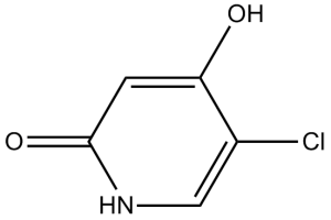
| 化学名 |
5-chloro-2-hydroxypyridin-4(1H)-one
|
|
| 别名 |
|
|
| HS Tariff Code |
2934.99.9001
|
|
| 存储方式 |
Powder -20°C 3 years 4°C 2 years In solvent -80°C 6 months -20°C 1 month |
|
| 运输条件 |
Room temperature (This product is stable at ambient temperature for a few days during ordinary shipping and time spent in Customs)
|
| 溶解度 (体外实验) |
|
|||
|---|---|---|---|---|
| 溶解度 (体内实验) |
配方 1 中的溶解度: ≥ 2.5 mg/mL (17.18 mM) (饱和度未知) in 10% DMSO + 40% PEG300 + 5% Tween80 + 45% Saline (这些助溶剂从左到右依次添加,逐一添加), 澄清溶液。
例如,若需制备1 mL的工作液,可将100 μL 25.0 mg/mL澄清DMSO储备液加入到400 μL PEG300中,混匀;然后向上述溶液中加入50 μL Tween-80,混匀;加入450 μL生理盐水定容至1 mL。 *生理盐水的制备:将 0.9 g 氯化钠溶解在 100 mL ddH₂O中,得到澄清溶液。 配方 2 中的溶解度: ≥ 2.5 mg/mL (17.18 mM) (饱和度未知) in 10% DMSO + 90% (20% SBE-β-CD in Saline) (这些助溶剂从左到右依次添加,逐一添加), 澄清溶液。 例如,若需制备1 mL的工作液,可将 100 μL 25.0 mg/mL澄清DMSO储备液加入900 μL 20% SBE-β-CD生理盐水溶液中,混匀。 *20% SBE-β-CD 生理盐水溶液的制备(4°C,1 周):将 2 g SBE-β-CD 溶解于 10 mL 生理盐水中,得到澄清溶液。 View More
配方 3 中的溶解度: ≥ 2.5 mg/mL (17.18 mM) (饱和度未知) in 10% DMSO + 90% Corn Oil (这些助溶剂从左到右依次添加,逐一添加), 澄清溶液。 1、请先配制澄清的储备液(如:用DMSO配置50 或 100 mg/mL母液(储备液)); 2、取适量母液,按从左到右的顺序依次添加助溶剂,澄清后再加入下一助溶剂。以 下列配方为例说明 (注意此配方只用于说明,并不一定代表此产品 的实际溶解配方): 10% DMSO → 40% PEG300 → 5% Tween-80 → 45% ddH2O (或 saline); 假设最终工作液的体积为 1 mL, 浓度为5 mg/mL: 取 100 μL 50 mg/mL 的澄清 DMSO 储备液加到 400 μL PEG300 中,混合均匀/澄清;向上述体系中加入50 μL Tween-80,混合均匀/澄清;然后继续加入450 μL ddH2O (或 saline)定容至 1 mL; 3、溶剂前显示的百分比是指该溶剂在最终溶液/工作液中的体积所占比例; 4、 如产品在配制过程中出现沉淀/析出,可通过加热(≤50℃)或超声的方式助溶; 5、为保证最佳实验结果,工作液请现配现用! 6、如不确定怎么将母液配置成体内动物实验的工作液,请查看说明书或联系我们; 7、 以上所有助溶剂都可在 Invivochem.cn网站购买。 |
| 制备储备液 | 1 mg | 5 mg | 10 mg | |
| 1 mM | 6.8710 mL | 34.3548 mL | 68.7096 mL | |
| 5 mM | 1.3742 mL | 6.8710 mL | 13.7419 mL | |
| 10 mM | 0.6871 mL | 3.4355 mL | 6.8710 mL |
1、根据实验需要选择合适的溶剂配制储备液 (母液):对于大多数产品,InvivoChem推荐用DMSO配置母液 (比如:5、10、20mM或者10、20、50 mg/mL浓度),个别水溶性高的产品可直接溶于水。产品在DMSO 、水或其他溶剂中的具体溶解度详见上”溶解度 (体外)”部分;
2、如果您找不到您想要的溶解度信息,或者很难将产品溶解在溶液中,请联系我们;
3、建议使用下列计算器进行相关计算(摩尔浓度计算器、稀释计算器、分子量计算器、重组计算器等);
4、母液配好之后,将其分装到常规用量,并储存在-20°C或-80°C,尽量减少反复冻融循环。
计算结果:
工作液浓度: mg/mL;
DMSO母液配制方法: mg 药物溶于 μL DMSO溶液(母液浓度 mg/mL)。如该浓度超过该批次药物DMSO溶解度,请首先与我们联系。
体内配方配制方法:取 μL DMSO母液,加入 μL PEG300,混匀澄清后加入μL Tween 80,混匀澄清后加入 μL ddH2O,混匀澄清。
(1) 请确保溶液澄清之后,再加入下一种溶剂 (助溶剂) 。可利用涡旋、超声或水浴加热等方法助溶;
(2) 一定要按顺序加入溶剂 (助溶剂) 。
| NCT Number | Recruitment | interventions | Conditions | Sponsor/Collaborators | Start Date | Phases |
| NCT06255379 | Not yet recruiting | Drug: Fuquinitinib+Tegafur Gimeracil Oteracil |
Metastasis Colorectal Cancer Colon Cancer |
Guangzhou University of Traditional Chinese Medicine |
March 22, 2024 | Phase 2 |
| NCT04310774 | Not yet recruiting | Drug: Tegafur, Gimeracil and Oteracil Potassium Capsules (one drug) |
Cervical Cancer Chemotherapy |
Peking Union Medical College Hospital | April 15, 2020 | Phase 1 Phase 2 |
| NCT03267121 | Completed | Drug: Tegafur Gimeracil Oteracil Potassium Capsules |
Head and Neck Squamous Cell Carcinoma |
Shanghai Ninth People's Hospital Affiliated to Shanghai Jiao Tong University |
October 1, 2017 | Phase 2 |
| NCT03192735 | Active, not recruiting | Drug: ApatinibMesylateTablets | Apatinib Combined SOX |
Chang-Ming Huang, Prof. | September 1, 2017 | Phase 2 |
InvivoChem的所有产品仅用于作科学研究,不面向患者销售
Copyright 2020 InvivoChem LLC | All Rights Reserved 粤ICP备20063088号-1


 463611831
