| 规格 | 价格 | 库存 | 数量 |
|---|---|---|---|
| 5mg |
|
||
| 10mg |
|
||
| 25mg |
|
||
| 50mg |
|
||
| 100mg |
|
||
| 250mg |
|
||
| 500mg |
|
||
| Other Sizes |
|
| 靶点 |
SGLT; hSGLT2 (IC50 = 7.4 nM); hSGLT1 (IC50 = 1876 nM); rSGLT2 (IC50 = 6.73 nM); rSGLT1 (IC50 = 1166 nM); mSGLT2 (IC50 = 5.64 nM); mSGLT1 (IC50 = 1380 nM)
|
|---|---|
| 体外研究 (In Vitro) |
Ipragliflozin (1-50 μM) 以剂量依赖性方式显着抑制人乳腺癌细胞系 MCF-7 的增殖。 Ipragliflozin 最初会降低乳腺癌细胞增殖,但当使用 siRNA 敲低 SGLT2 表达时,这种效应被完全消除。这表明 Ipragliflozin 抑制 SGLT2 以最大限度地减少乳腺癌细胞增殖。 BrdU 测定表明,高剂量(50 和 100 μM)的 Ipragliflozin 会显着降低 MCF-7 细胞中的 DNA 产量 [1]。
SGLT2和SGLT1抑制试验[1] Ipragliflozin浓度依赖性抑制小鼠、大鼠和人在纳摩尔浓度下的SGLT2活性(表1)。ipragliflozin对人SGLT2的抑制作用约为phlorizin的5倍,但对人SGLT1的抑制作用仅为phlorizin的1 / 9。根皮素对两种SGLT的抑制作用都很小。ipragliflozin、phlorizin和phloretin的选择性比(human SGLT1/SGLT2的IC50)分别为254、6和3。此外,ipragliflozin对小鼠和大鼠SGLT2具有高选择性。达格列净也能有效地、选择性地抑制小鼠和人SGLT2活性。 特异性测定[1] 为了确认特异性,使用放射性配体结合和酶分析检测了Ipragliflozin对几种代表性受体、通道和转运体的影响。伊普列净不与多种受体、离子通道和转运体相互作用,如肾上腺素能(α1、α2和β)、毒碱碱(M1、M2和非选择性)、血管紧张素(AT1和AT2)、钙通道(l型和n型)、钾通道(KATP和SKCa)、钠通道(2号位点)、胆囊收缩素(CCKA和CCKB)、多巴胺(D1、D2和转运体)、内皮素(ETA和ETB)、γ -氨基丁酸(GABAA和GABAB)、谷氨酸(AMPA、kainate和NMDA)、血清素(5-HT1、5HT2B、组胺(H1、H2和H3)和神经激肽(NK1、NK2和NK3), IC50值为0 ~ 3000 nM。 Ipragliflozin对小鼠肠道葡萄糖苷酶的稳定性[1] 采用小鼠小肠粘膜匀浆法测定了异格列净和苯连菌素对葡萄糖苷酶的体外生物稳定性。虽然ipragliflozin完全没有被降解(图2a),但在小鼠粘膜匀浆中,根连素被迅速降解为其糖基,根连素(Fig. 2b)。 癌症是目前2型糖尿病患者死亡的主要原因之一。我们之前报道了胰高血糖素样肽-1受体激动剂exendin-4对前列腺癌和乳腺癌的有益作用。在本研究中,我们通过乳腺癌模型检测了钠-葡萄糖共转运蛋白2 (SGLT2)抑制剂Ipragliflozin的抗癌作用。在人乳腺癌MCF-7细胞中,采用RT-PCR和免疫组织化学检测SGLT2的表达。Ipragliflozin在1 ~ 50 μM浓度下显著且剂量依赖性地抑制MCF-7细胞的生长。BrdU实验还显示,ipragliflozin以剂量依赖的方式减弱MCF-7细胞的增殖。因为ipragliflozin对乳腺癌细胞的作用通过敲除SGLT2而被完全取消,所以Ipragliflozin可以通过抑制SGLT2来起作用。接下来,我们使用膜片钳技术测量了膜电位和全细胞电流。用ipragliflozin或无糖培养基处理MCF-7细胞时,观察到膜超极化。此外,无葡萄糖培养基和siRNA敲除SGLT2抑制了葡萄糖诱导的MCF-7细胞的全细胞电流,表明ipragliflozin抑制了钠和葡萄糖通过SGLT2的共转运。ipragliflozin显著增加了JC-1绿色荧光,提示线粒体膜电位发生了变化。这些发现表明,SGLT2抑制剂ipragliflozin通过膜超极化和线粒体膜不稳定来减弱乳腺癌细胞的增殖。[2] |
| 体内研究 (In Vivo) |
Ipragliflozin 具有降血糖特性。血糖水平的升高受到伊格列净 (0.1-1 mg/kg) 剂量依赖性的抑制。这种效应在 STZ 诱导的 1 型糖尿病大鼠中在 0.3 和 1 mg/kg 剂量下显着,在 KK-Ay 2 型糖尿病小鼠中在所有测试剂量下均显着[1]。在链脲佐菌素诱导的 1 型糖尿病大鼠中,重复剂量的伊格列净(0.3 和 1 mg/kg)显示出抗糖尿病作用 [1]。
对新型SGLT2选择性抑制剂伊格列净(Ipragliflozin, ASP1941; (1S)-1,5-脱水-1-C-{3-[(1-苯并噻吩-2-基)甲基]-4-氟苯基}-D: -葡萄糖醇与L: -脯氨酸(1:1)的化合物)的药理学特征进行了研究。在体外,评估了伊格列净抑制SGLT2和SGLT1的效力以及稳定性。在体内,在正常小鼠、链脲佐菌素诱导的1型糖尿病大鼠和KK-Ay 2型糖尿病小鼠中研究了伊格列净的药代动力学和药理学特征。伊格列净在纳摩尔范围内有效且选择性地抑制人、大鼠和小鼠的SGLT2,并表现出对肠道葡萄糖苷酶的稳定性。口服给药后,伊格列净显示出良好的药代动力学特性,并剂量依赖性地增加尿葡萄糖排泄,该效应在正常小鼠中持续超过12小时。单次给药伊格列净在两种糖尿病模型中均产生剂量依赖性和持续的抗高血糖作用。此外,在两种糖尿病模型中,每日一次伊格列净治疗4周改善了高血糖,同时伴随着尿葡萄糖排泄的增加。相比之下,在药理学剂量下,伊格列净不影响正常血糖(如格列本脲的情况),并且不影响肠道葡萄糖吸收和电解质平衡。这些结果表明,伊格列净是一种口服有效的SGLT2选择性抑制剂,它通过抑制肾脏葡萄糖重吸收来诱导尿葡萄糖排泄持续增加,随后产生抗高血糖作用且低血糖风险低。因此,伊格列净通过增加葡萄糖向尿液中的排泄,具有治疗糖尿病高血糖的治疗潜力。[1] 伊格列净对正常小鼠尿葡萄糖排泄的影响 [1] 在正常小鼠中,Ipragliflozin/伊格列净(0.01–10 mg/kg)剂量依赖性地显著增加尿葡萄糖排泄,并且在剂量≥0.3 mg/kg时,该效应在给药后12–18小时仍然明显(图4a)。在3和10 mg/kg剂量下,尿量也显著增加(图4b)。 单次给药伊格列净对糖尿病动物的影响[1] 在STZ诱导的1型糖尿病大鼠和KK-Ay 2型糖尿病小鼠中,伊格列净(0.1−1 mg/kg)剂量依赖性地降低血糖水平,并且该效应在所有测试剂量下均显著(图5a和图6a)。在给药后12小时进行口服葡萄糖耐量试验(OGTT)期间,伊格列净(0.1–1 mg/kg)剂量依赖性地抑制血糖水平的升高。在STZ诱导的1型糖尿病大鼠中,该效应在0.3和1 mg/kg剂量下显著(图5b),而在KK-Ay 2型糖尿病小鼠中,该效应在所有测试剂量下均显著(图6b)。 重复给药伊格列净对STZ诱导的1型糖尿病大鼠的影响 [1] 与正常对照组大鼠相比,在非禁食条件下,STZ诱导的1型糖尿病大鼠的HbA1c、血糖和尿葡萄糖排泄的平均水平显著更高,血浆胰岛素水平和胰腺胰岛素含量显著更低(表2)。重复给药伊格列净(0.3和1 mg/kg)4周显著降低了HbA1c和血糖水平。血浆胰岛素水平未发生显著变化,但胰腺胰岛素含量在1 mg/kg剂量下显著增加。尿葡萄糖排泄呈剂量依赖性增加,并且在1 mg/kg剂量下显著增加。在整个研究期间,伊格列净不影响体重或食物摄入量(数据未显示)。 重复给药伊格列净对KK-Ay 2型糖尿病小鼠的影响 [1] 重复给药伊格列净(0.3和1 mg/kg)4周降低了HbA1c和血糖水平,同时伴随着尿葡萄糖排泄的增加(表3)。此外,尿白蛋白排泄显著减少。在整个研究期间,伊格列净治疗不影响体重或食物摄入量(数据未显示)。 伊格列净对正常小鼠空腹血糖水平的影响 [1] 在正常小鼠中,伊格列净(0.03–100 mg/kg)剂量依赖性地抑制葡萄糖负荷后血糖水平的升高,并且该效应在剂量≥0.1 mg/kg时显著(图7a)。格列本脲(0.3–300 mg/kg)也剂量依赖性地抑制血糖水平的升高;该效应在剂量≥3 mg/kg时显著(图7c)。在过夜禁食的小鼠中,伊格列净(0.03–100 mg/kg)剂量依赖性地降低血糖水平,但该效应仅在剂量≥10 mg/kg(比OGTT中的剂量高100倍)时才显著(图7b)。格列本脲(0.3–300 mg/kg)在与OGTT相同的剂量下也剂量依赖性地降低空腹血糖水平(图7d)。在空腹条件下,伊格列净不改变血浆胰岛素水平,但显著降低了葡萄糖负荷条件下血浆胰岛素水平的升高。相比之下,格列本脲在两种条件下均显著增加血浆胰岛素水平(数据未显示)。 伊格列净对正常小鼠胃肠道碳水化合物含量的影响 [1] 在正常小鼠中进行液体膳食负荷后,胃肠道二糖(蔗糖和麦芽糖)和单糖(葡萄糖和果糖)含量显著增加(图8)。伏格列波糖(1 mg/kg)显著增加胃肠道二糖含量(图8a, b),降低单糖含量(图8c, d),并显著抑制血糖水平的升高(数据未显示)。相比之下,伊格列净(0.3–30 mg/kg)即使在最高剂量下也未显著影响胃肠道二糖含量。此外,伊格列净未显著影响胃肠道果糖含量,虽然它确实剂量依赖性地增加了葡萄糖含量,但该效应仅在30 mg/kg的最大剂量时才显著。伊格列净剂量依赖性地抑制血糖水平的升高,并且该效应在所有测试剂量下均显著(数据未显示)。 伊格列净对KK-Ay 2型糖尿病小鼠血浆和尿液参数的影响 [1] 在2型糖尿病小鼠中,袢利尿剂呋塞米(10 mg/kg)显著增加尿电解质(Na+, K+, 和 Cl−)排泄和尿量,同时伴随着尿渗透压的降低(表4)。这种由电解质排泄引起的显著利尿作用还导致血浆电解质浓度显著降低和血浆渗透压显著升高。加压素V1A/V2受体拮抗剂YM471(3 mg/kg)在不增加电解质排泄的情况下显著增加尿量,并显著降低尿渗透压。这种显著的利尿作用导致血浆电解质浓度和渗透压显著升高。相比之下,伊格列净(1 mg/kg)显著增加尿葡萄糖排泄,同时伴随着尿量的轻微增加,并显著降低血糖水平,但不显著影响血浆或尿电解质平衡。 |
| 酶活实验 |
SGLT2和SGLT1抑制试验[1]
克隆了人类、大鼠和小鼠SGLT2和SGLT1全长互补脱氧核糖核酸序列,并使用先前描述的标准技术稳定地转染到中国仓鼠卵巢(CHO)细胞中(Katsuno et al. 2007)。细胞接种于含10%胎牛血清的Ham’s F12培养基中,以3 × 10⁴个/孔的密度接种于96孔板中。细胞在镀后1天使用。测试化合物首先溶解在二甲亚砜中,然后用钠缓冲液(140 mM NaCl, 2 mM KCl, 1 mM CaCl2, 1 mM MgCl2, 10 mM n -2-羟乙基哌嗪- n ' -2-乙磺酸,5 mM Tris-HCl, pH 7.4)稀释到所需浓度。取出培养基后,细胞在100μl preincubated胆碱分析缓冲区(氯化钠在测定钠缓冲替换相同浓度的氯化胆碱)在37°C 20分钟。然后他们被孵化的测试化合物的解决方案(25μl)包含14 c-amg(2.2μCi /毫升)和nonlabeled AMG(最终浓度55μM) 37°C 2 h。200μl细胞被洗两次冰冷的清洗缓冲(胆碱分析缓冲区包含10毫米AMG),然后随着0.5%十二烷基硫酸钠(SDS)解决方案(25μl)。将细胞裂解液与75 μl MicroScint MS-40混合,用Top Count微孔板闪烁计数器测定放射性。 |
| 细胞实验 |
细胞活力测定[2]
细胞类型: MCF-7 人乳腺癌细胞系 测试浓度: 1、10、50 μM <孵育持续时间:24、48、72、96小时 实验结果:以剂量依赖性方式减少MCF-7细胞的数量。 细胞培养和细胞增殖试验[2] MCF-7和MDA-MB-231人乳腺癌细胞系购自美国类型培养库。所有细胞在添加10%胎牛血清(FBS)和1%青霉素/链霉素的Dulbecco 's Modified Eagle 's Medium (DMEM)中维持。细胞增殖实验按照前面所述进行,并进行了轻微修改。简单地说,将细胞置于12孔组织培养板中,并在0-50 μM Ipragliflozin的完整培养基中维持。0-4天后,用血细胞计进行细胞计数分析细胞增殖情况。 溴脱氧尿苷(BrdU)测定[2] 使用细胞增殖ELISA试剂盒进行BrdU掺入试验。简单地说,将MCF-7细胞以5000个/孔的速度在96孔培养皿中完全培养液中进行培养。达到60%-70%的一致性后,细胞用含有10% FBS (0-100 μM Ipragliflozin)的培养基处理24小时。在刺激的最后2小时加入BrdU溶液(10 μM)。将细胞干燥并固定,用FixDenat溶液在室温下去除细胞DNA 30分钟。在培养板中加入过氧化物酶偶联的小鼠抗brdu单克隆抗体,室温孵育90 min。加入四甲基联苯胺底物,在室温下孵育15分钟。样品的吸光度用酶标仪在450 - 620 nm处测定。平均数据表示为相对于对照(未处理)细胞增殖的比率。 SGLT2小干扰(si)RNA敲低及细胞增殖试验[2] 为了敲除SGLT2,我们使用了SGLT-2 siRNA,这是针对人类SGLT2设计的;siRNA作为阴性对照。将MCF-7细胞以2 × 10⁵个/孔的比例在六孔板中进行细胞层析,使用MISSION siRNA转染试剂转染10 nmol/L SGLT-2 siRNA或阴性对照siRNA。转染72小时后,进行细胞增殖试验。简单地说,将细胞分离并重新镀于24孔组织培养板中,在含或不含10 μM Ipragliflozin的完整培养基中。在治疗后0-4天,收集细胞并用血细胞计计数。 线粒体通透性电位[2] 根据公司的说明书,使用JC-1线粒体膜电位检测试剂盒检测线粒体膜电位(ΔΨm)。MCF-7细胞经10 μM Ipragliflozin处理或不处理后,用阳离子染料JC-1染色,在线粒体中表现出电位依赖性积累。在低膜电位下,JC-1作为单体存在并产生绿色荧光(527 nm发射)。在高膜电位和极化下,JC-1形成J聚集体并产生红色荧光(发射波长为590nm)。 |
| 动物实验 |
动物/疾病模型:单次给药[1]链脲佐菌素(STZ;50 mg/kg)诱导的1型糖尿病大鼠和KK-Ay 2型糖尿病小鼠
剂量:0.1-1 mg/kg 给药途径:在饱食状态下单次口服给药。随后在空腹状态下测量8小时的血糖水平。 实验结果:血糖水平呈剂量依赖性降低,且在所有测试剂量下均具有显著性差异。 动物/疾病模型:重复给药[1]链脲佐菌素(STZ;50 mg/kg)诱导的1型糖尿病大鼠 剂量:0.3和1 mg/kg 给药途径:每日一次(睡前)口服,持续4周。 实验结果:显著降低了HbA1c和血糖水平。1 mg/kg剂量下,胰腺胰岛素含量显著增加。尿糖排泄量呈剂量依赖性增加,在 1 mg/kg 剂量下显著增加。 对小鼠肠道葡萄糖苷酶的稳定性[1] 在乙醚麻醉下,从隔夜禁食的正常小鼠中取出小肠,用冷生理盐水冲洗,切除,并用磷酸盐缓冲液(48 mM NaCl、5.4 mM KCl、28 mM Na2HPO4、43 mM NaH2PO4、35 mM 甘露醇、10 mM 葡萄糖,pH 6.5)冲洗。用载玻片轻轻刮取黏膜组织,用磷酸盐缓冲液匀浆,用于稳定性研究。测试化合物最初以 5 mM 的浓度溶解于乙腈中,然后用磷酸盐缓冲液稀释至 100 μM。将黏膜匀浆(5 mg/ml,100 μl)在微量离心管中于 37°C 预孵育。随后,加入各化合物溶液(100 μl,终浓度 50 μM),并在 37°C 下孵育不同时间。加入冰冷的乙腈(200 μl)终止反应,然后加入 200 μl 甲基叔丁基醚,并将混合物离心(15,000 rpm,10 min)。将上清液转移至离心管中,并在真空离心浓缩仪中蒸发。将残余物溶解于流动相中,用作测定样品。使用高效液相色谱法 (HPLC) 分析测定样品中化合物的浓度,色谱柱为 4.6 × 250 mm 反相 ODS-80Ts 色谱柱,配备紫外检测器(伊格列净检测波长为 265 nm,根皮苷和根皮素检测波长为 280 nm)。柱温维持在 60°C,流动相为 20 mM 乙酸铵/乙腈 [20:80 (v/v)],流速为 1.5 ml/min。 药代动力学 [1] 对未禁食的正常小鼠口服伊格列净 (3 mg/kg) 或根皮苷 (100 mg/kg) 后,在乙醚麻醉下从腹腔静脉抽取血液。采用高效液相色谱法(HPLC)测定血浆中伊格列净或根皮苷的浓度。向血浆样品(100 μl)中加入乙腈(100 μl)和甲基叔丁基醚(100 μl),混匀,然后离心(15,000 rpm,10 min)。将上清液转移至离心管中,并在真空离心浓缩仪中蒸发浓缩。将残余物溶解于HPLC流动相(0.1%甲酸溶液/乙腈[55:45 (v/v)])中,作为分析样品。如上所述,测定了试验样品中伊格列净或根皮苷的浓度。 伊格列净对正常小鼠尿糖排泄的影响[1] 向未禁食的正常小鼠给予伊格列净(0.01–10 mg/kg),并在给药后24小时内收集小鼠在代谢笼中自然排泄的尿液。测量尿量后,使用葡萄糖CII试剂盒测定尿液中的葡萄糖浓度。 单次给予伊格列净对糖尿病动物的影响[1] 为了研究其降血糖作用,在喂食状态下,对链脲佐菌素(STZ)诱导的1型糖尿病大鼠和KK-Ay 2型糖尿病小鼠分别给予伊格列净(0.1–1 mg/kg)。随后在禁食状态下测量8小时的血糖水平,以消除实验期间喂食的影响。为了评估其持续作用,对两种类型的糖尿病动物均给予伊格列净(0.1–1 mg/kg),然后禁食12小时(过夜)。随后口服葡萄糖溶液(2 g/kg),并按上述方法测量血糖水平。 重复给予伊格列净对链脲佐菌素(STZ)诱导的1型糖尿病大鼠的影响[1] 对STZ诱导的1型糖尿病大鼠,每日一次(睡前)给予伊格列净(0.3和1 mg/kg),持续4周。每周测量体重和食物摄入量。第26天给药后,将大鼠转移至代谢笼,收集24小时的自发排尿。第28天最后一次给药后的第二天早上,在非空腹状态下采集血样,并在乙醚麻醉下分离胰腺。按上述方法测量血液和尿液中的葡萄糖浓度。将胰腺组织加入酸性乙醇溶液(75%乙醇、23.5%纯水和1.5%浓盐酸)匀浆,并在4℃下孵育1小时以提取胰岛素。随后,将培养物离心,取上清液作为测定样品。采用酶联免疫吸附试验(ELISA)试剂盒测定血浆和胰腺胰岛素浓度。采用DCA2000系统测定糖化血红蛋白(HbA1c)水平。 重复给予伊格列净对KK-Ay 2型糖尿病小鼠的影响[1] 每天一次(睡前)给KK-Ay 2型糖尿病小鼠给予伊格列净(0.3和1 mg/kg),持续4周。每周测量体重和食物摄入量。在第28天给药后的第二天早晨,在非空腹状态下采集血样。在第30天给药后,将小鼠转移至代谢笼中,并收集24小时的自发排尿。如上所述,测定血液和尿液中的葡萄糖浓度、糖化血红蛋白(HbA1c)和血浆胰岛素水平。使用小鼠白蛋白ELISA法测定尿白蛋白浓度。 伊格列净对正常小鼠空腹血糖水平的影响[1] 为了研究伊格列净(0.03–100 mg/kg)或格列本脲(0.3–300 mg/kg)对餐后高血糖的影响,对隔夜禁食的正常小鼠分别给予伊格列净(0.03–100 mg/kg)或格列本脲(0.3–300 mg/kg)。30分钟后,口服葡萄糖溶液(2 g/kg),并测定血糖水平。为了研究伊格列净(0.03–100 mg/kg)或格列本脲(0.3–300 mg/kg)对低血糖的影响,对禁食过夜的正常小鼠进行给药,并测量血糖水平。 伊格列净对正常小鼠胃肠道碳水化合物含量的影响[1] 小鼠禁食24小时后,分别给予伊格列净(0.3–30 mg/kg)或伏格列波糖(1 mg/kg)。15分钟后,以20 ml/kg的剂量口服给予液体餐(ENSURE·H:碳水化合物206 mg/ml,脂肪53 mg/ml,蛋白质53 mg/ml)。对照组小鼠给予纯净水代替液体餐。给予液体餐或水后1小时,测量血糖水平,并在乙醚麻醉下分离胃肠道(胃、小肠上段和下段、盲肠和大肠下段)。将分离的胃肠道组织用纯水(5 ml)匀浆,并离心(3000 rpm,10 min)收集上清液。葡萄糖浓度按上述方法测定。蔗糖和麦芽糖浓度采用先前描述的方法并稍作修改进行测定(Dörner 1977)。使用果糖测定试剂盒测定果糖浓度。 伊格列净对KK-Ay 2型糖尿病小鼠血浆和尿液参数的影响[1] 将伊格列净(1 mg/kg)、呋塞米(10 mg/kg)或YM471(3 mg/kg)分别给予未禁食的糖尿病小鼠,然后将其转移至代谢笼中。小鼠可自由摄取食物和水,并收集8小时的自发排尿样本。之后,从尾静脉采集血样以测定血糖水平。在乙醚麻醉下,从膀胱收集尿液,并从腹腔静脉采集血样。测量自发排尿量与膀胱内尿液总量之和。血液和尿液样本经离心(15,000 rpm,10 分钟)后,取上清液用于测定多项参数。血浆和尿液渗透压采用冰点降低渗透压计测定。血浆和尿液电解质(Na⁺、K⁺ 和 Cl⁻)浓度采用火焰光度计测定,尿电解质排泄量计算为尿电解质浓度乘以尿量。 |
| 药代性质 (ADME/PK) |
药代动力学[1]
正常小鼠口服伊格列净(3 mg/kg)后,血浆伊格列净浓度在1小时达到峰值,随后逐渐下降(图3)。给药8小时后仍可检测到明显的血浆浓度。相比之下,口服根皮苷(100 mg/kg)后,血浆药物浓度较低且迅速清除。 |
| 参考文献 | |
| 其他信息 |
伊格列净是一种糖苷类药物。
伊格列净目前正在研究用于治疗2型糖尿病和2型糖尿病。 SGLT2特异性表达于肾脏,在肾脏葡萄糖重吸收中发挥重要作用(Jabbour和Goldstein,2008)。相反,SGLT1在小肠中高表达,介导膳食葡萄糖的吸收(Pajor和Wright,1992)。为了阐明伊格列净对肠道SGLT1的体内作用,我们研究了其对胃肠道碳水化合物吸收的影响。α-葡萄糖苷酶抑制剂,例如伏格列波糖,可抑制小肠二糖酶,即蔗糖酶和麦芽糖酶(Matsuo等,1992)。这会减缓小肠对碳水化合物的吸收,从而降低餐后高血糖(Baron 1998)。在本研究中,伏格列波糖显著增加了肠道二糖含量(通过延缓二糖消化),从而抑制了血糖水平的升高。因此,α-葡萄糖苷酶抑制剂可通过此机制有效预防餐后高血糖。然而,肠道二糖含量积累引起的渗透性水潴留可导致软便或腹泻等胃肠道症状(Vichayanrat等,2002)。然而,伊格列净不影响肠道二糖或果糖含量,但在30 mg/kg的最大剂量下,它确实显著增加了肠道葡萄糖含量。已知胃肠道中SGLT的表达和葡萄糖的吸收主要依赖于SGLT1(Turk等,1991),且口服给药后胃肠道药物浓度立即达到极高水平(Masaoka等,2006)。因此,尽管伊格列净对小鼠SGLT2的选择性比SGLT1高245倍,但最高剂量伊格列净引起的胃肠道葡萄糖含量显著升高被认为是由小肠中SGLT1介导的葡萄糖吸收抑制所致。然而,胃肠道葡萄糖升高仅在剂量比显著降低餐后高血糖的剂量高100倍时才具有统计学意义。因此,可以认为治疗剂量的伊格列净不会抑制肠道SGLT1,也不会影响肠道碳水化合物的消化和吸收。因此,胃肠道症状的风险应该较低。在急性及慢性实验过程中,任何剂量下均未观察到提示肠道SGLT1显著抑制的胃肠道副作用。 较高剂量的伊格列净可轻微增加尿量,同时显著增加尿糖排泄。由于钠离子转运伴随SGLT2促进的葡萄糖转运,我们评估了伊格列净对血浆和尿液电解质平衡的影响。在本实验中,袢利尿剂呋塞米可引起显著利尿,同时增加尿电解质排泄,并降低包括钠在内的血浆电解质浓度。这种呋塞米引起的低钠血症可能是一种严重的不良反应(Sonnenblick等,1993)。血管加压素V1A/V2受体拮抗剂YM471也表现出强效的利尿作用,并升高了血浆电解质浓度。相比之下,伊格列净(1 mg/kg)显著增加了尿糖排泄,同时尿量略有增加,但并未影响血浆或尿液电解质平衡。这些结果表明,在药理剂量下,伊格列净可以增加尿糖排泄,而不会引起明显的渗透性利尿或电解质紊乱。 SGLT2基因突变会导致一种罕见疾病——家族性肾性糖尿症,该病会导致肾脏对葡萄糖的重吸收严重受损,大量葡萄糖从尿液中排出(Francis等,2004;Magen等,2005)。尽管存在严重的糖尿,但患者病情似乎良好,未出现严重不良事件或肾功能问题。家族性肾性糖尿也与低血糖无关,通常无明显的临床表现(Santer 等,2003)。本研究中,长期服用药理剂量的伊格列净并未在糖尿病模型中诱发显著不良反应。基于这些发现,伊格列净似乎是一种治疗高血糖的有效且安全的药物。总之,本研究表明,伊格列净是一种强效的选择性SGLT2抑制剂,具有良好的药代动力学特征,可通过增加尿糖排泄发挥持续的降血糖作用,且不会引起低血糖或胰岛素分泌。这些结果表明,伊格列净具有作为糖尿病患者高血糖治疗药物的潜在应用价值,值得进一步开发。目前正在进行伊格列净的临床试验,概念验证研究应能阐明伊格列净治疗糖尿病的适用性(Kashiwagi 等,2010)。[1] 在人乳腺癌细胞中表达,而正常乳腺组织中不表达。我们的研究结果表明,SGLT2 抑制剂伊格列净可抑制乳腺癌细胞增殖和 DNA 合成(图 1)。抑制乳腺癌细胞增殖的伊格列净剂量为 1–10 μM,与其在血清中的药理浓度相似,表明我们的数据与临床情况基本一致。此外,口服伊格列净在腺体组织中的分布浓度与血清浓度相似或更高(安斯泰来制药公司未发表的数据)。尽管低剂量伊格列净也能抑制细胞生长(图2A),但高剂量(50–100 μM)的伊格列净在BrdU检测中降低了DNA合成(图2C)。这些结果表明,伊格列净不仅通过抑制DNA合成,还通过其他机制(例如细胞死亡,包括凋亡)来减弱乳腺癌细胞的增殖。我们采用TUNEL法检测了细胞凋亡,但未观察到可重复的凋亡现象。需要使用其他方法进行进一步研究。我们重点关注SGLT2介导的钠转运,因为钠摄取正逐渐成为包括乳腺癌在内的癌症生物学机制之一。伊格列净通过SGLT2抑制钠摄取,并诱导MCF-7细胞膜超极化。我们还发现,伊格列净诱导了线粒体膜电位(ΔΨm)的不稳定,这可能导致宿主细胞的凋亡和坏死。 SGLT2诱导线粒体膜不稳定的机制可能涉及葡萄糖或钠转运的抑制。细胞内钠可通过钠钙交换器和钠氢交换器改变线粒体膜电位(ΔΨm)。然而,低葡萄糖水平可能不会调节ΔΨm;葡萄糖转运蛋白1也在乳腺癌细胞中表达,并且是重要的能量调节因子和治疗靶点。进一步的实验可能会揭示SGLT2抑制剂对癌细胞的其他作用,并探索SGLT2抑制剂、二甲双胍和GLP-1的联合治疗。一项荟萃分析和病例报告提示SGLT2抑制剂具有抗癌作用。总之,在本研究中,我们发现SGLT2抑制剂伊格列净通过膜超极化和线粒体膜不稳定来减弱乳腺癌细胞增殖[2]。 |
| 分子式 |
C21H21FOS
|
|
|---|---|---|
| 分子量 |
404.45
|
|
| 精确质量 |
404.109
|
|
| 元素分析 |
C, 62.36; H, 5.23; F, 4.70; O, 19.78; S, 7.93
|
|
| CAS号 |
761423-87-4
|
|
| 相关CAS号 |
Ipragliflozin (L-Proline);951382-34-6
|
|
| PubChem CID |
10453870
|
|
| 外观&性状 |
White to off-white solid powder
|
|
| 密度 |
1.5±0.1 g/cm3
|
|
| 沸点 |
628.8±55.0 °C at 760 mmHg
|
|
| 闪点 |
334.1±31.5 °C
|
|
| 蒸汽压 |
0.0±1.9 mmHg at 25°C
|
|
| 折射率 |
1.684
|
|
| LogP |
5.59
|
|
| tPSA |
118.39
|
|
| 氢键供体(HBD)数目 |
4
|
|
| 氢键受体(HBA)数目 |
7
|
|
| 可旋转键数目(RBC) |
4
|
|
| 重原子数目 |
28
|
|
| 分子复杂度/Complexity |
525
|
|
| 定义原子立体中心数目 |
5
|
|
| SMILES |
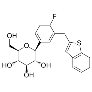
FC1=CC=C([C@H]2[C@H](O)[C@@H](O)[C@H](O)[C@@H](CO)O2)C=C1CC3=CC(C=CC=C4)=C4S3
|
|
| InChi Key |
AHFWIQIYAXSLBA-RQXATKFSSA-N
|
|
| InChi Code |
InChI=1S/C21H21FO5S/c22-15-6-5-12(21-20(26)19(25)18(24)16(10-23)27-21)7-13(15)9-14-8-11-3-1-2-4-17(11)28-14/h1-8,16,18-21,23-26H,9-10H2/t16-,18-,19+,20-,21+/m1/s1
|
|
| 化学名 |
(2S,3R,4R,5S,6R)-2-[3-(1-benzothiophen-2-ylmethyl)-4-fluorophenyl]-6-(hydroxymethyl)oxane-3,4,5-triol
|
|
| 别名 |
|
|
| HS Tariff Code |
2934.99.9001
|
|
| 存储方式 |
Powder -20°C 3 years 4°C 2 years In solvent -80°C 6 months -20°C 1 month |
|
| 运输条件 |
Room temperature (This product is stable at ambient temperature for a few days during ordinary shipping and time spent in Customs)
|
| 溶解度 (体外实验) |
|
|||
|---|---|---|---|---|
| 溶解度 (体内实验) |
配方 1 中的溶解度: ≥ 2.5 mg/mL (6.18 mM) (饱和度未知) in 10% DMSO + 40% PEG300 + 5% Tween80 + 45% Saline (这些助溶剂从左到右依次添加,逐一添加), 澄清溶液。
例如,若需制备1 mL的工作液,可将100 μL 25.0 mg/mL澄清DMSO储备液加入到400 μL PEG300中,混匀;然后向上述溶液中加入50 μL Tween-80,混匀;加入450 μL生理盐水定容至1 mL。 *生理盐水的制备:将 0.9 g 氯化钠溶解在 100 mL ddH₂O中,得到澄清溶液。 配方 2 中的溶解度: ≥ 2.5 mg/mL (6.18 mM) (饱和度未知) in 10% DMSO + 90% (20% SBE-β-CD in Saline) (这些助溶剂从左到右依次添加,逐一添加), 澄清溶液。 例如,若需制备1 mL的工作液,可将 100 μL 25.0 mg/mL澄清DMSO储备液加入900 μL 20% SBE-β-CD生理盐水溶液中,混匀。 *20% SBE-β-CD 生理盐水溶液的制备(4°C,1 周):将 2 g SBE-β-CD 溶解于 10 mL 生理盐水中,得到澄清溶液。 View More
配方 3 中的溶解度: ≥ 2.5 mg/mL (6.18 mM) (饱和度未知) in 10% DMSO + 90% Corn Oil (这些助溶剂从左到右依次添加,逐一添加), 澄清溶液。 1、请先配制澄清的储备液(如:用DMSO配置50 或 100 mg/mL母液(储备液)); 2、取适量母液,按从左到右的顺序依次添加助溶剂,澄清后再加入下一助溶剂。以 下列配方为例说明 (注意此配方只用于说明,并不一定代表此产品 的实际溶解配方): 10% DMSO → 40% PEG300 → 5% Tween-80 → 45% ddH2O (或 saline); 假设最终工作液的体积为 1 mL, 浓度为5 mg/mL: 取 100 μL 50 mg/mL 的澄清 DMSO 储备液加到 400 μL PEG300 中,混合均匀/澄清;向上述体系中加入50 μL Tween-80,混合均匀/澄清;然后继续加入450 μL ddH2O (或 saline)定容至 1 mL; 3、溶剂前显示的百分比是指该溶剂在最终溶液/工作液中的体积所占比例; 4、 如产品在配制过程中出现沉淀/析出,可通过加热(≤50℃)或超声的方式助溶; 5、为保证最佳实验结果,工作液请现配现用! 6、如不确定怎么将母液配置成体内动物实验的工作液,请查看说明书或联系我们; 7、 以上所有助溶剂都可在 Invivochem.cn网站购买。 |
| 制备储备液 | 1 mg | 5 mg | 10 mg | |
| 1 mM | 2.4725 mL | 12.3625 mL | 24.7249 mL | |
| 5 mM | 0.4945 mL | 2.4725 mL | 4.9450 mL | |
| 10 mM | 0.2472 mL | 1.2362 mL | 2.4725 mL |
1、根据实验需要选择合适的溶剂配制储备液 (母液):对于大多数产品,InvivoChem推荐用DMSO配置母液 (比如:5、10、20mM或者10、20、50 mg/mL浓度),个别水溶性高的产品可直接溶于水。产品在DMSO 、水或其他溶剂中的具体溶解度详见上”溶解度 (体外)”部分;
2、如果您找不到您想要的溶解度信息,或者很难将产品溶解在溶液中,请联系我们;
3、建议使用下列计算器进行相关计算(摩尔浓度计算器、稀释计算器、分子量计算器、重组计算器等);
4、母液配好之后,将其分装到常规用量,并储存在-20°C或-80°C,尽量减少反复冻融循环。
计算结果:
工作液浓度: mg/mL;
DMSO母液配制方法: mg 药物溶于 μL DMSO溶液(母液浓度 mg/mL)。如该浓度超过该批次药物DMSO溶解度,请首先与我们联系。
体内配方配制方法:取 μL DMSO母液,加入 μL PEG300,混匀澄清后加入μL Tween 80,混匀澄清后加入 μL ddH2O,混匀澄清。
(1) 请确保溶液澄清之后,再加入下一种溶剂 (助溶剂) 。可利用涡旋、超声或水浴加热等方法助溶;
(2) 一定要按顺序加入溶剂 (助溶剂) 。
Non-inferiority of iPragliflozin and metformin on glucose metabolism, pleiotropic effects and safety in type2 diabetes
CTID: UMIN000018979
Phase: Status: Recruiting
Date: 2015-09-11
Stability ofaipragliflozin andbphlorizin in mouse intestinal mucosal homogenates.Naunyn Schmiedebergs Arch Pharmacol.2012 Apr;385(4):423-36. |
|---|
Effects of ipragliflozin onaurinary glucose excretion andburine volume in normal mice.Naunyn Schmiedebergs Arch Pharmacol.2012 Apr;385(4):423-36. |
Effects of ipragliflozin on blood glucose levels in streptozotocin-induced type 1 diabetic rats.Naunyn Schmiedebergs Arch Pharmacol.2012 Apr;385(4):423-36. |
Effects of ipragliflozin on blood glucose levels in KK-Aytype 2 diabetic mice.Naunyn Schmiedebergs Arch Pharmacol.2012 Apr;385(4):423-36. |
|---|
Effects of ipragliflozin and glibenclamide on fasting blood glucose levels in normal mice.Naunyn Schmiedebergs Arch Pharmacol.2012 Apr;385(4):423-36. |
InvivoChem的所有产品仅用于作科学研究,不面向患者销售
Copyright 2020 InvivoChem LLC | All Rights Reserved 粤ICP备20063088号-1


 COA
COA


 463611831
