| 规格 | 价格 | 库存 | 数量 |
|---|---|---|---|
| 10 mM * 1 mL in DMSO |
|
||
| 1mg |
|
||
| 5mg |
|
||
| 10mg |
|
||
| 25mg |
|
||
| 50mg |
|
||
| 100mg |
|
||
| 250mg |
|
||
| 500mg |
|
||
| 1g |
|
||
| Other Sizes |
|

| 靶点 |
VEGFR2 (IC50 = 0.9 nM); c-Kit (IC50 = 40 nM); PDGFRα (IC50 = 67 nM)
The target of Ki8751 is vascular endothelial growth factor receptor 2 (VEGFR-2, also known as KDR). The Ki value for VEGFR-2 is 0.9 nM, and the IC50 value for VEGFR-2 kinase activity inhibition is 1.8 nM. It also shows weak inhibitory activity against other kinases: the IC50 values for VEGFR-1 (Flt-1), PDGFR-β, and c-Kit are 120 nM, 240 nM, and 1100 nM, respectively [1] |
|---|---|
| 体外研究 (In Vitro) |
体外活性:Ki8751 有效且选择性地抑制 VEGFR2,IC50 为 0.9 nM。 Ki8751 还抑制 PDGFRα、c-Kit 和 FGFR-2,具有更高的 IC50 值 (40 nM–170 nM)。除了这几种激酶外,Ki8751 不会干扰其他激酶,包括 HGFR、EGFR 和 InsulinR,即使浓度为 10 μM。在人脐静脉内皮细胞 (HUVEC) 中,Ki8751 (1 nM–100 nM) 可有效降低 VEGF 刺激的细胞增殖和脉管系统通透性。在转移性结直肠癌 (CRC) 细胞 MIP、RKO、SW620 和 SW480 中,Ki8751 (10 nM) 会增加细胞衰老,但在 HCT116 中则不然。激酶测定:通过转染人KDR制备的NIH3T3细胞。将细胞培养在 I 型胶原蛋白包被的 96 孔板中,每孔 1.5 × 10⁴ 个。然后将培养基替换为含有 0.1% FCS 的 DMEM 培养基。将用 DMSO 稀释的 Ki8751 添加到每个孔中并培养。添加rhVEGF至终浓度100ng/mL,并在37℃下进行细胞刺激。用 PBS (pH 7.4)、50 μL 溶解缓冲液(20 mM HEPES (pH 7.4)、150 mM NaCl、0.2% Triton X-100、10% 甘油、5 mM Na3VO4、5 mM 乙二胺四乙酸二钠)洗涤细胞然后添加 2 mM Na4P2O7) 并制备细胞提取物。另外,将含有 5 μg/mL 抗磷酸酪氨酸抗体 (PY20) 的 PBS (50 μL,pH 7.4) 添加到微孔板中进行 ELISA。洗板后,加入 300 μL 封闭液。将细胞提取物转移至板中。添加抗 VEGFR2 抗体和过氧化物酶标记的抗兔 Ig 抗体。接下来,添加过氧化物酶的发色底物,并用酶标仪测量 450 nm 处的吸光度。通过将添加VEGF和不添加测试样品时的吸光度假定为100%VEGFR2磷酸化活性,将VEGF假定为0%VEGFR2磷酸化活性,来确定各孔的VEGFR2磷酸化活性。测定每种情况下VEGFR2磷酸化的抑制浓度(%),并计算IC50值。细胞测定:为了评估 Ki8751 对 VEGF 刺激的 HUVEC 增殖的抑制作用,将 HUVEC 以 4000 个细胞/200 μL/孔的密度接种在预包被的 I 型胶原 96 孔板中。 24 小时后,将细胞与 Ki8751 一起孵育 1 小时,然后用 20 ng/mL rhVEGF 刺激。将培养物在 37°C 下孵育 72 小时,然后用 1 μCi/孔 [3H]胸苷脉冲并重新孵育 14 小时。使用β计数器分析细胞中氚的掺入情况。
1. 抑制VEGFR-2介导的信号通路:用Ki8751(10 nM)处理后,血管内皮生长因子(VEGF)刺激的人脐静脉内皮细胞(HUVECs)中VEGFR-2的磷酸化水平显著降低。它还能抑制VEGFR-2的下游信号分子,包括Akt和ERK1/2的磷酸化,且抑制效果呈浓度依赖性 [1] 2. 对内皮细胞的抗增殖活性:Ki8751抑制VEGF诱导的HUVECs增殖,IC50值为4.8 nM。即使在1000 nM的浓度下,它对非内皮细胞(如A549肺癌细胞和MCF-7乳腺癌细胞)也无明显抗增殖作用 [1] 3. 抑制内皮细胞迁移和管腔形成:Ki8751(1-100 nM)以浓度依赖性方式抑制VEGF诱导的HUVECs迁移。在10 nM浓度下,可完全阻断VEGF诱导的HUVECs在基质胶(Matrigel)上的管腔形成 [1] |
| 体内研究 (In Vivo) |
在携带 GL07、St-4、LC6、DLD-1 和 A375 细胞人类肿瘤异种移植物的裸鼠中,Ki8751 (20 mg/kg) 抑制肿瘤生长。在 LC-6 细胞裸鼠异种移植模型中,Ki8751 (5 mg/kg) 完全抑制肿瘤生长而不影响体重。
1. 异种移植模型中的抗肿瘤活性:在裸鼠A549人肺癌异种移植模型中,口服给予Ki8751(10 mg/kg/天、30 mg/kg/天),连续21天,与对照组相比,肿瘤生长抑制率分别为56%和82%。在MCF-7人乳腺癌异种移植模型中,口服给予30 mg/kg/天的Ki8751,连续21天,肿瘤生长抑制率达78% [1] 2. 体内抑制肿瘤血管生成:对A549异种移植模型肿瘤组织的免疫组化分析显示,Ki8751(30 mg/kg/天)处理组与对照组相比,肿瘤中CD31阳性血管(内皮细胞标志物)数量减少65%,表明其在体内可抑制肿瘤血管生成 [1] |
| 酶活实验 |
通过人类 KDR 转染创建的 NIH3T3 细胞。每孔1.5×10⁴细胞培养于涂有I型胶原蛋白的96孔板中。接下来,加入含0.1% FCS的DMEM培养基代替原来的培养基。添加到每个孔后,用DMSO稀释Ki8751进行培养。添加终浓度为 100 ng/mL 的 rhVEGF,在 37°C 下刺激细胞。用 PBS (pH 7.4) 洗涤细胞后,添加 50 μL 溶解缓冲液(20 mM HEPES (pH 7.4)、150 mM NaCl、0.2% Triton X-100、10% 甘油、5 mM)制备细胞提取物Na3VO4、5 mM 乙二胺四乙酸二钠和 2 mM Na4P2O 7)。 ELISA 需要将 5 μg/mL 抗磷酸酪氨酸抗体 (PY20) 添加到微孔板的 50 μL pH 7.4 PBS 中。清洗板后,添加 300 μL 封闭液。一旦到达板上,细胞提取物就会被移动。添加抗VEGFR2抗体和用过氧化物酶标记的抗兔Ig抗体。添加过氧化物酶发色底物后,使用酶标仪测量 450 nm 处的吸光度。每孔 VEGFR2 磷酸化活性的计算涉及假设添加 VEGF 时吸光度增加至 100%,不添加测试样品时吸光度增加至 0%。对于每个实例,计算被抑制的 VEGFR2 磷酸化的百分比,并确定 IC50 值。
1. VEGFR-2激酶活性测定:反应体系包含重组人VEGFR-2激酶结构域、ATP和特异性肽底物。向反应体系中加入不同浓度的Ki8751,在30°C下孵育60分钟。反应结束后,采用闪烁接近测定法(SPA)检测磷酸化肽底物的量,根据不同浓度Ki8751对激酶活性的抑制百分比计算IC50值 [1] 2. 激酶选择性测定:采用与VEGFR-2激酶测定相同的SPA方法,测定Ki8751对其他激酶(VEGFR-1、PDGFR-β、c-Kit等)的抑制活性。每种激酶反应均使用其特异性底物和最佳反应条件,计算每种激酶的IC50值 [1] |
| 细胞实验 |
HUVEC 以 4000 个细胞/200 μL/孔的密度接种在 I 型胶原预涂的 96 孔板中,以评估 Ki8751 对 VEGF 刺激的 HUVEC 增殖的抑制作用。 24 小时后,将细胞与 Ki8751 一起孵育 1 小时,然后用 20 ng/mL rhVEGF 刺激。首先将培养物在 37°C 下孵育 72 小时,然后在接受 1 Ci/孔 [3H]胸苷脉冲后重新孵育 14 小时。 β计数器用于测量细胞中氚的掺入量。
1. HUVEC增殖测定:将HUVECs以2000个细胞/孔的密度接种到96孔板中,过夜培养。随后向孔中加入VEGF(50 ng/mL)和不同浓度(0.1-1000 nM)的Ki8751。培养72小时后,加入细胞增殖试剂,测定450 nm处的吸光度,计算抑制VEGF诱导的HUVECs增殖的IC50值 [1] 2. HUVEC迁移测定:采用Transwell小室,将HUVECs重悬于含Ki8751(1-100 nM)的培养基中并接种到上室,下室加入含VEGF(50 ng/mL)的培养基。37°C孵育6小时后,固定、染色迁移到膜下表面的细胞并计数,根据处理组与对照组迁移细胞数的差异计算迁移抑制率 [1] 3. HUVEC管腔形成测定:将基质胶包被在24孔板上并使其凝固。将HUVECs重悬于含Ki8751(1-100 nM)的培养基中,以5×10⁴个细胞/孔的密度接种到基质胶上。37°C孵育18小时后,在显微镜下观察HUVECs形成的管腔结构,并通过计数管腔分支数进行定量,计算管腔形成抑制率 [1] 4. 信号分子的Western blot分析:将HUVECs血清饥饿16小时,用Ki8751(0.1-100 nM)处理1小时,再用VEGF(50 ng/mL)刺激10分钟。裂解细胞后,将裂解液进行SDS-PAGE电泳,转膜后,用抗磷酸化VEGFR-2、磷酸化Akt、磷酸化ERK1/2以及总VEGFR-2、Akt、ERK1/2的一抗孵育膜,随后加入辣根过氧化物酶偶联的二抗,采用增强化学发光(ECL)系统检测信号 [1] |
| 动物实验 |
小鼠:采用裸鼠人源肿瘤异种移植模型,测试 Ki8751 对多种肿瘤(包括人源黑色素瘤 (A375)、人源胃癌 (St-4)、人源肺癌 (LC-6) 和人源结肠癌 (DLD-1))生长的影响。实验组小鼠连续 9 天每日口服一次 5 mg/kg Ki8751,对照组小鼠则给予溶剂。每两周检查一次肿瘤体积[1]。
1. 肿瘤异种移植模型的建立:将 A549 或 MCF-7 肿瘤细胞(5×10⁶ 个细胞/只小鼠)皮下注射到 6 周龄裸鼠的右侧腹部。当肿瘤体积达到约 100 mm³ 时,将小鼠随机分为三组:对照组(口服赋形剂)、低剂量组(口服 10 mg/kg/天的 Ki8751)和高剂量组(口服 30 mg/kg/天的 Ki8751)[1] 2. 给药和肿瘤体积测量:将 Ki8751 溶解于由 0.5% 甲基纤维素和 0.2% Tween 80 组成的赋形剂中。连续 21 天,每天口服一次药物或赋形剂。每隔3天用游标卡尺测量肿瘤体积,并使用以下公式计算肿瘤体积:体积 = (长度 × 宽度²)/2 [1] 3. 肿瘤组织的免疫组织化学分析:治疗结束后,处死小鼠并切除肿瘤。将肿瘤组织用福尔马林固定,石蜡包埋,切成4 μm厚的切片。切片先与CD31一抗孵育,再与二抗孵育。然后用二氨基联苯胺(DAB)染色,并用苏木精复染。计数每个切片中五个随机高倍视野(×400)内的CD31阳性血管数量[1] |
| 药代性质 (ADME/PK) |
1. 小鼠口服生物利用度:小鼠单次口服Ki8751(30 mg/kg)后,最大血浆浓度(Cmax)为1.2 μg/mL,血浆浓度-时间曲线下面积(AUC0-24h)为8.6 μg·h/mL。静脉注射Ki8751(10 mg/kg)后,AUC0-24h为3.2 μg·h/mL。口服生物利用度计算为 88% [1]
2. 小鼠血浆半衰期:小鼠口服 30 mg/kg Ki8751 后,血浆消除半衰期 (t1/2) 为 4.2 小时 [1] 3. 小鼠组织分布:口服 30 mg/kg Ki8751 2 小时后,肝脏药物浓度最高 (8.5 μg/g),其次是肾脏 (3.2 μg/g) 和肿瘤组织 (2.1 μg/g)。此时血浆浓度为 0.9 μg/mL [1] |
| 毒性/毒理 (Toxicokinetics/TK) |
1. 小鼠急性毒性:单次口服剂量高达 300 mg/kg 的 Ki8751 后,小鼠未出现死亡或明显的毒性症状(如体重减轻、嗜睡或行为异常)[1]
2. 小鼠亚急性毒性:在为期 21 天的亚急性毒性研究(口服剂量分别为 10 mg/kg/天和 30 mg/kg/天)中,治疗组小鼠的体重与对照组无显著差异。组织学检查显示,治疗组小鼠的主要器官(肝、肾、心、肺和脾脏)均未见明显的病理变化[1] 3. 血浆蛋白结合率:采用平衡透析法测定,Ki8751 在人血浆中的血浆蛋白结合率为 92%[1] |
| 参考文献 | |
| 其他信息 |
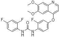
1-(2,4-二氟苯基)-3-[4-[(6,7-二甲氧基-4-喹啉基)氧基]-2-氟苯基]脲是一种芳香醚。
Ki8751属于N-苯基-N'-{4-(4-喹啉基氧基)苯基}脲类化合物。它被设计为一种选择性VEGFR-2抑制剂,靶向肿瘤血管生成,而肿瘤血管生成是肿瘤生长和转移的关键过程。构效关系(SAR)研究表明,Ki8751结构中的喹啉环和脲基对其对VEGFR-2的高亲和力和选择性至关重要。在喹啉环上引入特定的取代基可以增强其对VEGFR-2的抑制活性,同时降低对其他激酶的抑制作用[1]。 |
| 分子式 |
C24H18F3N3O4
|
|
|---|---|---|
| 分子量 |
469.41
|
|
| 精确质量 |
469.124
|
|
| 元素分析 |
C, 61.41; H, 3.87; F, 12.14; N, 8.95; O, 13.63
|
|
| CAS号 |
228559-41-9
|
|
| 相关CAS号 |
|
|
| PubChem CID |
11317348
|
|
| 外观&性状 |
White to off-white solid powder
|
|
| 密度 |
1.4±0.1 g/cm3
|
|
| 沸点 |
497.1±45.0 °C at 760 mmHg
|
|
| 熔点 |
239℃
|
|
| 闪点 |
254.4±28.7 °C
|
|
| 蒸汽压 |
0.0±1.3 mmHg at 25°C
|
|
| 折射率 |
1.657
|
|
| LogP |
5.91
|
|
| tPSA |
81.71
|
|
| 氢键供体(HBD)数目 |
2
|
|
| 氢键受体(HBA)数目 |
8
|
|
| 可旋转键数目(RBC) |
6
|
|
| 重原子数目 |
34
|
|
| 分子复杂度/Complexity |
677
|
|
| 定义原子立体中心数目 |
0
|
|
| SMILES |
FC1C([H])=C(C([H])=C([H])C=1N([H])C(N([H])C1C([H])=C([H])C(=C([H])C=1F)F)=O)OC1C([H])=C([H])N=C2C([H])=C(C(=C([H])C2=1)OC([H])([H])[H])OC([H])([H])[H]
|
|
| InChi Key |
LFKQSJNCVRGFCC-UHFFFAOYSA-N
|
|
| InChi Code |
InChI=1S/C24H18F3N3O4/c1-32-22-11-15-20(12-23(22)33-2)28-8-7-21(15)34-14-4-6-19(17(27)10-14)30-24(31)29-18-5-3-13(25)9-16(18)26/h3-12H,1-2H3,(H2,29,30,31)
|
|
| 化学名 |
1-(2,4-difluorophenyl)-3-[4-(6,7-dimethoxyquinolin-4-yl)oxy-2-fluorophenyl]urea
|
|
| 别名 |
|
|
| HS Tariff Code |
2934.99.03.00
|
|
| 存储方式 |
Powder -20°C 3 years 4°C 2 years In solvent -80°C 6 months -20°C 1 month |
|
| 运输条件 |
Room temperature (This product is stable at ambient temperature for a few days during ordinary shipping and time spent in Customs)
|
| 溶解度 (体外实验) |
|
|||
|---|---|---|---|---|
| 溶解度 (体内实验) |
配方 1 中的溶解度: 2.5 mg/mL (5.33 mM) in 10% DMSO + 40% PEG300 + 5% Tween80 + 45% Saline (这些助溶剂从左到右依次添加,逐一添加), 悬浮液;超声助溶。
例如,若需制备1 mL的工作液,可将100 μL 25.0 mg/mL澄清DMSO储备液加入到400 μL PEG300中,混匀;然后向上述溶液中加入50 μL Tween-80,混匀;加入450 μL生理盐水定容至1 mL。 *生理盐水的制备:将 0.9 g 氯化钠溶解在 100 mL ddH₂O中,得到澄清溶液。 配方 2 中的溶解度: 2.5 mg/mL (5.33 mM) in 10% DMSO + 90% (20% SBE-β-CD in Saline) (这些助溶剂从左到右依次添加,逐一添加), 悬浊液; 超声助溶。 例如,若需制备1 mL的工作液,可将 100 μL 25.0 mg/mL澄清DMSO储备液加入900 μL 20% SBE-β-CD生理盐水溶液中,混匀。 *20% SBE-β-CD 生理盐水溶液的制备(4°C,1 周):将 2 g SBE-β-CD 溶解于 10 mL 生理盐水中,得到澄清溶液。 View More
配方 3 中的溶解度: ≥ 2.5 mg/mL (5.33 mM) (饱和度未知) in 10% DMSO + 90% Corn Oil (这些助溶剂从左到右依次添加,逐一添加), 澄清溶液。 配方 4 中的溶解度: 4% DMSO+corn oil: 2.5mg/mL 1、请先配制澄清的储备液(如:用DMSO配置50 或 100 mg/mL母液(储备液)); 2、取适量母液,按从左到右的顺序依次添加助溶剂,澄清后再加入下一助溶剂。以 下列配方为例说明 (注意此配方只用于说明,并不一定代表此产品 的实际溶解配方): 10% DMSO → 40% PEG300 → 5% Tween-80 → 45% ddH2O (或 saline); 假设最终工作液的体积为 1 mL, 浓度为5 mg/mL: 取 100 μL 50 mg/mL 的澄清 DMSO 储备液加到 400 μL PEG300 中,混合均匀/澄清;向上述体系中加入50 μL Tween-80,混合均匀/澄清;然后继续加入450 μL ddH2O (或 saline)定容至 1 mL; 3、溶剂前显示的百分比是指该溶剂在最终溶液/工作液中的体积所占比例; 4、 如产品在配制过程中出现沉淀/析出,可通过加热(≤50℃)或超声的方式助溶; 5、为保证最佳实验结果,工作液请现配现用! 6、如不确定怎么将母液配置成体内动物实验的工作液,请查看说明书或联系我们; 7、 以上所有助溶剂都可在 Invivochem.cn网站购买。 |
| 制备储备液 | 1 mg | 5 mg | 10 mg | |
| 1 mM | 2.1303 mL | 10.6517 mL | 21.3033 mL | |
| 5 mM | 0.4261 mL | 2.1303 mL | 4.2607 mL | |
| 10 mM | 0.2130 mL | 1.0652 mL | 2.1303 mL |
1、根据实验需要选择合适的溶剂配制储备液 (母液):对于大多数产品,InvivoChem推荐用DMSO配置母液 (比如:5、10、20mM或者10、20、50 mg/mL浓度),个别水溶性高的产品可直接溶于水。产品在DMSO 、水或其他溶剂中的具体溶解度详见上”溶解度 (体外)”部分;
2、如果您找不到您想要的溶解度信息,或者很难将产品溶解在溶液中,请联系我们;
3、建议使用下列计算器进行相关计算(摩尔浓度计算器、稀释计算器、分子量计算器、重组计算器等);
4、母液配好之后,将其分装到常规用量,并储存在-20°C或-80°C,尽量减少反复冻融循环。
计算结果:
工作液浓度: mg/mL;
DMSO母液配制方法: mg 药物溶于 μL DMSO溶液(母液浓度 mg/mL)。如该浓度超过该批次药物DMSO溶解度,请首先与我们联系。
体内配方配制方法:取 μL DMSO母液,加入 μL PEG300,混匀澄清后加入μL Tween 80,混匀澄清后加入 μL ddH2O,混匀澄清。
(1) 请确保溶液澄清之后,再加入下一种溶剂 (助溶剂) 。可利用涡旋、超声或水浴加热等方法助溶;
(2) 一定要按顺序加入溶剂 (助溶剂) 。
InvivoChem的所有产品仅用于作科学研究,不面向患者销售
Copyright 2020 InvivoChem LLC | All Rights Reserved 粤ICP备20063088号-1


 COA
COA
 463611831
