| 规格 | 价格 | 库存 | 数量 |
|---|---|---|---|
| 10 mM * 1 mL in DMSO |
|
||
| 1mg |
|
||
| 5mg |
|
||
| 10mg |
|
||
| 25mg |
|
||
| 50mg |
|
||
| 100mg |
|
||
| 250mg |
|
||
| 500mg |
|
||
| Other Sizes |
|

| 靶点 |
VEGFR1 (IC50 = 170 nM); VEGFR2 (IC50 = 160 nM); VEGFR3 (IC50 = 125 nM)
KRN 633 inhibits vascular endothelial growth factor receptor 2 (VEGFR2) tyrosine kinase (IC₅₀ = 0.02 μM) and VEGFR1 tyrosine kinase (IC₅₀ = 0.2 μM) [1] KRN 633 also shows inhibitory activity against platelet-derived growth factor receptor β (PDGFRβ) (IC₅₀ = 0.3 μM) [3] |
|---|---|
| 体外研究 (In Vitro) |
KRN 633 是一种新型喹唑啉脲衍生物,强烈抑制 VEGFR1、VEGFR2 和 VEGFR3 受体,IC50 值分别为 170 nM、160 nM 和 125 nM。它对非 RTK 表现出较低的抑制活性,例如 PDGF 受体(PDGFRα 和 β、c-Kit、乳腺肿瘤激酶和内膜内皮细胞激酶酪氨酸激酶)(IC50 = 965、9850、4330、9200 和 9900 nM, KRN 633 有效抑制 HUVEC 中配体 VEGF 诱导的 VEGFR2 磷酸化,IC50 为 1.16 nM。KRN 633 还抑制内皮细胞中 VEGF 依赖性而非 bFGF 依赖性 MAP 激酶磷酸化,IC50 值为 3.51 ERK1 和 ERK2 分别为 nM 和 6.08 nM。KRN633 还被证明可以抑制 VEGF 驱动的 HUVEC 增殖,IC50 为 14.9 nM,但仅在 3 μM 时微弱地抑制 FGF 驱动的增殖。KRN 633 抑制缺氧-通过抑制 Akt 和 ERK 磷酸化信号通路,以浓度依赖性方式诱导 HIF-1α 转录激活,IC50 为 3.79 μM。 激酶测定:进行无细胞激酶测定以获得针对多种药物的 IC50 值重组VEGF受体。 KRN633 的测试浓度范围为 0.3 nM 至 10 μM。所有测定均使用 1 μM ATP 一式四份进行。细胞测定:将癌细胞(A549、Ls174T、DU145、HT29、LNCap 和 PC-3 细胞系)接种在含有 10% FBS 和抗生素的培养基中,其密度已知可在测定期间呈指数生长。将细胞培养 24 小时,然后添加 KRN633(0.01 至 10 μM)或仅添加载体(培养基中含有 0.1% DMSO),然后再生长 96 小时。使用 WST-1 试剂测量细胞活力。
KRN 633剂量依赖性抑制血管内皮生长因子(VEGF)诱导的人脐静脉内皮细胞(HUVECs)增殖,IC₅₀为0.05μM。0.5μM时,可抑制VEGF介导的HUVECs迁移约85%、管腔形成约90%;浓度≥0.1μM时,可阻断HUVECs中VEGF诱导的VEGFR2及下游信号分子(Akt和ERK1/2)的磷酸化[1] KRN 633抑制多种肿瘤细胞系增殖,包括A549肺癌细胞和HT-29结直肠癌细胞,IC₅₀分别为3.2μM和4.5μM。2μM时可诱导A549细胞发生G1期周期阻滞[2] KRN 633抑制血小板衍生生长因子-BB(PDGF-BB)诱导的大鼠主动脉平滑肌细胞(RASMCs)增殖,IC₅₀为0.4μM。0.5μM时可阻断RASMCs中PDGFRβ磷酸化及下游信号(STAT3)[3] |
| 体内研究 (In Vivo) |
尽管KRN633在体外对多种癌细胞没有细胞毒性,但由于其对肿瘤血管形成和血管通透性的抑制作用,在体内表现出优异的抗肿瘤活性。每日一次以 100 mg/kg/d 剂量施用 KRN633 可在 A549、LC-6-LCK、HT29、Ls174T、LNCap 和 Du145 细胞中产生显着的肿瘤生长抑制作用,而每日两次以 100 mg/kg 剂量施用 KRN633 可诱导约 90 HT29 肿瘤的生长抑制百分比。用 KRN 633(300 mg/kg,口服)治疗中期妊娠小鼠会减少胎儿组织的血液供应,因为胎盘和胎儿器官的血管化减少,从而增加诱发宫内生长受限 (IUGR) 的风险。
KRN 633以30mg/kg/天的剂量口服给药21天,可抑制裸鼠HT-29结直肠癌异种移植瘤的生长和血管生成。与对照组相比,肿瘤体积减少约65%,通过CD31免疫染色检测,瘤内微血管密度降低约70%[1] KRN 633抑制C57BL/6小鼠体内B16-F10黑色素瘤细胞的肺转移。每周两次腹腔注射10mg/kg,持续14天,肺转移结节数量减少约80%[2] KRN 633抑制大鼠颈动脉损伤模型的新生内膜增生。以20mg/kg/天的剂量口服给药28天,与溶媒对照组相比,新生内膜面积减少约55%[3] |
| 酶活实验 |
为了找到针对各种重组 VEGF 受体的 IC50 值,进行了无细胞激酶测定。 KRN633 的检测浓度范围为 0.3 nM 至 10 μM。每个测定均使用一微克 ATP 进行四次重复。
将重组VEGFR1、VEGFR2和PDGFRβ激酶结构域分别与ATP及特异性多肽底物在系列稀释的KRN 633存在下孵育,反应在37°C下进行60分钟,采用均相时间分辨荧光(HTRF)法检测磷酸化底物。通过与溶媒对照组的荧光强度对比计算抑制率,从量效曲线中得出IC₅₀值[1] 采用比色法进一步验证VEGFR2激酶活性。将重组VEGFR2与KRN 633、ATP和显色底物孵育,45分钟后终止反应,通过测量吸光度定量磷酸化水平,确定IC₅₀以验证与HTRF结果的一致性[3] |
| 细胞实验 |
含有 10% FBS 和抗生素的培养基用于以已知在检测期间允许指数生长的密度铺板癌细胞。将细胞孵育 24 小时后,用 KRN633(0.01 至 10 μM)或仅用载体(培养基中含有 0.1% DMSO)处理它们,然后让它们再生长 96 小时。 WST-1试剂用于测量细胞的活力。
将HUVECs以5×10³个细胞/孔接种到96孔板中,过夜培养。加入KRN 633(0.01-1μM)预处理1小时后,用VEGF(50ng/mL)刺激细胞。72小时后,采用四唑盐法检测细胞活性并计算增殖抑制的IC₅₀值。蛋白质印迹分析中,用药物(0.1-1μM)和VEGF处理HUVECs,裂解后与抗磷酸化VEGFR2、Akt、ERK1/2和GAPDH的抗体孵育[1] 将A549和HT-29细胞接种到96孔板中,用KRN 633(0.1-10μM)处理72小时,采用相同的四唑盐法检测细胞活性。A549细胞用2μM KRN 633处理24小时后,固定、碘化丙啶染色,通过流式细胞术分析细胞周期[2] 将RASMCs接种到96孔板中,血清饥饿24小时。加入KRN 633(0.05-2μM)预处理1小时后,用PDGF-BB(20ng/mL)刺激细胞。48小时后,通过BrdU掺入法评估细胞增殖;采用蛋白质印迹法检测磷酸化PDGFRβ和STAT3[3] |
| 动物实验 |
大鼠:将人源肿瘤异种移植瘤植入BALB/cA和Jcl-nu无胸腺大鼠的后侧腹部。当肿瘤达到指定平均大小(162至657 mm³)时,将大鼠随机分为5组,并分别以指定剂量每日一次(qd)或两次(bid)给予KRN-633或载体对照治疗。在末次治疗后第14天,计算肿瘤生长抑制率,并与载体对照组进行比较[1]。
小鼠:当肿瘤达到平均大小500至667 mm³或103至260 mm³时,将小鼠随机分为5组。随后,以10至100 mg/kg的剂量每日一次(qd)或两次(bid)给予KRN-633或载体对照治疗。在最后一次治疗后的第二天,计算肿瘤生长抑制率(TGI)相对于载体对照组的百分比[1]。 妊娠期间血管内皮生长因子(VEGF)信号通路的抑制会导致多种病理性妊娠,例如高血压、子痫前期和宫内生长受限,但其对胎儿的影响尚未得到充分研究。为了确定VEGF信号通路抑制如何影响妊娠中期胎儿血管发育,我们在妊娠13.5至15.5天期间,每天给妊娠小鼠灌胃给予VEGF受体2(VEGFR-2)酪氨酸激酶抑制剂KRN633(300 mg/kg)或载体。在妊娠16.5天,通过荧光免疫组织化学方法观察胎盘和胎儿多个器官的血管床。所有接受KRN633治疗的小鼠均表现健康,每窝胎儿总数未受影响。然而,KRN633治疗组小鼠的胎盘和胎儿重量低于载体对照组。胎盘和胎儿均未观察到外部畸形和出血,但免疫组织化学分析显示,KRN633治疗损害了胎盘迷路区和所检查的胎儿器官(皮肤、胰腺、肾脏和肺)的血管发育。这些结果表明,妊娠中期抑制VEGF信号通路会抑制胎盘和胎儿的血管生长,而不会对母鼠的健康造成明显损害,但会增加宫内生长受限的风险。[3] 携带HT-29结直肠癌异种移植瘤(100-150 mm³)的裸鼠被随机分为对照组和治疗组。将KRN 633悬浮于0.5%羧甲基纤维素溶液中,以30 mg/kg/天的剂量口服给药,持续21天。每3天测量一次肿瘤体积,并处死小鼠以收集肿瘤组织进行CD31免疫染色[1]。 将B16-F10黑色素瘤细胞经尾静脉注射到C57BL/6小鼠体内。两天后,小鼠接受KRN 633腹腔注射治疗,剂量为10 mg/kg,每周两次,持续14天。治疗结束后,处死小鼠并收集肺组织以计数转移结节[2]。 雄性Sprague-Dawley大鼠接受颈动脉球囊损伤。损伤后24小时开始,大鼠接受KRN 633口服治疗,剂量为20 mg/kg/天,持续28天。将大鼠实施安乐死,并收集颈动脉进行新生内膜增生的组织病理学分析[3] |
| 药代性质 (ADME/PK) |
在小鼠中,单次口服 30 mg/kg 剂量的 KRN 633 的生物利用度约为 40%。血浆半衰期约为 6.5 小时,给药后 1.5 小时达到最大血浆浓度 (Cmax) 为 2.8 μg/mL [1]。在大鼠中,口服 20 mg/kg 剂量的 KRN 633 后,24 小时 AUC₀-24h 为 22.4 μg·h/mL。该药物广泛分布于肝脏、肾脏和肺脏,肿瘤组织浓度约为血浆浓度的 2.5 倍 [3]。
|
| 毒性/毒理 (Toxicokinetics/TK) |
小鼠以 30 mg/kg/天的剂量接受 KRN 633 治疗 21 天后,体重略有下降(约 7%),但未见明显的器官毒性。血清中丙氨酸氨基转移酶 (ALT)、天冬氨酸氨基转移酶 (AST) 和肌酐水平均在正常范围内 [1]。大鼠以 20 mg/kg/天的剂量接受 KRN 633 治疗 28 天后,未见明显的血液学异常或肾毒性。10% 的动物出现轻度胃肠道刺激(腹泻),无需干预即可自行缓解 [3]。通过平衡透析法测定,KRN 633 在人血浆中的血浆蛋白结合率约为 92% [2]。
|
| 参考文献 | |
| 其他信息 |
血管内皮生长因子 (VEGF) 及其受体 VEGFR-2 在血管生成中发挥着核心作用,而血管生成是实体瘤生长和转移所必需的。因此,VEGFR-2 酪氨酸激酶的特异性抑制剂被认为可用于治疗癌症。我们发现喹唑啉脲衍生物 KRN633 可抑制人脐静脉内皮细胞中 VEGFR-2 的酪氨酸磷酸化(IC50 = 1.16 nmol/L)。利用重组酪氨酸激酶进行的选择性分析表明,KRN633 对 VEGFR-1、-2 和 -3 具有高度选择性。KRN633 还能阻断 VEGF 激活丝裂原活化蛋白激酶,并抑制人脐静脉内皮细胞的增殖和管状结构形成。KRN633 对多种癌细胞系的体外增殖没有抑制作用。然而,在多种不同组织来源的体内肿瘤异种移植模型中,包括肺癌、结肠癌和前列腺癌,KRN633 口服给药可抑制裸鼠和裸鼠体内肿瘤的生长。KRN633 还能使一些已形成的肿瘤以及停药后复发的肿瘤消退。在这些模型中,KRN633 的谷浓度比其峰值浓度对肿瘤活性的影响更为显著。KRN633 耐受性良好,对动物的体重或整体健康状况无显著影响。对 KRN633 治疗的肿瘤异种移植模型进行组织学分析显示,非坏死区域的内皮细胞数量减少,血管通透性降低。这些数据表明,KRN633可能对实体瘤和其他依赖于病理性血管生成的疾病的治疗具有潜在价值。[1]
缺氧诱导因子(HIF)是一种异二聚体碱性螺旋-环-螺旋转录因子,活化的HIF在包括炎症和癌症在内的多种病理状态下发挥着关键作用。在许多常见的人类癌症中,包括脑癌、乳腺癌、结肠癌、肺癌、卵巢癌和前列腺癌,均观察到HIF-1α的过表达。HIF介导的基因,例如血管内皮生长因子(VEGF)、诱导型一氧化氮合酶(iNOS)和胰岛素样生长因子(IGF)-1,与肿瘤血管生成、转移和侵袭密切相关。因此,促癌蛋白HIF是癌症治疗的一个新靶点。我们研究了VEGFR抑制剂AAL993、SU5416和KRN633在缺氧条件下对HIF-1α积累抑制的影响。我们发现,VEGFR酪氨酸激酶抑制剂AAL993、SU5416和KRN633具有双重作用:在缺氧条件下抑制VEGFR信号传导和HIF-1α表达。详细的机制研究表明,SU5416 和 KRN633 通过抑制 Akt 和 ERK 磷酸化信号通路来抑制 HIF-1α 的表达,而 AAL993 则通过抑制 ERK 来抑制 HIF-1α 的表达,而不影响 Akt 的磷酸化。[2] KRN-633 是一种小分子药物,目前处于 I 期临床试验阶段。 KRN 633 是一种小分子抑制剂,靶向 VEGFR1、VEGFR2 和 PDGFRβ,具有抗肿瘤、抗血管生成和抗新生内膜增生的作用。[1] KRN 633 对肿瘤转移的抑制活性表明其在治疗高转移风险的晚期癌症方面具有潜在的应用价值。[2] 由于其能够抑制肿瘤转移,KRN 633 可能是一种有前景的血管介入术后预防再狭窄的候选药物。抑制平滑肌细胞增殖[3] |
| 分子式 |
C20H21CLN4O4
|
|
|---|---|---|
| 分子量 |
416.86
|
|
| 精确质量 |
416.125
|
|
| 元素分析 |
C, 57.62; H, 5.08; Cl, 8.50; N, 13.44; O, 15.35
|
|
| CAS号 |
286370-15-8
|
|
| 相关CAS号 |
|
|
| PubChem CID |
9549295
|
|
| 外观&性状 |
White to off-white solid powder
|
|
| 密度 |
1.3±0.1 g/cm3
|
|
| 沸点 |
545.6±50.0 °C at 760 mmHg
|
|
| 熔点 |
229 °C
|
|
| 闪点 |
283.7±30.1 °C
|
|
| 蒸汽压 |
0.0±1.5 mmHg at 25°C
|
|
| 折射率 |
1.629
|
|
| LogP |
4.14
|
|
| tPSA |
98.09
|
|
| 氢键供体(HBD)数目 |
2
|
|
| 氢键受体(HBA)数目 |
6
|
|
| 可旋转键数目(RBC) |
7
|
|
| 重原子数目 |
29
|
|
| 分子复杂度/Complexity |
529
|
|
| 定义原子立体中心数目 |
0
|
|
| SMILES |
ClC1C([H])=C(C([H])=C([H])C=1N([H])C(N([H])C([H])([H])C([H])([H])C([H])([H])[H])=O)OC1C2=C([H])C(=C(C([H])=C2N=C([H])N=1)OC([H])([H])[H])OC([H])([H])[H]
|
|
| InChi Key |
VPBYZLCHOKSGRX-UHFFFAOYSA-N
|
|
| InChi Code |
InChI=1S/C20H21ClN4O4/c1-4-7-22-20(26)25-15-6-5-12(8-14(15)21)29-19-13-9-17(27-2)18(28-3)10-16(13)23-11-24-19/h5-6,8-11H,4,7H2,1-3H3,(H2,22,25,26)
|
|
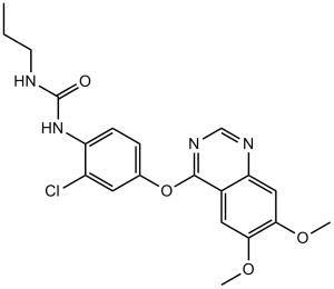
| 化学名 |
1-[2-chloro-4-(6,7-dimethoxyquinazolin-4-yl)oxyphenyl]-3-propylurea
|
|
| 别名 |
KRN633; KRN-633; K00589a; VEGF receptor tyrosine kinase inhibitor III; 1-(2-chloro-4-(6,7-dimethoxyquinazolin-4-yloxy)phenyl)-3-propylurea; 1-{2-chloro-4-[(6,7-dimethoxyquinazolin-4-yl)oxy]phenyl}-3-propylurea; KRN 633
|
|
| HS Tariff Code |
2934.99.9001
|
|
| 存储方式 |
Powder -20°C 3 years 4°C 2 years In solvent -80°C 6 months -20°C 1 month |
|
| 运输条件 |
Room temperature (This product is stable at ambient temperature for a few days during ordinary shipping and time spent in Customs)
|
| 溶解度 (体外实验) |
|
|---|
| 制备储备液 | 1 mg | 5 mg | 10 mg | |
| 1 mM | 2.3989 mL | 11.9944 mL | 23.9889 mL | |
| 5 mM | 0.4798 mL | 2.3989 mL | 4.7978 mL | |
| 10 mM | 0.2399 mL | 1.1994 mL | 2.3989 mL |
1、根据实验需要选择合适的溶剂配制储备液 (母液):对于大多数产品,InvivoChem推荐用DMSO配置母液 (比如:5、10、20mM或者10、20、50 mg/mL浓度),个别水溶性高的产品可直接溶于水。产品在DMSO 、水或其他溶剂中的具体溶解度详见上”溶解度 (体外)”部分;
2、如果您找不到您想要的溶解度信息,或者很难将产品溶解在溶液中,请联系我们;
3、建议使用下列计算器进行相关计算(摩尔浓度计算器、稀释计算器、分子量计算器、重组计算器等);
4、母液配好之后,将其分装到常规用量,并储存在-20°C或-80°C,尽量减少反复冻融循环。
计算结果:
工作液浓度: mg/mL;
DMSO母液配制方法: mg 药物溶于 μL DMSO溶液(母液浓度 mg/mL)。如该浓度超过该批次药物DMSO溶解度,请首先与我们联系。
体内配方配制方法:取 μL DMSO母液,加入 μL PEG300,混匀澄清后加入μL Tween 80,混匀澄清后加入 μL ddH2O,混匀澄清。
(1) 请确保溶液澄清之后,再加入下一种溶剂 (助溶剂) 。可利用涡旋、超声或水浴加热等方法助溶;
(2) 一定要按顺序加入溶剂 (助溶剂) 。
InvivoChem的所有产品仅用于作科学研究,不面向患者销售
Copyright 2020 InvivoChem LLC | All Rights Reserved 粤ICP备20063088号-1


 COA
COA
 463611831
