| 规格 | 价格 | 库存 | 数量 |
|---|---|---|---|
| 5mg |
|
||
| 10mg |
|
||
| 25mg |
|
||
| 50mg |
|
||
| 100mg |
|
||
| 250mg |
|
||
| 500mg |
|
||
| Other Sizes |
|

| 靶点 |
PI3Kα (IC50 = 4 nM); PI3Kβ (IC50 = 0.5 nM); PI3Kγ (IC50 = 0.594 μM);PI3Kδ (IC50 = 0.1 nM); DNA-PK (IC50 = 8.6 nM)
KU-0060648 exhibits differential effects on growth inhibition, but is not profoundly cytotoxic in a panel of human cancer cell lines. When compared to SW620 cells, MCF7 cells exhibit more potent inhibition of DNA-PK and PI-3K. In MCF7 cells, exposure to 1 mM KU-0060648 for five days significantly reduces cell proliferation by more than 95%, but only by 55% in SW620 cells. In clonogenic survival assays, KU-0060648 increases etoposide and doxorubicin's cytotoxicity across a panel of DNA-PKcs-proficient cells, but not in DNA-PKcs-deficient cells, demonstrating that the increased cytotoxicity of the topoisomerase II poisons is caused by DNA-PK inhibition. [1] |
|---|---|
| 体外研究 (In Vitro) |
KU-0060648 对生长抑制表现出不同的作用,但在一组人类癌细胞系中没有明显的细胞毒性。与 SW620 细胞相比,MCF7 细胞表现出更有效的 DNA-PK 和 PI-3K 抑制作用。在 MCF7 细胞中,暴露于 1 mM KU-0060648 5 天,细胞增殖显着降低 95% 以上,但在 SW620 细胞中仅降低 55%。在克隆生存测定中,KU-0060648 在一组 DNA-PKcs 熟练的细胞中增加依托泊苷和阿霉素的细胞毒性,但在 DNA-PKcs 缺陷的细胞中则不然,这表明拓扑异构酶 II 毒物的细胞毒性增加是由 DNA-PK 引起的抑制。 [1]
KU-0060648 浓度依赖性地抑制细胞中DNA-PK在Ser2056位点的自磷酸化,在MCF7乳腺癌细胞中的IC₅₀为0.02 µM,在SW620结肠癌细胞中的IC₅₀为0.2 µM。[1] 它也抑制PI-3K介导的AKT在Ser473位点的磷酸化,在MCF7细胞中的IC₅₀为0.04 µM。然而,其在SW620细胞中对PI-3K的抑制活性很弱 (IC₅₀ >10 µM),表明其抑制具有细胞系依赖性。[1] 连续暴露5天可抑制一系列人乳腺癌(MCF7, T47D, MDA-MB-231)和结肠癌(LoVo, SW620)细胞系的增殖。生长抑制 (GI₅₀) 值范围从0.21 µM (LoVo) 到1 µM (MDA-MB-231 和 SW620)。[1] KU-0060648 (1 µM,暴露16小时) 单药细胞毒性较低(在大多数细胞系中存活率 >80%,MDA-MB-231除外,为41%)。[1] 它显著增强了拓扑异构酶II毒物依托泊苷和多柔比星的细胞毒性。在DNA-PKcs正常的细胞系 (V3-YAC, M059-Fus-1) 中,该化合物将依托泊苷的细胞毒性增强了4至13倍,将多柔比星的细胞毒性增强了高达32倍。而在DNA-PKcs缺陷的细胞系 (V3, M059J) 中,这种增强作用微乎其微,证实化学增敏作用主要归因于DNA-PK的抑制。[1] 在一组人类癌细胞系中,KU-0060648 (1 µM) 将依托泊苷的细胞毒性最高增强了105倍(在SW620细胞中),将多柔比星的细胞毒性最高增强了107倍(在MCF7细胞中)。[1] |
| 体内研究 (In Vivo) |
KU-0060648 可增加 MCF7 和 SW620 异种移植模型中依托泊苷的抗肿瘤活性,并且在 MCF7 异种移植模型中具有单药活性。 [1]
在体内,腹腔注射KU-0060648显著抑制了裸鼠体内HepG2异种移植物的生长。AKT-mTOR活化在异种移植肿瘤中也受到抑制。最后,我们发现DNA-PKcs在人HCC组织中的表达显著上调。[2] 在携带MCF7乳腺癌异种移植瘤的小鼠中,单用KU-0060648治疗 (10 mg/kg 腹腔注射,每日两次,持续14天) 导致中位肿瘤生长延迟30天。在SW620结肠癌异种移植瘤中未观察到显著的单一药物活性。[1] KU-0060648 与依托泊苷磷酸酯联用显著增强了抗肿瘤活性。在MCF7移植瘤中,联合用药导致中位生长延迟55天,而单独使用依托泊苷为38天,单独使用KU-0060648为30天。[1] 在SW620移植瘤中,当KU-0060648与依托泊苷联用时,特别是在抑制剂每日给药两次的情况下,观察到活性增强的趋势。[1] |
| 酶活实验 |
KU-0060648抗DNA-PK和PI-3K细胞活性的测定[1]
在X射线照射(10 Gy)前,在暴露于不同浓度KU-0060648 1小时的细胞中测定DNA-PK自磷酸化。30分钟后,根据制造商的说明,使用磷安全提取试剂制备细胞裂解物。通过蛋白质印迹法测定Ser2056处DNA-PKcs相对于未磷酸化DNA-PKcs的自磷酸化水平。为了测定PI-3K活性,在用50ng/ml胰岛素样生长因子-1处理30分钟之前,将细胞暴露于不同浓度的KU-0060648中1小时。通过蛋白质印迹法测定PI-3K依赖性AKT磷酸化(Ser473)相对于非磷酸化AKT的水平。 |
| 细胞实验 |
细胞毒性和生长抑制研究[1]
通过克隆试验测量细胞毒性。在6孔板中生长的细胞在收获和接种到直径10cm的无药物培养基中的皮氏培养皿之前,暴露于含有或不含KU-0060648(1μM)的依托泊苷或阿霉素16小时。10至14天后,用结晶紫对菌落进行染色,并用自动菌落计数器计数。如前所述,通过SRB测定法测定连续暴露于KU-0060648 5天后的细胞生长抑制。GI50是导致50%细胞生长抑制的浓度。 DNA-PK和PI-3K细胞活性测定: 细胞用一系列浓度的KU-0060648处理1小时。对于DNA-PK抑制评估,随后用X射线照射细胞(10 Gy),30分钟后裂解。对于PI-3K抑制评估,在1小时预孵育后,用胰岛素样生长因子-1 (50 ng/ml) 处理细胞30分钟。制备细胞裂解液,使用特异性抗体通过蛋白质印迹法测定磷酸化DNA-PKcs (Ser2056) 或磷酸化AKT (Ser473) 相对于总蛋白的水平。进行光密度分析以计算抑制百分比和IC₅₀值。[1] 克隆形成存活实验: 为了评估细胞毒性和化学增敏作用,将培养板中的细胞暴露于依托泊苷或多柔比星,同时使用或不使用KU-0060648 (1 µM),持续16小时。然后收集细胞,在无药培养基中接种到培养皿中,并使其形成克隆10-14天。对克隆进行染色和计数。[1] 生长抑制 (SRB) 实验: 细胞连续暴露于KU-0060648 5天。然后使用磺酰罗丹明B (SRB) 比色法测量细胞密度以确定生长抑制 (GI₅₀)。[1] |
| 动物实验 |
人源肿瘤SW620或MCF7异种移植模型
10 mg/kg,每日两次 i.p. KU-0060648不同给药途径后的血浆药代动力学[1] 所有体内实验均经相关机构动物福利委员会审查批准,并按照国家法律进行。我们测定了雌性Balb C小鼠分别以10 mg/kg剂量静脉注射(iv)、腹腔注射(ip)或口服(po)给药后KU-0060648的血浆药代动力学。 KU-0060648 配制于等摩尔磷酸溶剂中,用无菌生理盐水定容至最终 pH 值为 5。小鼠在注射 KU-0060648 后,每隔一段时间处死一次,最长达 360 分钟,并按照先前描述的方法,通过 LC-MS/MS 分析测定血浆中 KU-0060648 的浓度。KU-0060648 在肿瘤异种移植模型中的分布[1] 雌性无胸腺小鼠在特定病原体清除 (SPF) 条件下饲养和处理,用于组织分布和疗效研究。将 KU-0060648 (12.5 mg/kg,静脉注射) 注射到携带 MCF7 或 SW620 肿瘤(650 mm³)的小鼠体内,分别在 60 分钟或 240 分钟后处死小鼠。将肿瘤切除后,置于冰上,用搅拌式均质器在PBS(1:3 w/v)中以10秒为单位进行均质。采用LC-MS/MS分析测定血浆和肿瘤中KU-0060648的浓度,方法如前所述。 DNA-PK离体药效学试验[1] 将KU-0060648(2.5或25 mg/kg)或载体单独静脉注射给携带SW620肿瘤的小鼠。1小时或4小时后,处死动物,切除肿瘤并进行均质处理。通过 ELISA 检测测定 p53 肽底物 (Ser15) 的 DNA-PK 依赖性磷酸化,确定肿瘤匀浆中的 DNA-PK 活性,如前所述。 抗肿瘤疗效研究[1] 当肿瘤可触及时(植入后 8-10 天,约 5 mm × 5 mm),对携带 SW620 或 MCF7 异种移植瘤的小鼠(每组 n = 5)进行治疗。动物每天接受一次生理盐水腹腔注射(对照组),单药 KU-0060648 10 mg/kg 腹腔注射,每天两次,SW620 荷瘤小鼠连续 5 天(2 × d × 5),MCF7 荷瘤小鼠连续 14 天(2 × d × 14),每天两次给药间隔 8 小时,或每天一次腹腔注射依托泊苷磷酸盐(11.35 mg/kg 生理盐水,相当于 10 mg/kg 游离依托泊苷,腹腔注射,d × 5)。对于联合用药,KU-0060648 每日腹腔注射一次或两次,持续 5 天(SW620 细胞)或每日一次,持续 14 天(MCF7 细胞),首次给药紧接依托泊苷磷酸酯给药之前。 体内抗肿瘤疗效测定[2] 将大量 HepG2 细胞(500 万个/只小鼠)皮下注射到 6-8 周龄雌性裸鼠的右侧腹部。当肿瘤体积达到约 100 mm³ 时,将小鼠随机分为三组,每组 12 只:载体对照组(生理盐水)、KU-0060648 10 mg/kg 组(每日腹腔注射,持续 21 天)和 KU-0060648 50 mg/kg 组(每日腹腔注射,持续 21 天)。当肿瘤形成(体积约 100 mm³)时开始注射。每周记录肿瘤体积,并使用既定公式计算:体积 (mm³) = (d² × D)/2,其中 d 和 D 分别为肿瘤的最短直径和最长直径。首次给予 KU-0060648 两周后,分离每组两只小鼠的异种移植瘤,并进行蛋白质印迹和免疫组织化学 (IHC) 染色分析。采用人道终点以最大程度减少动物痛苦。首次给予 KU-0060648 五周后,通过手术分离 HepG2 异种移植瘤并称重。药代动力学研究:KU-0060648 配制于等摩尔磷酸溶剂中,并用无菌生理盐水调节 pH 至 5。雌性 Balb C 小鼠通过静脉注射 (iv)、腹腔注射 (ip) 或口服 (po) 途径单次给予 10 mg/kg 的 KU-0060648。在长达 360 分钟的不同时间点采集血液样本,用于血浆浓度分析。[1] 肿瘤分布研究:将携带 MCF7 或 SW620 皮下异种移植瘤(约 650 mm³)的雌性无胸腺小鼠单次静脉注射 KU-0060648(12.5 mg/kg)。分别于给药后 60 分钟或 240 分钟处死小鼠。切除肿瘤,在 PBS 中匀浆,并与血浆一起分析药物浓度。[1] 体外 DNA-PK 活性测定:将携带 SW620 肿瘤的小鼠单次静脉注射 KU-0060648(2.5 或 25 mg/kg)或载体。分别于给药后 1 小时或 4 小时处死小鼠。切除肿瘤并匀浆。采用基于 ELISA 的检测方法,通过测定 DNA-PK 依赖的 p53 肽底物 (Ser15) 的磷酸化水平来确定匀浆中的 DNA-PK 活性。[1] 异种移植瘤疗效研究:将已建立的 SW620 或 MCF7 皮下异种移植瘤小鼠在肿瘤长至约 5×5 mm 时进行治疗。 SW620 模型:动物分别接受赋形剂(对照组)、单独使用 KU-0060648(10 mg/kg,腹腔注射,每日一次或两次,持续 5 天)、单独使用依托泊苷磷酸酯(11.35 mg/kg,腹腔注射,每日一次,持续 5 天)或联合治疗。 MCF7 模型:动物分别接受赋形剂、单独使用 KU-0060648(10 mg/kg,腹腔注射,每日两次,持续 14 天)、单独使用依托泊苷磷酸酯(11.35 mg/kg,腹腔注射,每日一次,持续 5 天)或联合治疗。 (以mg/kg剂量腹腔注射,每日一次,连续5天),或联合用药,其中KU-0060648的首剂应在依托泊苷给药前立即给予。[1] |
| 药代性质 (ADME/PK) |
表2列出了通过不同途径向Balb/C小鼠给予10 mg/kg KU-0060648后测定的血浆药代动力学参数。口服给药后KU-0060648的生物利用度≥100%。腹腔注射给药后KU-0060648的药代动力学参数与静脉注射给药相似,生物利用度为78%。[1]
静脉注射给药后KU-0060648在MCF7和SW620荷瘤小鼠体内的组织分布[1] 向携带MCF7或SW620异种移植瘤的小鼠静脉注射KU-0060648(12.5 mg/kg)后,KU-0060648广泛分布于肿瘤组织,并在血浆清除后仍保留在肿瘤组织中(表2)。 KU-0060648 在肿瘤中的浓度维持在 1 μM 以上(该浓度在体外可产生化疗增敏作用),且至少持续 4 小时。小鼠单次静脉注射 10 mg/kg 剂量后,KU-0060648 的血浆峰浓度 (Cmax) 为 19 µg/ml(5 分钟),消除半衰期 (T₁/₂) 为 102 分钟,清除率 (CL) 为 41 ml/kg/min。[1] 口服给药 (10 mg/kg po) 的生物利用度接近 100% (119%),Tmax 为 120 分钟,T₁/₂ 为 142 分钟。[1] 腹腔注射给药 (10 mg/kg ip) 的生物利用度为 78%,Tmax 为 30 分钟。半衰期为 106 分钟。[1] 在荷瘤小鼠中静脉注射 12.5 mg/kg 剂量后,KU-0060648 广泛分布于 MCF7 和 SW620 异种移植瘤中。血浆清除后,肿瘤浓度至少维持在 1 µM 以上(体外化疗增敏的有效浓度)。[1] |
| 毒性/毒理 (Toxicokinetics/TK) |
在MCF7异种移植疗效研究中,KU-0060648单药治疗的毒性可忽略不计(最大体重下降3%)。与依托泊苷磷酸酯联合用药时,最大体重下降7%,该结果被认为是可接受的。[1]
在SW620异种移植研究中,KU-0060648单药治疗或依托泊苷磷酸酯单药治疗均未引起显著的体重下降。二者联合用药时,最大体重下降11%。[1] |
| 参考文献 | |
| 其他信息 |
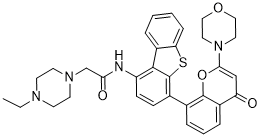
2-(4-乙基-1-哌嗪基)-N-[4-[2-(4-吗啉基)-4-氧代-1-苯并吡喃-8-基]-1-二苯并噻吩基]乙酰胺是二苯并噻吩类化合物。
KU-0060648 是一种双重 ATP 竞争性 DNA-PK 和 PI-3K 抑制剂,由 LY294002 药效团开发而来。它对其他PIKK家族成员(如ATM、ATR和mTOR)具有选择性。[1] 它对拓扑异构酶II抑制剂(依托泊苷、阿霉素)的化疗增敏作用主要归因于DNA-PK的抑制,从而削弱了DNA双链断裂的非同源末端连接(NHEJ)修复途径。[1] 在MCF7异种移植瘤(携带PIK3CA突变)中观察到了单药抗肿瘤活性,但在SW620异种移植瘤中未观察到,这表明PI-3K通路激活的肿瘤可能对其敏感。[1] 该研究为DNA-PK和PI-3K双重抑制的治疗策略提供了原理验证。[1] |
| 分子式 |
C33H34N4O4S
|
|---|---|
| 分子量 |
582.7125
|
| 精确质量 |
582.23
|
| 元素分析 |
C, 68.02; H, 5.88; N, 9.61; O, 10.98; S, 5.50
|
| CAS号 |
881375-00-4
|
| 相关CAS号 |
881375-00-4
|
| PubChem CID |
11964036
|
| 外观&性状 |
Off-white to brown solid powder
|
| 密度 |
1.3±0.1 g/cm3
|
| 沸点 |
819.9±65.0 °C at 760 mmHg
|
| 闪点 |
449.7±34.3 °C
|
| 蒸汽压 |
0.0±3.0 mmHg at 25°C
|
| 折射率 |
1.694
|
| LogP |
5.56
|
| tPSA |
106.5
|
| 氢键供体(HBD)数目 |
1
|
| 氢键受体(HBA)数目 |
8
|
| 可旋转键数目(RBC) |
6
|
| 重原子数目 |
42
|
| 分子复杂度/Complexity |
1010
|
| 定义原子立体中心数目 |
0
|
| SMILES |
O=C1C2C=CC=C(C=2OC(N2CCOCC2)=C1)C1C2SC3C(C=2C(NC(CN2CCN(CC)CC2)=O)=CC=1)=CC=CC=3
|
| InChi Key |
AATCBLYHOUOCTO-UHFFFAOYSA-N
|
| InChi Code |
InChI=1S/C33H34N4O4S/c1-2-35-12-14-36(15-13-35)21-29(39)34-26-11-10-23(33-31(26)25-6-3-4-9-28(25)42-33)22-7-5-8-24-27(38)20-30(41-32(22)24)37-16-18-40-19-17-37/h3-11,20H,2,12-19,21H2,1H3,(H,34,39)
|
| 化学名 |
2-(4-ethylpiperazin-1-yl)-N-[4-(2-morpholin-4-yl-4-oxochromen-8-yl)dibenzothiophen-1-yl]acetamide
|
| 别名 |
KU0060648; KU 0060648; LM6DZS6PYA; 2-(4-ethylpiperazin-1-yl)-N-[4-(2-morpholin-4-yl-4-oxochromen-8-yl)dibenzothiophen-1-yl]acetamide; CHEMBL1086377; 2-(4-ethylpiperazin-1-yl)-N-(4-(2-morpholino-4-oxo-4H-chromen-8-yl)dibenzo[b,d]thiophen-1-yl)acetamide; KU-0060648
|
| HS Tariff Code |
2934.99.9001
|
| 存储方式 |
Powder -20°C 3 years 4°C 2 years In solvent -80°C 6 months -20°C 1 month |
| 运输条件 |
Room temperature (This product is stable at ambient temperature for a few days during ordinary shipping and time spent in Customs)
|
| 溶解度 (体外实验) |
DMSO: 2~2.8 mg/mL (3.4~4.8 mM)
|
|---|---|
| 溶解度 (体内实验) |
配方 1 中的溶解度: ≥ 0.28 mg/mL (0.48 mM) (饱和度未知) in 10% DMSO + 40% PEG300 + 5% Tween80 + 45% Saline (这些助溶剂从左到右依次添加,逐一添加), 澄清溶液。
例如,若需制备1 mL的工作液,可将100 μL 2.8 mg/mL澄清DMSO储备液加入400 μL PEG300中,混匀;然后向上述溶液中加入50 μL Tween-80,混匀;加入450 μL生理盐水定容至1 mL。 *生理盐水的制备:将 0.9 g 氯化钠溶解在 100 mL ddH₂O中,得到澄清溶液。 配方 2 中的溶解度: ≥ 0.28 mg/mL (0.48 mM) (饱和度未知) in 10% DMSO + 90% (20% SBE-β-CD in Saline) (这些助溶剂从左到右依次添加,逐一添加), 澄清溶液。 例如,若需制备1 mL的工作液,可将 100 μL 2.8mg/mL澄清的DMSO储备液加入到900μL 20%SBE-β-CD生理盐水中,混匀。 *20% SBE-β-CD 生理盐水溶液的制备(4°C,1 周):将 2 g SBE-β-CD 溶解于 10 mL 生理盐水中,得到澄清溶液。 View More
配方 3 中的溶解度: ≥ 0.28 mg/mL (0.48 mM) (饱和度未知) in 10% DMSO + 90% Corn Oil (这些助溶剂从左到右依次添加,逐一添加), 澄清溶液。 配方 4 中的溶解度: 30% propylene glycol, 5% Tween 80, 65% D5W: 20 mg/mL 1、请先配制澄清的储备液(如:用DMSO配置50 或 100 mg/mL母液(储备液)); 2、取适量母液,按从左到右的顺序依次添加助溶剂,澄清后再加入下一助溶剂。以 下列配方为例说明 (注意此配方只用于说明,并不一定代表此产品 的实际溶解配方): 10% DMSO → 40% PEG300 → 5% Tween-80 → 45% ddH2O (或 saline); 假设最终工作液的体积为 1 mL, 浓度为5 mg/mL: 取 100 μL 50 mg/mL 的澄清 DMSO 储备液加到 400 μL PEG300 中,混合均匀/澄清;向上述体系中加入50 μL Tween-80,混合均匀/澄清;然后继续加入450 μL ddH2O (或 saline)定容至 1 mL; 3、溶剂前显示的百分比是指该溶剂在最终溶液/工作液中的体积所占比例; 4、 如产品在配制过程中出现沉淀/析出,可通过加热(≤50℃)或超声的方式助溶; 5、为保证最佳实验结果,工作液请现配现用! 6、如不确定怎么将母液配置成体内动物实验的工作液,请查看说明书或联系我们; 7、 以上所有助溶剂都可在 Invivochem.cn网站购买。 |
| 制备储备液 | 1 mg | 5 mg | 10 mg | |
| 1 mM | 1.7161 mL | 8.5806 mL | 17.1612 mL | |
| 5 mM | 0.3432 mL | 1.7161 mL | 3.4322 mL | |
| 10 mM | 0.1716 mL | 0.8581 mL | 1.7161 mL |
1、根据实验需要选择合适的溶剂配制储备液 (母液):对于大多数产品,InvivoChem推荐用DMSO配置母液 (比如:5、10、20mM或者10、20、50 mg/mL浓度),个别水溶性高的产品可直接溶于水。产品在DMSO 、水或其他溶剂中的具体溶解度详见上”溶解度 (体外)”部分;
2、如果您找不到您想要的溶解度信息,或者很难将产品溶解在溶液中,请联系我们;
3、建议使用下列计算器进行相关计算(摩尔浓度计算器、稀释计算器、分子量计算器、重组计算器等);
4、母液配好之后,将其分装到常规用量,并储存在-20°C或-80°C,尽量减少反复冻融循环。
计算结果:
工作液浓度: mg/mL;
DMSO母液配制方法: mg 药物溶于 μL DMSO溶液(母液浓度 mg/mL)。如该浓度超过该批次药物DMSO溶解度,请首先与我们联系。
体内配方配制方法:取 μL DMSO母液,加入 μL PEG300,混匀澄清后加入μL Tween 80,混匀澄清后加入 μL ddH2O,混匀澄清。
(1) 请确保溶液澄清之后,再加入下一种溶剂 (助溶剂) 。可利用涡旋、超声或水浴加热等方法助溶;
(2) 一定要按顺序加入溶剂 (助溶剂) 。
 |
|---|
 |
 |
 DNA-PK-IN-15
 Lys(CO-C3-p-I-Ph)-OMe
 DNA-PK-IN-13
 DNA-PK-IN-14
InvivoChem的所有产品仅用于作科学研究,不面向患者销售
Copyright 2020 InvivoChem LLC | All Rights Reserved 粤ICP备20063088号-1


 COA
COA
 463611831
