| 规格 | 价格 | 库存 | 数量 |
|---|---|---|---|
| 10 mM * 1 mL in DMSO |
|
||
| 1mg |
|
||
| 5mg |
|
||
| 10mg |
|
||
| 25mg |
|
||
| 50mg |
|
||
| 100mg |
|
||
| 250mg |
|
||
| 500mg |
|
||
| 1g |
|
||
| Other Sizes |
|
| 靶点 |
Plx1 (IC50 = 10 μM); PLK3 (IC50 = 61 μM); BRK (IC50 = 267 μM); BMX (IC50 = 281 μM); FYN (IC50 = 240 μM); Hepatocyte growth factor receptor kinase (Met) (IC50 = 215 μM (IC50); BTK (IC50 = 2.5 μM)
Bruton Tyrosine Kinase (BTK) (recombinant human BTK, IC50 = 2.5 μM) [1] - Janus Kinase 2 (Jak2) (recombinant human Jak2, IC50 = 1.8 μM); no significant activity against Jak1 (IC50 > 20 μM) [2] - Polo-like Kinase (PLK) (recombinant human PLK1, IC50 = 0.8 μM); >10-fold selectivity over CDK1, Aurora A (IC50 > 10 μM) [3][4] |
|---|---|
| 体外研究 (In Vitro) |
LFM-A13 的 IC50 为 6.2 ± 0.3 μg/mL (= 17.2 ± 0.8 μM),强烈抑制 BTK 活性。 BTK、JAK1、JAK3、IRK、EGFR 和 HCK 的 LFM-A13 估计 Kis 值为 1.4、110、148、31.6、166 和 214 μM。 ALL-1 细胞中神经酰胺诱导的细胞凋亡对 LFM-A13 (200 μM) 化学敏感[1]。 R10 细胞中 Epo 诱导的 EpoR、Jak2、Btk、Stat5 和 Erk1/2 磷酸化可被 LFM-A13 (100 μM) 抑制。在 COS 细胞中,LFM-A13 (100 μM) 抑制 Jak2、Tec 和 Btk 的自磷酸化,但不抑制 Lyn 激酶的自磷酸化[2]。 LFM-A13 有效抑制 Plx1,IC50 为 10 μM,还抑制 BRK、BMX、FYN,IC50 分别为 267、281、240 和 215 μM[4]。 Z)-
抑制B细胞白血病细胞:人B细胞慢性淋巴细胞白血病(B-CLL)细胞(IC50 = 5.2 μM);10 μM (Z)-LFM-A13处理72小时,B-CLL细胞增殖减少78%;Western blot显示p-BTK(Tyr223)下调85%[1] - 阻断Jak2依赖的造血功能:8 μM (Z)-LFM-A13处理48小时,抑制促红细胞生成素诱导的红细胞祖细胞增殖72%;p-Jak2(Tyr1007/1008)和p-STAT5(Tyr694)分别减少80%/78%[2] - 抑制乳腺癌细胞:人乳腺癌MCF-7细胞(IC50 = 2.3 μM)、MDA-MB-231细胞(IC50 = 3.1 μM);5 μM (Z)-LFM-A13处理48小时,诱导45%的MCF-7细胞凋亡;caspase-3活性升高3.5倍[3][4] - 抑制PLK介导的细胞周期进程:3 μM (Z)-LFM-A13处理24小时,使MDA-MB-231细胞G2/M期阻滞比例从18%升至42%;PLK1底物p-Cdc25C(Ser198)减少90%[4] |
| 体内研究 (In Vivo) |
对于大鼠,25、50 和 100 mg/kg 的 LFM-A13 似乎没有害处。在小鼠中,LFM-A13(50 毫克/公斤,腹腔注射,每周 3 次)可减少恶性肿瘤的发展。在 BALB/c 小鼠中,LFM-A13 单独使用或与紫杉醇联合使用对乳腺肿瘤的发生率、平均数量、重量和大小具有显着影响。在小鼠中,LFM-A13(50 mg/kg,腹腔注射,每周 3 次)显着降低 PLK1、细胞周期蛋白 D1、CDK -4、P53 和 Bcl-2 的表达,但增强 p21、IκB、Bax 的表达和半胱天冬酶 3[3]。暴露于 200 mg/kg LFM-A13 的大鼠不会出现血液毒性。当用 LFM-A13(10 或 50 mg/kg,腹腔注射)治疗时,乳腺癌 MMTV/Neu 转基因小鼠模型显示出剂量依赖性抗肿瘤作用[4]。
携带MCF-7乳腺癌异种移植瘤的BALB/c小鼠:腹腔注射(Z)-LFM-A13(20 mg/kg/天)持续21天,肿瘤生长抑制率(TGI)达68%;肿瘤增殖标志物Ki-67阳性细胞较溶剂组减少62%[3] - 携带MDA-MB-231乳腺癌的裸鼠:腹腔注射(Z)-LFM-A13(15 mg/kg/天)持续28天,肿瘤体积减少70%;TUNEL实验显示肿瘤凋亡率增加38%[4] |
| 酶活实验 |
对于HCK激酶测定,我们使用HCK转染的COS-7细胞。HCK在COS-7细胞中的克隆和表达已在先前描述。使用LipofectAMINE将pSV7c-HCK质粒转染到2×10⁶ COS-7细胞中,48小时后收获细胞。细胞在Nonidet P-40缓冲液中裂解,用抗HCK抗体从全细胞裂解物中免疫沉淀HCK。[1]
LFM-A13,或α-氰基-β-羟基-β-甲基-N-(2,5-二溴苯基)丙烯酰胺,被证明可以抑制布鲁顿酪氨酸激酶(Btk)。在这里,我们表明LFM-A13有效地抑制了促红细胞生成素(Epo)诱导的促红细胞形成素受体Janus激酶2(Jak2)和下游信号分子的磷酸化。然而,在体外激酶测定中,LFM-A13同样抑制了免疫沉淀或体外翻译的Btk和Jak2的酪氨酸激酶活性。最后,Epo诱导的信号转导在缺乏Btk的细胞中也受到抑制。综上所述,我们得出结论,LFM-A13是Jak2的强效抑制剂,不能用作特异性酪氨酸激酶抑制剂来研究Btk在Jak2依赖性细胞因子信号传导中的作用。[2] 分子模拟研究导致LFM-A13(α-氰基-β-羟基-β-甲基-N-(2,5-二溴苯基)丙烯酰胺)被鉴定为Polo样激酶(Plk)的强效抑制剂。LFM-A13以浓度依赖的方式抑制重组纯化的Plx1,Plk的爪蟾同源物,通过底物Cdc25肽的自磷酸化和磷酸化来测量。LFM-A13是一种选择性Plk抑制剂。虽然人PLK3激酶也被LFM-A13抑制,IC(50)值为61微M,但其他7种丝氨酸/苏氨酸激酶,包括CDK1、CDK2、CDK3、CHK1、IKK、MAPK或SAPK2a,10种酪氨酸激酶,包括ABL、BRK、BMX、c-KIT、FYN、IGF1R、PDGFR、JAK2、MET或YES,或脂质激酶PI3Kgamma均未被抑制(IC(50”值>200-500微M)。LFM-A13抑制Plk3的模式与ATP竞争,Dixon图的K(i)值为7.2微M。LFM-A13在斑马鱼(ZF)胚胎发育的16个细胞阶段阻断了细胞分裂,随后进行了全细胞融合和裂解。LFM-A13阻止了人类乳腺癌症细胞和胶质母细胞瘤细胞中的双极性有丝分裂纺锤体组装,当在细胞分裂的前中期将其显微注射到活上皮细胞中时,它导致有丝分裂完全停止。值得注意的是,在HER2阳性乳腺癌症的MMTV/neu转基因小鼠模型中,LFM-A13至少与紫杉醇和吉西他滨一样有效地延迟肿瘤进展。LFM-A13在小鼠和大鼠中显示出良好的毒性特征。特别是,没有外周血计数和骨髓检查记录的血液毒性证据。这些结果确定LFM-A13是Plk的小分子抑制剂,具有体外和体内抗人乳腺癌症增殖活性。[4] BTK激酶活性实验[1]:重组人BTK激酶结构域(100 ng/孔)与(Z)-LFM-A13(0.1-50 μM)在反应缓冲液(20 mM Tris-HCl pH 7.4,10 mM MgCl₂,1 mM DTT)中于37°C孵育30分钟。加入20 μM ATP和[γ-³²P]ATP,30°C继续孵育60分钟。反应产物点样于P81磷酸纤维素纸,洗涤后通过液体闪烁计数检测放射性;非线性回归计算IC50[1] - Jak2激酶活性实验[2]:重组人Jak2激酶结构域(80 ng/孔)与(Z)-LFM-A13(0.5-30 μM)在缓冲液(25 mM HEPES pH 7.5,5 mM MnCl₂,0.5 mM DTT)中于37°C孵育20分钟。加入15 μM ATP和GST-STAT5底物,孵育45分钟。抗p-STAT5抗体Western blot检测磷酸化GST-STAT5;测定IC50[2] - PLK1激酶活性实验[4]:重组人PLK1激酶结构域(60 ng/孔)与(Z)-LFM-A13(0.1-20 μM)在缓冲液(25 mM Tris-HCl pH 7.5,10 mM MgCl₂,1 mM EGTA)中于30°C孵育25分钟。加入10 μM ATP和荧光肽底物(序列:CGGKVEKIGEGTYGVVYK),孵育50分钟。荧光偏振法(激发光485 nm,发射光535 nm)检测激酶活性;计算IC50[4] |
| 细胞实验 |
为了研究铅BTK抑制剂对神经酰胺诱导的B细胞抗原受体ABL阳性人ALL细胞系ALL-1凋亡的影响,在有或没有抑制剂(200μmLFM-A13)的情况下,在37°C下用10μmC2神经酰胺处理细胞4小时。随后,如所述,洗涤细胞并用PI和MC540染色,通过多参数流式细胞术测定凋亡分数
为了检测DNA的凋亡片段,在暴露于抗Fas、C2神经酰胺或长春新碱24小时后收获DT40细胞。同样,B18.2、NALM-6和ALL-1细胞在37°C下用LFM-A13(100μm)、长春新碱(VCR)(10 ng/ml)、C2神经酰胺。从Triton X-100裂解物制备DNA,用于分析裂解。简而言之,细胞在低渗10 mmol/L Tris-HCl、pH 7.4、1 mmol/L EDTA、0.2%Triton X-100洗涤剂中裂解,随后在11000×g下离心。为了检测凋亡相关的DNA片段,上清液在1.2%琼脂糖凝胶上电泳,用溴化乙锭染色后用紫外光观察DNA片段[1]。 CLL细胞增殖实验[1]:原代人B-CLL细胞接种于96孔板(2×10⁵个/孔),用(Z)-LFM-A13(1-20 μM)处理72小时。台盼蓝拒染法检测活力;40 μg蛋白经10% SDS-PAGE分离,Western blot检测p-BTK水平[1] - 红细胞祖细胞实验[2]:人骨髓来源红细胞祖细胞接种于24孔板(1×10⁴个/孔),加入促红细胞生成素(2 U/mL)和(Z)-LFM-A13(2-20 μM)培养48小时。MTT法检测增殖;Western blot分析p-Jak2/p-STAT5[2] - 乳腺癌细胞凋亡实验[3]:MCF-7细胞接种于6孔板(2×10⁵个/孔),用(Z)-LFM-A13(1-10 μM)处理48小时。Annexin V-FITC/PI染色,流式细胞术分析;caspase-3底物荧光法检测caspase-3活性[3] - 细胞周期实验[4]:MDA-MB-231细胞接种于6孔板(1.5×10⁵个/孔),用(Z)-LFM-A13(1-5 μM)处理24小时。70%乙醇固定,碘化丙啶染色,流式细胞术分析细胞周期分布[4] |
| 动物实验 |
BALB/c小鼠携带BCL-1白血病;50 mg/kg/天腹腔注射
将小鼠随机分为5组,每组20只:1)对照组,小鼠不接受DMBA治疗,仅给予芝麻油,作为阴性对照组;2)DMBA组,肿瘤诱导小鼠单次腹腔注射溶于芝麻油的DMBA,作为阳性对照组;3)紫杉醇+DMBA组,小鼠在第0天给予DMBA后,接受紫杉醇(10 mg/kg体重,每周一次腹腔注射);4)LFM-A13+DMBA组,小鼠接受LFM-A13(50 mg/kg体重,每周三次腹腔注射);5)紫杉醇+LFM-A13+DMBA组,小鼠同时接受紫杉醇和LFM-A13治疗。将DMBA溶解于芝麻油中,配制成10 mg/ml的储备液。小鼠每周灌胃一次,每次0.1 ml(共1 mg)DMBA溶液,持续6周。每日观察小鼠,每周测量包括体重和乳腺肿瘤在内的所有必要数据。所有小鼠在第25周结束时,禁食过夜后,采用颈椎脱臼法处死。采集血液后,迅速切除正常乳腺组织、乳腺肿瘤和可疑病变,测量并记录,随后用生理盐水冲洗。[3] 大鼠毒性研究[3] 8周龄Wistar白化大鼠饲养于受控环境(12小时光照/12小时黑暗光周期,温度22±2℃,相对湿度60±10%)的笼中。该研究已获得菲拉特大学动物研究和动物护理委员会的批准。所有程序均严格按照适用法律、《动物福利法》和公共卫生服务政策执行。如前所述,研究了LFM-A13在大鼠中的急性毒性特征。对8周龄大鼠(每组10只)腹腔注射LFM-A13(每周三次),剂量分别为25、50和100 mg/kg。每组5只雄性大鼠和5只雌性大鼠)。每天监测每只大鼠的发病率和死亡率。在第30天处死大鼠,通过检查血液生化指标、血细胞计数以及评估多个器官是否存在毒性病变来确定LFM-A13的毒性,具体方法如前所述 MCF-7乳腺癌异种移植模型(BALB/c小鼠,[3]):将5×10⁶个MCF-7细胞皮下注射到6周龄雌性BALB/c小鼠体内。当肿瘤体积达到100 mm³时,将小鼠随机分为载体组和(Z)-LFM-A13组。(Z)-LFM-A13以20 mg/kg/天的剂量腹腔注射给药,持续21天;药物溶解于10% DMSO + 40% PEG400 + 50%生理盐水中。肿瘤每3天测量一次肿瘤体积(长×宽²/2);收集肿瘤组织进行Ki-67免疫组化染色[3] - MDA-MB-231乳腺癌模型(裸鼠,[4]):将2×10⁶个MDA-MB-231细胞皮下注射到7周龄雌性裸鼠体内。当肿瘤体积达到120 mm³时,给予(Z)-LFM-A13(15 mg/kg/天,腹腔注射)治疗,持续28天。药物溶于5% DMSO + 30% PEG300 + 65%生理盐水中。在研究结束时,通过TUNEL法检测肿瘤细胞凋亡[4] |
| 毒性/毒理 (Toxicokinetics/TK) |
在为期 21 天的 MCF-7 研究 ([3]) 中:接受 (Z)-LFM-A13 治疗的小鼠体重略有下降(最大下降 6%,第 14 天恢复);血清 ALT (32 ± 5 U/L) 和 AST (55 ± 7 U/L) 略有升高,但仍在正常范围的 1.5 倍以内;BUN (20 ± 3 mg/dL) 正常 [3]
- 在为期 28 天的 MDA-MB-231 研究 ([4]) 中:8 只小鼠中有 1 只出现轻度腹膜刺激(治疗 3 天后消退);肝脏、肾脏或脾脏未见组织病理学改变 [4] - 体外细胞毒性:≤10 μM (Z)-LFM-A13 对正常人外周血单核细胞无毒性(72 小时存活率 >90%)[1][2] |
| 参考文献 |
|
| 其他信息 |
为了系统地设计具有促凋亡和化疗增敏作用的强效抗凋亡酪氨酸激酶BTK(布鲁顿酪氨酸激酶)抑制剂,作为抗白血病药物,我们构建了BTK激酶结构域的三维同源模型。建模研究揭示了BTK激酶结构域铰链区附近存在一个独特的矩形结合口袋,Leu460、Tyr476、Arg525和Asp539残基分别占据该矩形的四个角。该矩形的尺寸约为18 × 8 × 9 × 17 Å,口袋厚度约为7 Å。我们采用先进的分子对接方法,合理设计了与BTK激酶结构域催化位点具有高结合概率的来氟米特代谢物(LFM)类似物。我们计算得到的先导化合物LFM-A13的Ki值为1.4 μM,其体外抑制人BTK的IC50值为17.2 ± 0.8 μM。同样,LFM-A13对杆状病毒表达载体系统中表达的重组BTK的抑制IC50值为2.5 μM。LFM-A13在结合口袋中的能量有利位置使其芳香环靠近Tyr476,其取代基位于Arg525和Asp539残基之间。此外,LFM-A13能够通过Asp539和Arg525残基与BTK形成有利的氢键相互作用。除了在BTK激酶活性测定中表现出的显著活性外,LFM-A13还被发现是一种高度特异性的BTK抑制剂。即使浓度高达 100 微克/毫升(约 278 微摩尔),这种新型抑制剂也不影响其他蛋白酪氨酸激酶的酶活性,包括 JAK1、JAK3、HCK、表皮生长因子受体激酶和胰岛素受体激酶。与 BTK 的抗凋亡功能相符,用 LFM-A13 处理 BTK+ B 系白血病细胞可增强其对神经酰胺或长春新碱诱导的细胞凋亡的敏感性。据我们所知,LFM-A13是首个BTK特异性酪氨酸激酶抑制剂,也是首个靶向BTK的抗白血病药物。[11]本研究旨在明确LFM-A13(α-氰基-β-羟基-β-甲基-N-(2,5-二溴苯基)-丙烯酰胺)——一种强效的Polo样激酶(PLK)抑制剂——在7,12-二甲基苯并蒽(DMBA)诱导的小鼠乳腺癌模型中的抗癌活性,并探索其抗癌机制。我们还研究了LFM-A13抑制PLK是否能提高紫杉醇在体内对乳腺癌生长的抑制作用。为此,我们每周一次,连续6周,通过灌胃给予雌性BALB/c小鼠1 mg DMBA。 LFM-A13(50 mg/kg 体重)与 DMBA 联合腹腔注射,持续 25 周。我们发现 LFM-A13、紫杉醇及其联合用药均能显著降低 DMBA 诱导的乳腺肿瘤发生率、平均肿瘤数量、平均肿瘤重量和大小。在分子水平上,LFM-A13 通过调节 PLK1、细胞周期调控蛋白细胞周期蛋白 D1、细胞周期蛋白依赖性激酶 4 (CDK-4) 以及 CDK 抑制剂 p21 的表达,抑制乳腺癌的发生发展。此外,LFM-A13 治疗上调了乳腺肿瘤中 IκB、促凋亡蛋白 Bax 和 caspase-3 的水平,并下调了 p53 和抗凋亡蛋白 Bcl-2 的水平。LFM-A13 与紫杉醇联合用药的疗效优于单独使用任一药物。总的来说,这些结果表明 LFM-A13 在体内对乳腺癌具有抗增殖活性,并且 LFM-A13 与紫杉醇联合用药可能是一种治疗乳腺癌的策略。[3]
(Z)-LFM-A13 是一种多靶点激酶抑制剂,最初被鉴定为 B 细胞白血病的 BTK 抑制剂,后来发现它还能抑制 Jak2 和 PLK1,从而拓展了其在血液系统疾病和乳腺癌中的应用潜力。[1][2][3][4] - 其抗肿瘤机制因靶点而异:抑制 BTK 介导的 B 细胞活化(白血病)、阻断 Jak2-STAT5 信号通路(造血)以及抑制 PLK1 依赖的细胞周期进程(乳腺癌)[1][2][4] - 临床前数据支持其在乳腺癌中的疗效,但与选择性抑制剂相比,其多靶点特性可能会增加脱靶风险。[3][4] |
| 分子式 |
C11H8BR2N2O2
|
|---|---|
| 分子量 |
360.001420974731
|
| 精确质量 |
357.895
|
| 元素分析 |
C, 36.70; H, 2.24; Br, 44.39; N, 7.78; O, 8.89
|
| CAS号 |
244240-24-2
|
| 相关CAS号 |
LFM-A13;62004-35-7
|
| PubChem CID |
54676905
|
| 外观&性状 |
Light yellow to yellow solid powder
|
| 密度 |
1.9±0.1 g/cm3
|
| 沸点 |
487.9±45.0 °C at 760 mmHg
|
| 闪点 |
248.9±28.7 °C
|
| 蒸汽压 |
0.0±1.3 mmHg at 25°C
|
| 折射率 |
1.677
|
| LogP |
3.42
|
| tPSA |
73.12
|
| 氢键供体(HBD)数目 |
2
|
| 氢键受体(HBA)数目 |
3
|
| 可旋转键数目(RBC) |
2
|
| 重原子数目 |
17
|
| 分子复杂度/Complexity |
386
|
| 定义原子立体中心数目 |
0
|
| SMILES |
C/C(=C(\C#N)/C(=O)NC1=C(C=CC(=C1)Br)Br)/O
|
| InChi Key |
UVSVTDVJQAJIFG-VURMDHGXSA-N
|
| InChi Code |
InChI=1S/C11H8Br2N2O2/c1-6(16)8(5-14)11(17)15-10-4-7(12)2-3-9(10)13/h2-4,16H,1H3,(H,15,17)/b8-6-
|
| 化学名 |
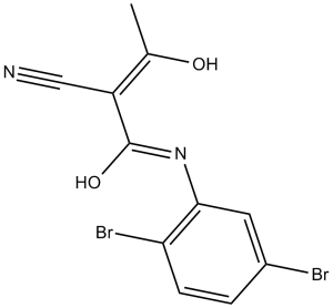
2-Cyano-N-(2,5-dibromophenyl)-3-hydroxy-2-butenamide
|
| 别名 |
LFM-A13; LFM A13; lfm-a13; 244240-24-2; (Z)-2-cyano-N-(2,5-dibromophenyl)-3-hydroxybut-2-enamide; CHEMBL228043; 62004-35-7; SMR001230714; alpha-Cyano-beta-hydroxy-beta-methyl-N-(2,5-dibromophenyl)propenamide; SR-01000075965; LFM A13
|
| HS Tariff Code |
2934.99.9001
|
| 存储方式 |
Powder -20°C 3 years 4°C 2 years In solvent -80°C 6 months -20°C 1 month |
| 运输条件 |
Room temperature (This product is stable at ambient temperature for a few days during ordinary shipping and time spent in Customs)
|
| 溶解度 (体外实验) |
DMSO: 72 mg/mL (200.0 mM)
Water:<1 mg/mL
Ethanol:<1 mg/mL
|
|---|---|
| 溶解度 (体内实验) |
配方 1 中的溶解度: ≥ 2.5 mg/mL (6.94 mM) (饱和度未知) in 10% DMSO + 40% PEG300 + 5% Tween80 + 45% Saline (这些助溶剂从左到右依次添加,逐一添加), 澄清溶液。
例如,若需制备1 mL的工作液,可将100 μL 25.0 mg/mL澄清DMSO储备液加入到400 μL PEG300中,混匀;然后向上述溶液中加入50 μL Tween-80,混匀;加入450 μL生理盐水定容至1 mL。 *生理盐水的制备:将 0.9 g 氯化钠溶解在 100 mL ddH₂O中,得到澄清溶液。 配方 2 中的溶解度: 2.5 mg/mL (6.94 mM) in 10% DMSO + 90% (20% SBE-β-CD in Saline) (这些助溶剂从左到右依次添加,逐一添加), 悬浊液; 超声助溶。 例如,若需制备1 mL的工作液,可将 100 μL 25.0 mg/mL澄清DMSO储备液加入900 μL 20% SBE-β-CD生理盐水溶液中,混匀。 *20% SBE-β-CD 生理盐水溶液的制备(4°C,1 周):将 2 g SBE-β-CD 溶解于 10 mL 生理盐水中,得到澄清溶液。 View More
配方 3 中的溶解度: ≥ 2.5 mg/mL (6.94 mM) (饱和度未知) in 10% DMSO + 90% Corn Oil (这些助溶剂从左到右依次添加,逐一添加), 澄清溶液。 1、请先配制澄清的储备液(如:用DMSO配置50 或 100 mg/mL母液(储备液)); 2、取适量母液,按从左到右的顺序依次添加助溶剂,澄清后再加入下一助溶剂。以 下列配方为例说明 (注意此配方只用于说明,并不一定代表此产品 的实际溶解配方): 10% DMSO → 40% PEG300 → 5% Tween-80 → 45% ddH2O (或 saline); 假设最终工作液的体积为 1 mL, 浓度为5 mg/mL: 取 100 μL 50 mg/mL 的澄清 DMSO 储备液加到 400 μL PEG300 中,混合均匀/澄清;向上述体系中加入50 μL Tween-80,混合均匀/澄清;然后继续加入450 μL ddH2O (或 saline)定容至 1 mL; 3、溶剂前显示的百分比是指该溶剂在最终溶液/工作液中的体积所占比例; 4、 如产品在配制过程中出现沉淀/析出,可通过加热(≤50℃)或超声的方式助溶; 5、为保证最佳实验结果,工作液请现配现用! 6、如不确定怎么将母液配置成体内动物实验的工作液,请查看说明书或联系我们; 7、 以上所有助溶剂都可在 Invivochem.cn网站购买。 |
| 制备储备液 | 1 mg | 5 mg | 10 mg | |
| 1 mM | 2.7778 mL | 13.8889 mL | 27.7778 mL | |
| 5 mM | 0.5556 mL | 2.7778 mL | 5.5556 mL | |
| 10 mM | 0.2778 mL | 1.3889 mL | 2.7778 mL |
1、根据实验需要选择合适的溶剂配制储备液 (母液):对于大多数产品,InvivoChem推荐用DMSO配置母液 (比如:5、10、20mM或者10、20、50 mg/mL浓度),个别水溶性高的产品可直接溶于水。产品在DMSO 、水或其他溶剂中的具体溶解度详见上”溶解度 (体外)”部分;
2、如果您找不到您想要的溶解度信息,或者很难将产品溶解在溶液中,请联系我们;
3、建议使用下列计算器进行相关计算(摩尔浓度计算器、稀释计算器、分子量计算器、重组计算器等);
4、母液配好之后,将其分装到常规用量,并储存在-20°C或-80°C,尽量减少反复冻融循环。
计算结果:
工作液浓度: mg/mL;
DMSO母液配制方法: mg 药物溶于 μL DMSO溶液(母液浓度 mg/mL)。如该浓度超过该批次药物DMSO溶解度,请首先与我们联系。
体内配方配制方法:取 μL DMSO母液,加入 μL PEG300,混匀澄清后加入μL Tween 80,混匀澄清后加入 μL ddH2O,混匀澄清。
(1) 请确保溶液澄清之后,再加入下一种溶剂 (助溶剂) 。可利用涡旋、超声或水浴加热等方法助溶;
(2) 一定要按顺序加入溶剂 (助溶剂) 。
|
|
|
InvivoChem的所有产品仅用于作科学研究,不面向患者销售
Copyright 2020 InvivoChem LLC | All Rights Reserved 粤ICP备20063088号-1


 COA
COA


 463611831
