| 规格 | 价格 | 库存 | 数量 |
|---|---|---|---|
| 5mg |
|
||
| 10mg |
|
||
| 25mg |
|
||
| 50mg |
|
||
| 100mg |
|
||
| 250mg |
|
||
| Other Sizes |
|

| 靶点 |
Protein Kinase C α (PKCα) (IC₅₀ = 0.8 nM) [2]
Protein Kinase C β1 (PKCβ1) (IC₅₀ = 0.9 nM) [2] Protein Kinase C β2 (PKCβ2) (IC₅₀ = 1.2 nM) [2] Protein Kinase C γ (PKCγ) (IC₅₀ = 1.5 nM) [2] Protein Kinase C δ (PKCδ) (IC₅₀ = 3.7 nM) [2] Protein Kinase C ε (PKCε) (IC₅₀ = 4.2 nM) [2] Protein Kinase C η (PKCη) (IC₅₀ = 5.1 nM) [2] Protein Kinase C θ (PKCθ) (IC₅₀ = 6.3 nM) [2] Protein Kinase C ζ (PKCζ) (IC₅₀ = 12.8 nM) [2] Other kinases (selectivity ≥100-fold vs. PKCα): EGFR (IC₅₀ = 150 nM), ERK1 (IC₅₀ = 210 nM), AKT1 (IC₅₀ = 320 nM), JNK1 (IC₅₀ = 280 nM) [2] |
|---|---|
| 体外研究 (In Vitro) |
毒素后,PKC 与蛋白质产物 C 头 Darovasertib (LXS196) 结合,后者抑制 PKC 并阻止 PKC 介导的信号放大器激活。在脆弱的肿瘤细胞中,这可能会导致细胞周期蜡烛和肝癌的产生。 PKC 是一种丝氨酸/苏氨酸蛋白失速,在某些癌细胞类型中过度表达,并与缺血、肿瘤细胞猝死、泌尿生殖道和猝死有关[1]。
1. 强效PKC激酶抑制活性:达罗伐替尼(Darovasertib,LXS-196;IDE-196)对所有经典型(α、β1、β2、γ)和新型(δ、ε、η、θ)PKC亚型均具有纳摩尔级抑制活性,IC₅₀值范围为0.8-6.3 nM;对非典型PKCζ抑制较弱(IC₅₀ = 12.8 nM),对非PKC激酶具有高选择性(相对于PKCα≥100倍),证实其PKC特异性靶向性[2] 2. PKC依赖性癌细胞抗增殖活性:达罗伐替尼以剂量依赖性方式抑制多种PKC信号失调的癌细胞系增殖。72小时MTT法检测IC₅₀值为:UM-UC-3(膀胱癌,2.3 μM)、MUM-2B(葡萄膜黑色素瘤,1.8 μM)、OCM-1A(葡萄膜黑色素瘤,1.5 μM)、PANC-1(胰腺癌,3.1 μM)、MDA-MB-231(乳腺癌,2.7 μM);对正常人成纤维细胞(NHF)无显著抗增殖作用(IC₅₀ > 50 μM)[2] 3. 抑制PKC下游信号通路:达罗伐替尼(0.5-5 μM)以剂量依赖性方式抑制MUM-2B细胞中PKC下游底物的磷酸化,包括ERK1/2(Thr202/Tyr204)、AKT(Ser473)和NF-κB(p65 Ser536)(Western blot检测);ERK1/2、AKT和p65总蛋白水平无变化,证实其特异性抑制PKC介导的信号传导[2] 4. 诱导凋亡与细胞周期阻滞:达罗伐替尼(1-10 μM)处理OCM-1A细胞48小时后,流式细胞术分析显示G2/M期细胞比例从16%升高至45%(5 μM剂量),诱导G2/M期阻滞;Annexin V-FITC/PI染色显示凋亡率从3%升高至38%(10 μM剂量)。Western blot检测到caspase-3、caspase-7和PARP的剪切片段,表明外源性和内源性凋亡通路均被激活[2] 5. 抑制癌细胞迁移与侵袭:达罗伐替尼(0.5-5 μM)以剂量依赖性方式抑制PANC-1细胞迁移(Transwell实验:5 μM剂量下迁移率降低72%)和侵袭(Matrigel Transwell实验:5 μM剂量下侵袭率降低68%),且与基质金属蛋白酶(MMP)-2和MMP-9的mRNA及蛋白表达下调相关(qPCR和Western blot检测)[2] |
| 体内研究 (In Vivo) |
darovasertib(LXS196;化合物 9)以剂量依赖性方式抑制 92.1 GNAQ 葡萄黑色素瘤异种移植模型中的肿瘤生长 [2]。具有 92.1 GNAQ 突变的葡萄膜黑色素瘤细胞被植入小鼠体内 [2]。
1. 葡萄膜黑色素瘤异种移植瘤模型抗肿瘤疗效:6-8周龄BALB/c nu/nu裸鼠皮下接种5×10⁶ MUM-2B细胞,肿瘤体积达100-150 mm³后,随机分为三组(每组n=6):溶媒组(DMSO/PEG400/生理盐水=1:4:5)、达罗伐替尼10 mg/kg组和20 mg/kg组。药物每日腹腔注射(i.p.)一次,持续21天。20 mg/kg组肿瘤体积较溶媒组缩小78%(P<0.001),肿瘤重量减轻65%(P<0.001);肿瘤组织Western blot证实ERK1/2和AKT磷酸化水平降低,剪切型caspase-3表达升高[2] 2. 胰腺癌异种移植瘤模型抗肿瘤疗效:皮下接种1×10⁷ PANC-1细胞的裸鼠接受达罗伐替尼(20 mg/kg,腹腔注射,每日一次)处理28天,肿瘤体积较溶媒组缩小71%(P<0.001),小鼠中位生存期从35天延长至52天(P<0.01)。肿瘤组织病理学分析显示凋亡细胞增多(TUNEL实验),Ki-67阳性增殖细胞减少[2] 3. 异种移植瘤模型中无全身毒性:21-28天治疗期间,达罗伐替尼(10-20 mg/kg,腹腔注射)未导致体重显著变化(平均体重下降<5%)、进食量减少或行为异常;血清ALT、AST、BUN和肌酐水平均在正常范围内,肝、肾、心、肺组织病理学检查未发现药物相关病变[2] |
| 酶活实验 |
1. 重组PKC激酶活性测定:通过异源表达和纯化制备人重组PKC亚型(α、β1、β2、γ、δ、ε、η、θ、ζ);构建含50 nM PKC亚型、10 μM ATP、50 μM荧光标记PKC特异性肽底物、10 mM MgCl₂和不同浓度达罗伐替尼(0.01-100 nM)的反应体系,缓冲液为25 mM Tris-HCl(pH 7.5)、0.1 mM EGTA、1 mM DTT、0.01% Triton X-100;30°C孵育45分钟后,加入50 mM EDTA终止反应,检测荧光强度(激发光485 nm,发射光535 nm)以反映底物磷酸化水平;以抑制剂浓度为横坐标、抑制百分比为纵坐标绘制曲线,计算IC₅₀值[2]
2. 激酶选择性面板实验:将1 μM 达罗伐替尼与97种人激酶(包括EGFR、ERK1、AKT1、JNK1等)共同孵育,采用高通量放射学方法([γ-³²P]ATP掺入)测定激酶活性;计算每种激酶的抑制百分比,生成选择性评分(非PKC激酶IC₅₀与PKCα IC₅₀的比值)[2] |
| 细胞实验 |
1. MTT细胞增殖测定:96孔板接种癌细胞(MUM-2B、OCM-1A、PANC-1、MDA-MB-231)和正常人成纤维细胞(NHF),密度为5×10³个细胞/孔,过夜贴壁后加入0.1-100 μM 达罗伐替尼(溶媒:DMSO+培养基),37°C、5% CO₂孵育72小时;每孔加入20 μL MTT溶液(5 mg/mL),孵育4小时后弃上清,加入150 μL DMSO溶解甲臜结晶,酶标仪测定570 nm吸光度,计算细胞活力和IC₅₀值[2]
2. PKC下游信号Western blot分析:6孔板接种MUM-2B或OCM-1A细胞(1×10⁶个细胞/孔),过夜贴壁后用0.5-5 μM 达罗伐替尼处理24小时;含蛋白酶和磷酸酶抑制剂的RIPA缓冲液裂解细胞,提取总蛋白并BCA定量;SDS-PAGE分离蛋白后转印至PVDF膜,一抗孵育(抗p-ERK1/2(Thr202/Tyr204)、总ERK1/2、p-AKT(Ser473)、总AKT、p-NF-κB p65(Ser536)、总NF-κB p65、剪切型caspase-3、剪切型PARP、微管蛋白(内参));HRP标记二抗孵育后化学发光显影,ImageJ软件定量条带强度[2] 3. 流式细胞术细胞周期与凋亡分析:细胞周期分析:6孔板接种OCM-1A细胞(5×10⁵个细胞/孔),1-10 μM药物处理48小时,70%乙醇固定,碘化丙啶(50 μg/mL)+ RNase A(100 μg/mL)染色,流式细胞术分析;凋亡分析:相同浓度药物处理48小时,Annexin V-FITC/PI染色,流式细胞术检测凋亡细胞[2] 4. Transwell迁移与侵袭实验:迁移实验:Transwell上室(8 μm孔径)接种PANC-1细胞(1×10⁴个细胞/孔),加入含达罗伐替尼(0.5-5 μM)的无血清培养基,下室加入含10% FBS的培养基,孵育24小时后甲醇固定下室细胞,结晶紫染色计数迁移细胞;侵袭实验:使用Matrigel包被的Transwell小室,其余步骤同迁移实验,孵育48小时[2] 5. MMP-2和MMP-9表达qPCR分析:0.5-5 μM 达罗伐替尼处理PANC-1细胞24小时,提取总RNA并逆转录为cDNA,用MMP-2、MMP-9和GAPDH(内参)特异性引物进行qPCR,采用2⁻ΔΔCt法计算相对基因表达量[2] |
| 动物实验 |
动物/疾病模型: 小鼠植入92.1 GNAQ突变型葡萄膜黑色素瘤细胞[2]。
剂量: 15、30、75、150 mg/kg 给药途径: 口服(每日两次),持续35天 实验结果: 呈剂量依赖性地抑制肿瘤生长。 1. MUM-2B葡萄膜黑色素瘤异种移植模型:使用6-8周龄雌性BALB/c裸鼠(每组n=6)。将5×10⁶个MUM-2B细胞悬浮于0.2 mL PBS:Matrigel(1:1)混合液中,皮下注射至小鼠右侧腹部。每日监测肿瘤生长情况;当肿瘤体积达到100-150 mm³时,开始治疗。将达罗伐塞替尼溶于DMSO(终体积10%),用PEG400(终体积40%)和生理盐水(终体积50%)稀释,配制成1 mg/mL和2 mg/mL的溶液。每日腹腔注射给药一次(10 mg/kg或20 mg/kg),持续21天;对照组注射不含药物的相同DMSO/PEG400/生理盐水混合物。每3天测量一次肿瘤体积(长×宽²/2)和体重。治疗结束后处死小鼠,解剖肿瘤进行Western blot和组织病理学分析[2] 2. PANC-1胰腺癌异种移植模型:使用6-8周龄雌性BALB/c裸鼠(每组n=8)。将 1×10⁷ 个 PANC-1 细胞悬浮于 0.2 mL PBS:Matrigel (1:1) 混合液中,皮下注射至小鼠右侧腹部。当肿瘤体积达到 100-150 mm³ 时,给予达罗伐塞替(Darovasertib,20 mg/kg,腹腔注射,每日一次)或载体对照,持续 28 天。每 3 天监测一次肿瘤体积和体重。每日记录小鼠存活情况。研究结束时,处死存活小鼠,解剖肿瘤组织进行 TUNEL 检测和 Ki-67 免疫组化分析 [2] |
| 药代性质 (ADME/PK) |
1. 口服吸收:单次口服 20 mg/kg 剂量后,达罗伐替尼 在大鼠中显示出中等的口服生物利用度 (42%)。血浆峰浓度 (Cₘₐₓ) 为 1.8 μg/mL,达峰时间为 1.2 小时 (Tₘₐₓ) [2]
2. 血浆蛋白结合率:体外人血浆蛋白结合率为 92-94%(浓度范围:0.1-10 μg/mL),无浓度依赖性结合 [2] 3. 半衰期:大鼠(静脉注射)末端消除半衰期 (t₁/₂) 为 6.8 小时,犬(静脉注射)末端消除半衰期为 8.3 小时 [2] 4. 组织分布:大鼠单次静脉注射 10 mg/kg 达罗伐塞替后,药物广泛分布于组织中,肝脏、肾脏和肿瘤组织中的浓度最高。脑/血浆浓度比为0.08,表明其血脑屏障穿透性有限[2] 5. 代谢:达罗伐塞替主要在肝脏中通过CYP3A4介导的氧化和葡萄糖醛酸化代谢。主要代谢物(M1)对PKC同工酶无活性(IC₅₀ > 100 nM)[2] 6. 排泄:在大鼠中,静脉注射剂量的65%在72小时内经粪便排出(30%为原药),28%经尿液排出(5%为原药)[2] |
| 毒性/毒理 (Toxicokinetics/TK) |
1. 急性毒性:达罗伐替尼(Darovasertib)在小鼠和大鼠中的半数致死剂量(LD₅₀)>200 mg/kg(口服),在大鼠中>100 mg/kg(腹腔注射),在犬中>50 mg/kg(静脉注射)[2]。
2. 亚慢性毒性:在一项为期4周的大鼠重复给药毒性研究中(剂量:10、30、100 mg/kg/天,口服),未观察到与治疗相关的死亡。在100 mg/kg/天剂量下观察到肝脏重量轻微增加,但未检测到组织病理学变化或肝功能指标改变。血液学参数(红细胞、白细胞、血小板)均在正常范围内[2] 3. 遗传毒性:达罗伐替尼在Ames试验、体外染色体畸变试验和体内微核试验中均为阴性,表明其无遗传毒性[2] 4. 药物相互作用:体外研究表明,在浓度高达10 μM时,达罗伐替尼对CYP450酶(CYP1A2、2C9、2C19、2D6、3A4)无抑制作用,且未诱导人肝细胞中CYP3A4 mRNA的表达[2] |
| 参考文献 | |
| 其他信息 |
IDE-196 正在进行临床试验 NCT03947385(IDE196 在携带 GNAQ/11 突变或 PRKC 融合的实体瘤患者中的研究)。
Darovasertib 是一种口服蛋白激酶 C (PKC) 抑制剂,具有潜在的免疫抑制和抗肿瘤活性。口服后,darovasertib 可靶向并抑制 PKC,从而阻止 PKC 介导的信号通路激活。这可能导致易感肿瘤细胞发生细胞周期阻滞和凋亡。 PKC 是一种丝氨酸/苏氨酸蛋白激酶,在某些类型的癌细胞中过度表达,参与肿瘤细胞的分化、增殖、侵袭和存活。 1. 化学和物理性质:达罗伐塞替 (LXS-196; IDE-196) 是一种小分子 ATP 竞争性 PKC 抑制剂,其化学名称为 (R)-3-(1-(4-(4-氟苯基)哌嗪-1-基)乙基)-2-氧代-2,3-二氢-1H-苯并[d]咪唑-5-腈。它是一种白色至类白色结晶性粉末,可溶于DMSO(≥50 mg/mL)和乙醇(≥10 mg/mL),微溶于水[2] 2. 作用机制:达罗伐替尼与PKC亚型的ATP结合口袋结合,阻止ATP结合和随后的激酶激活。通过抑制PKC介导的下游信号分子(ERK1/2、AKT、NF-κB)的磷酸化,它阻断癌细胞的增殖、存活、迁移和侵袭,并诱导细胞凋亡[2] 3. 治疗潜力:已开发用于治疗PKC依赖性实体瘤,包括葡萄膜黑色素瘤、胰腺癌、膀胱癌和乳腺癌。临床前数据支持其作为单药或与其他化疗药物(例如,吉西他滨用于治疗胰腺癌)联合使用[2] 4. 临床开发背景:该化合物受美国专利US20180179181保护,该专利描述了其合成、药理学特征和临床前疗效。该化合物已进入临床试验阶段,用于治疗晚期实体瘤,特别是葡萄膜黑色素瘤(一种治疗选择有限且高度依赖PKC信号通路的疾病)[2] |
| 分子式 |
C22H23F3N8O
|
|---|---|
| 分子量 |
472.47
|
| 精确质量 |
472.194
|
| CAS号 |
1874276-76-2
|
| 相关CAS号 |
1874276-76-2;LXS-196 HCl;
|
| PubChem CID |
118873253
|
| 外观&性状 |
Light yellow to yellow solid powder
|
| 密度 |
1.4±0.1 g/cm3
|
| 沸点 |
592.7±50.0 °C at 760 mmHg
|
| 闪点 |
312.3±30.1 °C
|
| 蒸汽压 |
0.0±1.7 mmHg at 25°C
|
| 折射率 |
1.622
|
| LogP |
3.7
|
| tPSA |
136
|
| 氢键供体(HBD)数目 |
3
|
| 氢键受体(HBA)数目 |
11
|
| 可旋转键数目(RBC) |
4
|
| 重原子数目 |
34
|
| 分子复杂度/Complexity |
702
|
| 定义原子立体中心数目 |
0
|
| InChi Key |
XXJXHXJWQSCNPX-UHFFFAOYSA-N
|
| InChi Code |
InChI=1S/C22H23F3N8O/c1-21(27)6-10-33(11-7-21)15-5-3-9-29-19(15)32-20(34)17-18(26)30-12-14(31-17)16-13(22(23,24)25)4-2-8-28-16/h2-5,8-9,12H,6-7,10-11,27H2,1H3,(H2,26,30)(H,29,32,34)
|
| 化学名 |
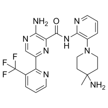
3-amino-N-[3-(4-amino-4-methylpiperidin-1-yl)pyridin-2-yl]-6-[3-(trifluoromethyl)pyridin-2-yl]pyrazine-2-carboxamide
|
| 别名 |
LXS196; NVP-LXS196; LXS-196; NVP-LXS-196
|
| HS Tariff Code |
2934.99.9001
|
| 存储方式 |
Powder -20°C 3 years 4°C 2 years In solvent -80°C 6 months -20°C 1 month |
| 运输条件 |
Room temperature (This product is stable at ambient temperature for a few days during ordinary shipping and time spent in Customs)
|
| 溶解度 (体外实验) |
DMSO : ~25 mg/mL (~52.91 mM)
|
|---|---|
| 溶解度 (体内实验) |
配方 1 中的溶解度: ≥ 2.5 mg/mL (5.29 mM) (饱和度未知) in 10% DMSO + 40% PEG300 + 5% Tween80 + 45% Saline (这些助溶剂从左到右依次添加,逐一添加), 澄清溶液。
例如,若需制备1 mL的工作液,可将100 μL 25.0 mg/mL澄清DMSO储备液加入到400 μL PEG300中,混匀;然后向上述溶液中加入50 μL Tween-80,混匀;加入450 μL生理盐水定容至1 mL。 *生理盐水的制备:将 0.9 g 氯化钠溶解在 100 mL ddH₂O中,得到澄清溶液。 配方 2 中的溶解度: ≥ 2.5 mg/mL (5.29 mM) (饱和度未知) in 10% DMSO + 90% (20% SBE-β-CD in Saline) (这些助溶剂从左到右依次添加,逐一添加), 澄清溶液。 例如,若需制备1 mL的工作液,可将 100 μL 25.0 mg/mL澄清DMSO储备液加入900 μL 20% SBE-β-CD生理盐水溶液中,混匀。 *20% SBE-β-CD 生理盐水溶液的制备(4°C,1 周):将 2 g SBE-β-CD 溶解于 10 mL 生理盐水中,得到澄清溶液。 View More
配方 3 中的溶解度: ≥ 2.5 mg/mL (5.29 mM) (饱和度未知) in 10% DMSO + 90% Corn Oil (这些助溶剂从左到右依次添加,逐一添加), 澄清溶液。 配方 4 中的溶解度: ≥ 1.67 mg/mL (3.53 mM) (饱和度未知) in 5% DMSO + 40% PEG300 + 5% Tween80 + 50% Saline (这些助溶剂从左到右依次添加,逐一添加), 澄清溶液。 *生理盐水的制备:将 0.9 g 氯化钠溶解在 100 mL ddH₂O中,得到澄清溶液。 配方 5 中的溶解度: ≥ 1.67 mg/mL (3.53 mM) (饱和度未知) in 5% DMSO + 95% (20% SBE-β-CD in Saline) (这些助溶剂从左到右依次添加,逐一添加), 澄清溶液。 *20% SBE-β-CD 生理盐水溶液的制备(4°C,1 周):将 2 g SBE-β-CD 溶解于 10 mL 生理盐水中,得到澄清溶液。 配方 6 中的溶解度: 0.33 mg/mL (0.70 mM) in 1% DMSO 99% Saline (这些助溶剂从左到右依次添加,逐一添加), 悬浊液; 超声助溶。 *生理盐水的制备:将 0.9 g 氯化钠溶解在 100 mL ddH₂O中,得到澄清溶液。 1、请先配制澄清的储备液(如:用DMSO配置50 或 100 mg/mL母液(储备液)); 2、取适量母液,按从左到右的顺序依次添加助溶剂,澄清后再加入下一助溶剂。以 下列配方为例说明 (注意此配方只用于说明,并不一定代表此产品 的实际溶解配方): 10% DMSO → 40% PEG300 → 5% Tween-80 → 45% ddH2O (或 saline); 假设最终工作液的体积为 1 mL, 浓度为5 mg/mL: 取 100 μL 50 mg/mL 的澄清 DMSO 储备液加到 400 μL PEG300 中,混合均匀/澄清;向上述体系中加入50 μL Tween-80,混合均匀/澄清;然后继续加入450 μL ddH2O (或 saline)定容至 1 mL; 3、溶剂前显示的百分比是指该溶剂在最终溶液/工作液中的体积所占比例; 4、 如产品在配制过程中出现沉淀/析出,可通过加热(≤50℃)或超声的方式助溶; 5、为保证最佳实验结果,工作液请现配现用! 6、如不确定怎么将母液配置成体内动物实验的工作液,请查看说明书或联系我们; 7、 以上所有助溶剂都可在 Invivochem.cn网站购买。 |
| 制备储备液 | 1 mg | 5 mg | 10 mg | |
| 1 mM | 2.1165 mL | 10.5827 mL | 21.1654 mL | |
| 5 mM | 0.4233 mL | 2.1165 mL | 4.2331 mL | |
| 10 mM | 0.2117 mL | 1.0583 mL | 2.1165 mL |
1、根据实验需要选择合适的溶剂配制储备液 (母液):对于大多数产品,InvivoChem推荐用DMSO配置母液 (比如:5、10、20mM或者10、20、50 mg/mL浓度),个别水溶性高的产品可直接溶于水。产品在DMSO 、水或其他溶剂中的具体溶解度详见上”溶解度 (体外)”部分;
2、如果您找不到您想要的溶解度信息,或者很难将产品溶解在溶液中,请联系我们;
3、建议使用下列计算器进行相关计算(摩尔浓度计算器、稀释计算器、分子量计算器、重组计算器等);
4、母液配好之后,将其分装到常规用量,并储存在-20°C或-80°C,尽量减少反复冻融循环。
计算结果:
工作液浓度: mg/mL;
DMSO母液配制方法: mg 药物溶于 μL DMSO溶液(母液浓度 mg/mL)。如该浓度超过该批次药物DMSO溶解度,请首先与我们联系。
体内配方配制方法:取 μL DMSO母液,加入 μL PEG300,混匀澄清后加入μL Tween 80,混匀澄清后加入 μL ddH2O,混匀澄清。
(1) 请确保溶液澄清之后,再加入下一种溶剂 (助溶剂) 。可利用涡旋、超声或水浴加热等方法助溶;
(2) 一定要按顺序加入溶剂 (助溶剂) 。
InvivoChem的所有产品仅用于作科学研究,不面向患者销售
Copyright 2020 InvivoChem LLC | All Rights Reserved 粤ICP备20063088号-1


 COA
COA
 463611831
