| 规格 | 价格 | 库存 | 数量 |
|---|---|---|---|
| 5mg |
|
||
| 10mg |
|
||
| 25mg |
|
||
| 50mg |
|
||
| 100mg |
|
||
| 250mg |
|
||
| 500mg |
|
||
| Other Sizes |
|
| 靶点 |
MELK (IC50 = 2 nM)
Maternal Embryonic Leucine Zipper Kinase (MELK) (IC₅₀=0.9 nM in recombinant kinase assay; Ki=0.3 nM by SPR binding assay) [1] |
|---|---|
| 体外研究 (In Vitro) |
当生化检测中 ATP 浓度从 20 μM 变为 2 mM 时,MELK-8a 仍然非常有效(IC50=140 nM)。催化结构域构建体和全长 MELK(5 nM 与 2 nM)之间的效力差异清晰可见。除 MELK 外,它仅抑制其他七种脱靶激酶,表现出优异的选择性,在 1 μM 时结合抑制率 >85%。
MELK-8a HCl 是一种高效、高选择性的ATP竞争性MELK抑制剂,对468种人源激酶面板的交叉反应性极低(选择性评分S₁₀=0.01,表明对MELK的选择性>99%)[1] - 在HTRF-based激酶实验中,强效抑制重组人MELK激酶活性,IC₅₀=0.9 nM;表面等离子体共振(SPR)结合分析测定其Ki=0.3 nM[1] - 对MELK过表达的肿瘤细胞系具有剂量依赖性抗增殖活性:GI₅₀值分别为0.12 μM(三阴性乳腺癌(TNBC)细胞系MDA-MB-468)、0.18 μM(TNBC细胞系BT-549)、0.25 μM(结直肠癌细胞系HCT-116)、0.31 μM(卵巢癌细胞系SKOV3)、0.45 μM(胰腺癌细胞系PANC-1);MELK低表达细胞(如MCF-7,GI₅₀=5.8 μM)敏感性降低[1] - 诱导MDA-MB-468细胞G₂/M期细胞周期停滞:0.2 μM MELK-8a HCl 处理24小时后,G₂/M期细胞比例从18%增至42%,同时下调Cyclin B1和Cdc25C的mRNA及蛋白水平[1] - 促进MELK过表达癌细胞凋亡:Annexin V/PI染色显示,0.5 μM处理48小时后,MDA-MB-468和BT-549细胞的凋亡率分别增至38%和32%(溶媒对照组分别为6%和5%);Western blot检测到Caspase-3和PARP的切割产物[1] - 抑制MELK介导的下游信号通路:0.1–1 μM MELK-8a HCl 剂量依赖性降低MDA-MB-468细胞中STAT3(Tyr705)和BAD(Ser112)的磷酸化水平,而总STAT3和BAD水平无变化[1] - 抑制MDA-MB-468细胞克隆形成:0.05–0.2 μM MELK-8a HCl 培养14天后,克隆形成效率较溶媒对照组降低40–75%[1] - siRNA敲低MDA-MB-468细胞中的MELK后,细胞对 MELK-8a HCl 的敏感性降低(GI₅₀从0.12 μM升至1.8 μM),证实其抗增殖活性依赖于MELK抑制[1] |
| 体内研究 (In Vivo) |
在 C57BL/6 小鼠中,皮下给予 30 mg/kg MELK-8a 的血浆暴露良好。峰值血浆浓度达到 6.6 M,化合物很快就会被吸收到体循环中(Tmax=0.4 小时)。根据对雌性无胸腺裸鼠进行的递增剂量 PK 研究,所有清除机制可在 240 mg/kg 时达到饱和,此时化合物释放速率达到最大。另一方面,当以 10 mg/kg 的剂量口服时,它在 C57BL/6 雄性小鼠模型中表现出非常差的 PK(口服生物利用度为 3.6%)[1]。
在MDA-MB-468(TNBC)移植瘤模型(BALB/c裸鼠)中,口服给予 MELK-8a HCl 25 mg/kg、50 mg/kg、100 mg/kg,每日两次(BID),连续21天,呈剂量依赖性诱导肿瘤生长抑制(TGI),抑制率分别为56%、78%和91%;100 mg/kg BID组6只小鼠中有3只实现部分肿瘤缓解(PR)[1] - 移植瘤药效动力学分析:50 mg/kg BID MELK-8a HCl 治疗7天后,与溶媒对照组相比,肿瘤组织中MELK激酶活性降低65%,p-STAT3(Tyr705)和Cyclin B1蛋白水平分别降低58%和62%[1] - 所有治疗组均未观察到显著体重下降(<5%),表明体内耐受性良好[1] |
| 酶活实验 |
重组MELK激酶活性实验(HTRF法):将重组人MELK激酶稀释于实验缓冲液(50 mM Tris-HCl pH 7.5、10 mM MgCl₂、1 mM EGTA、0.01% BSA、1 mM DTT)中。将 MELK-8a HCl 的系列3倍稀释液(0.001–10 nM)与激酶混合,室温预孵育30分钟。加入ATP(终浓度5 μM)和生物素化肽底物(终浓度2 μM)启动反应,37°C孵育60分钟。用50 mM EDTA终止反应,通过链霉亲和素偶联珠和抗磷酸酪氨酸抗体检测磷酸化底物。检测荧光强度,通过非线性回归计算IC₅₀值[1]
- SPR结合实验:将重组人MELK激酶域固定在传感器芯片上。在运行缓冲液(10 mM HEPES pH 7.4、150 mM NaCl、0.05% Tween 20)中,将 MELK-8a HCl 的系列2倍稀释液(0.01–100 nM)注入芯片表面。使用稳态亲和模型分析传感图,确定结合亲和力(Ki)[1] - 激酶选择性面板实验:在1 μM浓度下,通过放射活性激酶实验检测 MELK-8a HCl 对468种重组人源激酶的抑制活性。计算选择性评分S₁₀(抑制率>90%的激酶比例)以评估脱靶活性[1] |
| 细胞实验 |
MDA-MB-468 和 MCF7 细胞分别以 1000 和 4000 个细胞/孔接种在 96 孔生长培养基板中。铺板后 16 小时添加 MELK-8a,并孵育 7 天。将 ATPLite 试剂添加到每个孔中,然后孵育。多标签读板器用于测量发光[1]。
肿瘤细胞抗增殖实验:将MELK过表达和低表达的肿瘤细胞系以5×10³个/孔接种到96孔板,过夜孵育。加入 MELK-8a HCl 的系列3倍稀释液(0.001–10 μM),培养72小时。通过MTS实验检测细胞活力,计算GI₅₀值[1] - 细胞周期分析:MDA-MB-468细胞以2×10⁵个/孔接种到6孔板,用 MELK-8a HCl(0.05–0.5 μM)处理24小时。收集细胞,用70%乙醇固定,碘化丙啶(PI)染色,通过流式细胞术分析细胞周期分布[1] - 凋亡检测实验:MDA-MB-468和BT-549细胞用 MELK-8a HCl(0.1–1 μM)处理48小时,收集细胞并与Annexin V-FITC和PI染色,通过流式细胞术量化凋亡率[1] - 信号通路Western blot实验:肿瘤细胞用 MELK-8a HCl(0.05–1 μM)处理24小时后裂解,蛋白经SDS-PAGE分离,膜上用抗p-STAT3(Tyr705)、STAT3、p-BAD(Ser112)、BAD、Cyclin B1、Cdc25C、切割型Caspase-3、切割型PARP和β-肌动蛋白抗体进行免疫印迹[1] - 克隆形成实验:MDA-MB-468细胞以500个/孔接种到6孔板,过夜孵育。加入 MELK-8a HCl(0.05–0.2 μM),培养14天。克隆用甲醇固定,结晶紫染色后计数;相对于溶媒对照组计算克隆形成效率[1] - MELK siRNA敲低实验:MDA-MB-468细胞转染MELK特异性siRNA或 scramble siRNA。转染48小时后,用 MELK-8a HCl(0.01–10 μM)处理细胞72小时。通过MTS实验检测细胞活力,比较敲低组和对照组的GI₅₀值[1] |
| 动物实验 |
小鼠:用于药代动力学研究的静脉注射和口服剂量配制于含有 5% 乙醇、100% 丙二醇 (PG)、5% CremophorEL 和 80% PBS 的溶液中。皮下注射剂量的制剂为 10% PG 和 25% (20%, v/v) Solutol。在进行 MELK-8a 分析之前,按预定时间间隔采集血浆样本并冷冻保存 (20 °C)。采用 LC-MS/MS 技术定量血浆中 MELK-8a 的药物浓度[1]。MDA-MB-468 TNBC 异种移植模型:将 5×10⁶ 个 MDA-MB-468 细胞(悬浮于 50% Matrigel/PBS 中)皮下植入 6-8 周龄 BALB/c 裸鼠的右侧腹部。当肿瘤体积达到 100–150 mm³ 时,将小鼠随机分为载体对照组和治疗组(每组 n=6)。MELK-8a HCl 配制于 0.5% 羧甲基纤维素钠 (CMC-Na) + 0.1% Tween 80 溶液中,并以 25 mg/kg、50 mg/kg 或 100 mg/kg 的剂量,每日两次口服给药,持续 21 天。载体对照组小鼠接受相同体积的 0.5% CMC-Na + 0.1% Tween 80 溶液。每 3 天用游标卡尺测量肿瘤大小,肿瘤体积计算公式为:长×宽²×0.5 [1]
- 药效学取样:将携带 MDA-MB-468 异种移植瘤的小鼠用 MELK-8a HCl 50 mg/kg,每日两次给药,持续 7 天。研究结束时收集肿瘤组织,液氮速冻,并通过蛋白质印迹法分析p-STAT3、细胞周期蛋白B1和总MELK的表达水平;采用基于HTRF的激酶活性测定法测定MELK激酶活性[1] - 体内耐受性监测:每周测量小鼠体重两次;在整个研究期间每日观察小鼠的一般健康状况(活动、食物摄入量、腹泻情况)[1] |
| 药代性质 (ADME/PK) |
口服生物利用度:大鼠为 58%(口服 10 mg/kg),犬为 65%(口服 5 mg/kg)[1]
- 血浆药代动力学:大鼠口服 10 mg/kg 后,Cmax=2.8 μg/mL,AUC₀–24h=18.5 μg·h/mL,末端半衰期 (t₁/₂)=6.3 小时;静脉注射(2 mg/kg)显示分布容积 (Vd) = 2.1 L/kg,清除率 (CL) = 0.15 L/h/kg [1] - 在犬中,口服 5 mg/kg 后,血药峰浓度 (Cmax) = 1.9 μg/mL,24 小时曲线下面积 (AUC₀–24h) = 14.2 μg·h/mL,半衰期 (t₁/₂) = 8.7 小时 [1] - 血浆蛋白结合率:在人、大鼠和犬血浆中为 92–94%(平衡透析,0.1–10 μg/mL)[1] - 代谢:主要在人肝微粒体中通过细胞色素 P450 3A4 (CYP3A4) 代谢;已鉴定出一种主要代谢物 (M1),其对 MELK 的抑制效力比母体药物低 30 倍 [1] |
| 毒性/毒理 (Toxicokinetics/TK) |
急性毒性(小鼠):单次口服 300 mg/kg 的 MELK-8a HCl 未引起死亡或严重毒性;6 只小鼠中有 2 只出现轻微的短暂性腹泻 [1]
- 亚慢性毒性(大鼠,28 天):每日两次口服剂量高达 100 mg/kg,未见体重、食物摄入量或血液学/生化指标(ALT、AST、BUN、肌酐)的显著变化;主要器官(肝脏、肾脏、心脏、肺)未发现组织病理学异常 [1] - 在浓度高达 10 μM 时,未发现对 CYP450 酶(CYP1A2、CYP2C9、CYP2C19、CYP2D6、CYP3A4)的显著抑制作用 [1] |
| 参考文献 | |
| 其他信息 |
MELK-8a HCl 是一种高效且选择性极强的 MELK 小分子抑制剂,旨在验证 MELK 作为癌症治疗靶点的有效性 [1]
- MELK(母体胚胎亮氨酸拉链激酶)在多种人类癌症(例如三阴性乳腺癌、结直肠癌、卵巢癌)中过度表达,并在调节细胞周期进程、细胞凋亡和癌细胞干性方面发挥关键作用 [1] - 其作用机制涉及与 MELK 激酶结构域进行 ATP 竞争性结合,抑制 MELK 的催化活性及其下游信号通路(STAT3/BAD 和 Cyclin B1/Cdc25C),从而导致 MELK 过表达的癌细胞发生 G₂/M 期细胞周期阻滞和细胞凋亡 [1] - 对 MELK 的高选择性最大限度地减少了脱靶效应,并且具有良好的药代动力学特性(良好的口服生物利用度、适中的半衰期、高血浆蛋白结合率)支持其临床开发潜力[1] - TNBC异种移植模型中的临床前数据表明,MELK-8a HCl 具有强大的抗肿瘤疗效和良好的耐受性,提示其可能是一种有前景的MELK过表达癌症(尤其是三阴性乳腺癌)治疗药物[1] |
| 分子式 |
C25H33CLN6O
|
|
|---|---|---|
| 分子量 |
469.022124052048
|
|
| 精确质量 |
468.24
|
|
| 元素分析 |
C, 64.02; H, 7.09; Cl, 7.56; N, 17.92; O, 3.41
|
|
| CAS号 |
2096992-20-8
|
|
| 相关CAS号 |
MELK-8a;1922153-17-0
|
|
| PubChem CID |
126843227
|
|
| 外观&性状 |
Light yellow to yellow solid powder
|
|
| tPSA |
58.4
|
|
| 氢键供体(HBD)数目 |
2
|
|
| 氢键受体(HBA)数目 |
6
|
|
| 可旋转键数目(RBC) |
6
|
|
| 重原子数目 |
33
|
|
| 分子复杂度/Complexity |
557
|
|
| 定义原子立体中心数目 |
0
|
|
| SMILES |
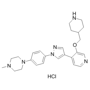
CN1CCN(CC1)C2=CC=C(C=C2)N3C=C(C=N3)C4=C(C=NC=C4)OCC5CCNCC5.Cl
|
|
| InChi Key |
AFGMSRRNYDSRPT-UHFFFAOYSA-N
|
|
| InChi Code |
InChI=1S/C25H32N6O.ClH/c1-29-12-14-30(15-13-29)22-2-4-23(5-3-22)31-18-21(16-28-31)24-8-11-27-17-25(24)32-19-20-6-9-26-10-7-20;/h2-5,8,11,16-18,20,26H,6-7,9-10,12-15,19H2,1H3;1H
|
|
| 化学名 |
1-methyl-4-[4-[4-[3-(piperidin-4-ylmethoxy)pyridin-4-yl]pyrazol-1-yl]phenyl]piperazine;hydrochloride
|
|
| 别名 |
|
|
| HS Tariff Code |
2934.99.9001
|
|
| 存储方式 |
Powder -20°C 3 years 4°C 2 years In solvent -80°C 6 months -20°C 1 month 注意: 请将本产品存放在密封且受保护的环境中,避免吸湿/受潮。 |
|
| 运输条件 |
Room temperature (This product is stable at ambient temperature for a few days during ordinary shipping and time spent in Customs)
|
| 溶解度 (体外实验) |
|
|||
|---|---|---|---|---|
| 溶解度 (体内实验) |
配方 1 中的溶解度: 50 mg/mL (106.61 mM) in PBS (这些助溶剂从左到右依次添加,逐一添加), 澄清溶液; 超声助溶。
请根据您的实验动物和给药方式选择适当的溶解配方/方案: 1、请先配制澄清的储备液(如:用DMSO配置50 或 100 mg/mL母液(储备液)); 2、取适量母液,按从左到右的顺序依次添加助溶剂,澄清后再加入下一助溶剂。以 下列配方为例说明 (注意此配方只用于说明,并不一定代表此产品 的实际溶解配方): 10% DMSO → 40% PEG300 → 5% Tween-80 → 45% ddH2O (或 saline); 假设最终工作液的体积为 1 mL, 浓度为5 mg/mL: 取 100 μL 50 mg/mL 的澄清 DMSO 储备液加到 400 μL PEG300 中,混合均匀/澄清;向上述体系中加入50 μL Tween-80,混合均匀/澄清;然后继续加入450 μL ddH2O (或 saline)定容至 1 mL; 3、溶剂前显示的百分比是指该溶剂在最终溶液/工作液中的体积所占比例; 4、 如产品在配制过程中出现沉淀/析出,可通过加热(≤50℃)或超声的方式助溶; 5、为保证最佳实验结果,工作液请现配现用! 6、如不确定怎么将母液配置成体内动物实验的工作液,请查看说明书或联系我们; 7、 以上所有助溶剂都可在 Invivochem.cn网站购买。 |
| 制备储备液 | 1 mg | 5 mg | 10 mg | |
| 1 mM | 2.1321 mL | 10.6605 mL | 21.3211 mL | |
| 5 mM | 0.4264 mL | 2.1321 mL | 4.2642 mL | |
| 10 mM | 0.2132 mL | 1.0661 mL | 2.1321 mL |
1、根据实验需要选择合适的溶剂配制储备液 (母液):对于大多数产品,InvivoChem推荐用DMSO配置母液 (比如:5、10、20mM或者10、20、50 mg/mL浓度),个别水溶性高的产品可直接溶于水。产品在DMSO 、水或其他溶剂中的具体溶解度详见上”溶解度 (体外)”部分;
2、如果您找不到您想要的溶解度信息,或者很难将产品溶解在溶液中,请联系我们;
3、建议使用下列计算器进行相关计算(摩尔浓度计算器、稀释计算器、分子量计算器、重组计算器等);
4、母液配好之后,将其分装到常规用量,并储存在-20°C或-80°C,尽量减少反复冻融循环。
计算结果:
工作液浓度: mg/mL;
DMSO母液配制方法: mg 药物溶于 μL DMSO溶液(母液浓度 mg/mL)。如该浓度超过该批次药物DMSO溶解度,请首先与我们联系。
体内配方配制方法:取 μL DMSO母液,加入 μL PEG300,混匀澄清后加入μL Tween 80,混匀澄清后加入 μL ddH2O,混匀澄清。
(1) 请确保溶液澄清之后,再加入下一种溶剂 (助溶剂) 。可利用涡旋、超声或水浴加热等方法助溶;
(2) 一定要按顺序加入溶剂 (助溶剂) 。
InvivoChem的所有产品仅用于作科学研究,不面向患者销售
Copyright 2020 InvivoChem LLC | All Rights Reserved 粤ICP备20063088号-1


 COA
COA
 463611831
