| 规格 | 价格 | 库存 | 数量 |
|---|---|---|---|
| 10 mM * 1 mL in DMSO |
|
||
| 1mg |
|
||
| 5mg |
|
||
| 10mg |
|
||
| 25mg |
|
||
| 50mg |
|
||
| 100mg |
|
||
| Other Sizes |
|
| 靶点 |
MELK (IC50 = 0.41 nM)
|
|---|---|
| 体外研究 (In Vitro) |
OTSSP167 阻断 MELK 过表达的癌细胞 A549、T47D、DU4475 和 22Rv1,IC50 值分别为 6.7、4.3、2.3 和 6.0 nM。 OTSSP167 阻止两种新型 MELK 底物 PSMA1(蛋白酶体 α 亚基 1 型)和 DBNL(类 Drebrin)的磷酸化,这两种底物对于干细胞特性和侵袭性至关重要。通过抑制 PSMA1 磷酸化,OTSSP167 可以防止乳腺癌细胞形成乳腺球。 [1]
|
| 体内研究 (In Vivo) |
在小鼠体内使用乳腺癌、肺癌、前列腺癌和胰腺癌细胞系进行的异种移植研究中,通过静脉内和口服给药,OTSSP167 表现出显着的肿瘤生长抑制作用。 OTSSP167以20mg/kg的剂量每两天一次静脉注射至MDA-MB-231模型,TGI为73%。每日一次口服剂量 10 mg/kg 时,TGI 为 72%。 OTSSP167 以剂量和 MELK 依赖性方式治疗各种癌症类型,且体重几乎没有减轻。 [1]
OTSSP167在异种移植小鼠模型中的生长抑制作用[1] 随后,我们通过使用MDA-MB-231细胞(MELK-阳性、三阴性乳腺癌症细胞)的异种移植模型研究了OTSSP167的体内抗肿瘤作用。在肿瘤大小达到约100mm3后,将该化合物给予携带异种移植物的小鼠14天。肿瘤大小被测量为药物反应的替代标志物(肿瘤生长抑制(TGI))。每两天静脉注射一次20mg/kg的OTSSP167,TGI为73%(图3A)。由于该化合物的生物利用度预计非常高(数据未显示),我们尝试口服该化合物。口服10mg/kg,每天一次,TGI为72%(图3B)。由于对各种癌症细胞系具有强烈的生长抑制作用,我们使用其他类型的癌症细胞系进一步研究了体内生长抑制效应,并发现OTSSP167以剂量依赖的方式对多种癌症类型显着抑制肿瘤生长,没有或有少量体重损失(图3和补充图S1)。例如,携带A549(肺癌)异种移植物的小鼠通过静脉内施用1、5和10 mg/kg OTSSP167每天一次治疗,其TGI分别为51%、91%和108%(图3C),而通过口服施用5和10 mg/kg每天一次的小鼠显示TGI分别为95%和124%(图3D)。此外,我们通过每天一次口服10 mg/kg检查了DU145(癌症)和MIAPaCa-2(癌症)异种移植物模型,并观察到TGI分别为106和87%(图3E和F)。为了进一步验证MELK特异性体内肿瘤抑制作用,我们检测了几乎无法检测到MELK表达的PC-14肺癌细胞(图3G)。口服10mg/kg OTSSP167,每天一次,持续14天,对PC-14异种移植物没有肿瘤生长抑制作用(图3H),进一步支持了OTSSP177的MELK依赖性抗肿瘤活性。 OTSSP16治疗在临床前GC患者衍生的异种移植物(PDX)小鼠模型中的疗效[2] 从我们建立的数据库中选择了两个MELK阳性和一个MELK阴性的GC-PDX模型,以评估MELK是否是GC体内的有效治疗靶点(图6A、6B和6C)。本实验使用第三代PDX小鼠。当肿瘤移植物体积达到100-200mm3时,PDX小鼠每隔一天静脉注射一次OTSSP167(15mg/kg)或赋形剂,持续两周。通过肿瘤生长抑制(TGI)定量对OTSSP167的反应。在两种MELK阳性模型中,给药结束时TGI值分别为106%和112%(图6D和6E,右图)。在MELK负模型中,TGI值仅为19%(图6F,右图)。随后通过IHC评估肿瘤移植物组织中的MELK表达水平。在这两种MELK阳性病例中,OTSSP167治疗后,肿瘤移植物中的MELK表达被消除,但载体治疗后没有消除(图6D和6E,中间图)。这些数据有力地表明MELK可能是治疗癌症的有效分子靶点。 |
| 酶活实验 |
将 MELK 重组蛋白 (0.4 μg) 与 5 μg 每种底物在 20 μL 激酶缓冲液中混合,该缓冲液含有 30 mM Tris-HCl (pH)、10 mM DTT、40 mM NaF、10 mM MgCl2、0.1 mM EGTA,50 μM 冷溶液-ATP 和 10 Ci 的 [γ-32P]ATP 在 30 °C 下持续 30 分钟。在 SDS-PAGE 之前,通过添加 SDS 样品缓冲液并煮沸 5 分钟来终止反应。在室温下,将凝胶干燥并用增感屏进行放射自显影。孵育前,将 DMSO 溶解的 OTSSP167(终浓度 10 nM)添加到激酶缓冲液中。
用于底物筛选的重组蛋白和体外激酶测定[1] 如前所述生成MELK重组蛋白。通过RT-PCR扩增每种MELK底物候选物的完整编码序列,并将其克隆到pGEX6p-1载体中。GST标记的重组蛋白在BL21密码子加RIL感受态细胞中表达,并根据供应商的说明使用谷胱甘肽琼脂糖4B珠纯化。根据供应商的说明,PreScission蛋白酶去除了GST标签。对于体外激酶测定,将MELK重组蛋白(0.4μg)与5μg每种底物在20μl激酶缓冲液中混合,该缓冲液含有30 mM Tris-HCl(pH)、10 mM DTT、40 mM NaF、10 mM MgCl2、0.1 mM EGTA、50μM冷ATP和10 Ci[γ-32P]ATP,在30°C下混合30分钟。通过加入SDS样品缓冲液终止反应,并在SDS-PAGE之前煮沸5分钟。将凝胶干燥,并在室温下用增感屏进行放射自显影OTSSP167(终浓度为10nM)溶解在DMSO中,并在孵育前加入激酶缓冲液。 体外激酶测定[3] 激酶作为从大肠杆菌纯化的重组蛋白或从有丝分裂细胞裂解物中纯化的免疫沉淀物提供。对于IP激酶测定,免疫沉淀物用补充有蛋白酶抑制剂(蛋白酶抑制剂鸡尾酒组III,不含EDTA)和磷酸酶抑制剂(100 mM NaF,1mM Na3VO4,60 mMβ-甘油磷酸)的细胞裂解缓冲液(1×PBS,10%甘油,0.5%NP-40)洗涤两次,用1×激酶缓冲液(25 mM Tris-HCl,pH 7.5,60mMß-甘油磷酸,10mM MgCl2)洗涤两遍。髓鞘碱性蛋白购自Sigma,组蛋白H3.3和H10购自新英格兰实验室作为底物。对于激酶反应,将4μl的5×激酶缓冲液与重组或免疫沉淀的激酶、底物、5μCi 32P-ATP或冷ATP混合。加入H2O,使最终体积为20μl。反应在30°C下孵育30分钟,然后加入20μl 2×SDS样品缓冲液终止。样品经过SDS-PAGE,然后转移到PVDF膜上。通过放射自显影或磷酸特异性抗体观察底物的磷酸化。 |
| 细胞实验 |
使用 Cell Counting Kit-8 通过比色测定来测量体外细胞活力。将细胞以 100 μL 的密度接种在 96 孔板中,以产生持续线性生长(A549,1×103 个细胞;T47D,3×103 个细胞;DU4475,4×103 个细胞;22Rv1,6×103 个细胞;和 HT1197) ,2×103 个细胞,每孔 100 μL)。让细胞粘附过夜,然后在 37°C 下暴露于 OTSSP167 72 小时。在 450 nm 波长下,分光光度计读取板的读数。每个测定进行三个副本。
|
| 动物实验 |
将MDA-MB-231细胞注射到NOD小鼠的乳腺脂肪垫中。CB17-Prkdcscid/J小鼠。雌性BALB/cSLC-nu/nu小鼠皮下注射1×10⁵个A549、MIAPaCa-2和PC-14细胞。雄性BALB/cSLC-nu/nu小鼠左侧腹部皮下注射DU145细胞。当MDA-MB-231、A549、DU145、MIAPaCa-2和PC-14异种移植瘤的平均体积分别达到100、210、110、250和250 mm³时,将动物随机分为6只一组(PC-14组每组3只)。 OTSSP167 和其他物质配制成含 0.5% 甲基纤维素的载体,用于口服给药,并按照规定的剂量和时间进行口服。化合物配制成 5% 葡萄糖溶液,用于静脉注射,并经尾静脉注射。两种给药途径的剂量均为每公斤体重 10 mL。每隔一天,使用游标卡尺测量肿瘤体积。
将 MDA-MB-231 细胞注射到 NOD.CB17-Prkdcscid/J 小鼠的乳腺脂肪垫中。将 A549、MIAPaCa-2 和 PC-14 细胞(1 × 10⁷ 个细胞)皮下注射到雌性 BALB/cSLC-nu/nu 小鼠的左侧腹部。将 DU145 细胞皮下注射到雄性 BALB/cSLC-nu/nu 小鼠的左侧腹部。当MDA-MB-231、A549、DU145、MIAPaCa-2和PC-14异种移植瘤的平均体积分别达到100、210、110、250和250 mm³时,将动物随机分为6只一组(PC-14组除外,该组每组3只)。对于口服给药,将化合物(如OTSSP167)配制于0.5%甲基纤维素溶液中,并按指定剂量和给药方案进行口服。对于静脉给药,将化合物配制于5%葡萄糖溶液中,并经尾静脉注射。两种给药途径的给药体积均为每公斤体重10毫升。浓度在正文和图中均有说明。每隔一天使用游标卡尺测量肿瘤体积。测量结果通过公式“长×宽²×1/2”转换为肿瘤体积(mm³)。小鼠体重在同一天测定,作为耐受性的指标。使用A549异种移植瘤的动物实验由一家CRO公司按照其《实验动物饲养和使用机构指南》进行。其他动物实验也由另一家CRO公司按照其《实验动物饲养和使用机构指南》进行。肿瘤生长抑制率(TGI)根据公式{1 – (T – T0) / (C – C0)}×100计算,其中T和T0分别为实验组第14天和第0天的平均肿瘤体积,C – C0为载体对照组的肿瘤体积。[1] 根据对原发性胃癌和肿瘤移植组织进行的免疫组化(IHC)结果,随机选择MELK阳性和阴性的GC-PDX小鼠模型。当肿瘤体积达到100-200 mm3时,将小鼠随机分配到治疗组和对照组,并开始给药。将OTSSP167以15 mg/kg的剂量,每两天静脉注射一次,持续两周,给予第三代小鼠。对照组以相同方式注射载体(PBS)。每两天用游标卡尺测量肿瘤大小。同时测量小鼠体重,作为治疗耐受性的指标。肿瘤生长抑制率(TGI)根据公式{1–(T–T0) / (C–C0)} × 100进行评估,其中T和T0分别为治疗组在给药结束时和第0天的平均肿瘤体积,C−C0为载体对照组的平均肿瘤体积。[2] |
| 参考文献 |
|
| 其他信息 |
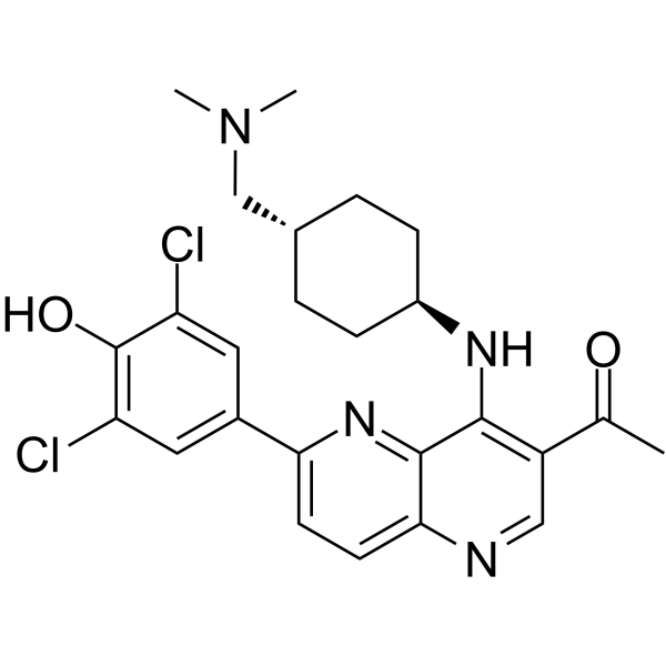
4-[7-乙酰基-8-[[4-[(二甲氨基)甲基]环己基]氨基]-1H-1,5-萘啶-2-亚基]-2,6-二氯-1-环己-2,5-二烯酮是一种萘啶衍生物。
MELK抑制剂OTS167是一种口服有效的母体胚胎亮氨酸拉链激酶(MELK)抑制剂,具有潜在的抗肿瘤活性。给药后,OTS167与MELK结合,阻止MELK的磷酸化和激活,从而抑制下游MELK底物的磷酸化。这可能导致表达MELK的肿瘤细胞的增殖和存活受到抑制。MELK是一种丝氨酸/苏氨酸激酶,参与癌细胞的存活、侵袭以及癌症干细胞的形成和维持; MELK(母体胚胎亮氨酸拉链激酶)在多种癌细胞中高度上调,而在正常健康细胞中则不存在。我们此前报道过MELK是乳腺癌的一种新型治疗靶点。MELK也被报道在多种人类癌症中高度上调,提示其在癌细胞存活中发挥着不可或缺的作用,并参与维持肿瘤起始细胞。我们对化合物库进行了高通量筛选,并开展了构效关系研究,成功获得了一种高效的MELK抑制剂OTSSP167,其IC₅₀值为0.41 nM。OTSSP167抑制了PSMA1(蛋白酶体α亚基1型)和DBNL(类drebrin蛋白)的磷酸化,我们发现PSMA1和DBNL是MELK的新型底物,对干细胞特性和侵袭性至关重要。该化合物抑制了乳腺癌细胞的乳腺球形成,并在小鼠异种移植模型中,通过静脉注射和口服给药,均能显著抑制乳腺癌、肺癌、前列腺癌和胰腺癌细胞系的肿瘤生长。这种MELK抑制剂有望成为一种抑制肿瘤起始细胞生长并用于治疗多种人类癌症的有效化合物。[1]母体胚胎亮氨酸拉链激酶(MELK)在多种人类肿瘤中表达上调,被认为是癌症治疗的一个有吸引力的分子靶点。我们对胃癌(GC)中MELK的表达进行了表征,并检测了降低MELK mRNA水平和蛋白活性对GC生长的影响。MELK在原发性GC中经常过表达,且MELK水平越高,临床预后越差。降低MELK表达或抑制激酶活性可抑制GC细胞的生长,导致G2/M期阻滞、细胞凋亡,并抑制其在体外和体内的侵袭能力。 MELK基因敲低导致上皮间质转化(EMT)相关蛋白的改变。此外,在胃癌患者来源的异种移植(PDX)模型中,使用OTSSP167进行靶向治疗显示出抗癌效果。因此,MELK通过抑制细胞凋亡、促进G2/M期转换和EMT来促进胃癌细胞的生长和侵袭。这些结果表明,MELK可能是一个有前景的胃癌治疗靶点。[2] OTSSP167最近被证实是一种有效的母体胚胎亮氨酸拉链激酶(MELK)抑制剂,目前正在进行针对其他治疗无效的实体瘤的I期临床试验。本文报道,OTSSP167在抑制MELK的浓度下可以消除有丝分裂检查点。这种消除作用无法通过RNAi介导的MELK沉默在细胞中重现。尽管OTSSP167确实能抑制MELK,但它在体外和细胞中均表现出对Aurora B激酶的脱靶活性。此外,OTSSP167还能抑制BUB1和Haspin激酶,降低组蛋白H2AT120和H3T3的磷酸化水平,导致Aurora B及其相关的染色体乘客复合物从着丝粒/动粒错位。这些结果表明,OTSSP167可能具有其他杀伤癌细胞的作用机制,因此在生化和细胞实验中应谨慎使用OTSSP167作为MELK特异性激酶抑制剂。[3] 鼠蛋白丝氨酸/苏氨酸激酶38 (MPK38),又称母体胚胎亮氨酸拉链激酶 (MELK),与多种人类癌症相关,并在癌症干细胞的形成中发挥重要作用。 OTSSP167 是一种 MELK 选择性抑制剂,具有很强的体外活性,IC50 值为 0.41 nM,并且对多种人类癌症异种移植模型具有体内抑制作用。本文报道了活性突变体 MPK38 (T167E) 与 OTSSP167 复合物的晶体结构,并详细描述了其蛋白质-抑制剂相互作用。与先前解析的 MELK 与纳摩尔级抑制剂结合的结构相比,OTSSP167 能有效地结合到活性位点,从而为基于结构的 MELK 抑制剂的开发和优化提供了契机。[4] |
| 分子式 |
C25H28CL2N4O2
|
|
|---|---|---|
| 分子量 |
487.42
|
|
| 精确质量 |
486.158
|
|
| 元素分析 |
C, 61.60; H, 5.79; Cl, 14.55; N, 11.49; O, 6.56
|
|
| CAS号 |
1431697-89-0
|
|
| 相关CAS号 |
OTSSP167 hydrochloride;1431698-10-0
|
|
| PubChem CID |
135398499
|
|
| 外观&性状 |
Light yellow to yellow solid powder
|
|
| 密度 |
1.3±0.1 g/cm3
|
|
| 沸点 |
619.0±55.0 °C at 760 mmHg
|
|
| 闪点 |
328.2±31.5 °C
|
|
| 蒸汽压 |
0.0±1.9 mmHg at 25°C
|
|
| 折射率 |
1.644
|
|
| LogP |
6.41
|
|
| tPSA |
81.32
|
|
| 氢键供体(HBD)数目 |
2
|
|
| 氢键受体(HBA)数目 |
6
|
|
| 可旋转键数目(RBC) |
6
|
|
| 重原子数目 |
33
|
|
| 分子复杂度/Complexity |
648
|
|
| 定义原子立体中心数目 |
0
|
|
| SMILES |
ClC1=CC(C2=CC=C(N=CC(C(C)=O)=C3N[C@H]4CC[C@H](CN(C)C)CC4)C3=N2)=CC(Cl)=C1O
|
|
| InChi Key |
DKZYXHCYPUVGAF-UHFFFAOYSA-N
|
|
| InChi Code |
InChI=1S/C25H28Cl2N4O2/c1-14(32)18-12-28-22-9-8-21(16-10-19(26)25(33)20(27)11-16)30-24(22)23(18)29-17-6-4-15(5-7-17)13-31(2)3/h8-12,15,17,33H,4-7,13H2,1-3H3,(H,28,29)
|
|
| 化学名 |
1-[6-(3,5-dichloro-4-hydroxyphenyl)-4-[[4-[(dimethylamino)methyl]cyclohexyl]amino]-1,5-naphthyridin-3-yl]ethanone
|
|
| 别名 |
|
|
| HS Tariff Code |
2934.99.9001
|
|
| 存储方式 |
Powder -20°C 3 years 4°C 2 years In solvent -80°C 6 months -20°C 1 month |
|
| 运输条件 |
Room temperature (This product is stable at ambient temperature for a few days during ordinary shipping and time spent in Customs)
|
| 溶解度 (体外实验) |
|
|||
|---|---|---|---|---|
| 溶解度 (体内实验) |
注意: 如下所列的是一些常用的体内动物实验溶解配方,主要用于溶解难溶或不溶于水的产品(水溶度<1 mg/mL)。 建议您先取少量样品进行尝试,如该配方可行,再根据实验需求增加样品量。
注射用配方
注射用配方1: DMSO : Tween 80: Saline = 10 : 5 : 85 (如: 100 μL DMSO → 50 μL Tween 80 → 850 μL Saline)(IP/IV/IM/SC等) *生理盐水/Saline的制备:将0.9g氯化钠/NaCl溶解在100 mL ddH ₂ O中,得到澄清溶液。 注射用配方 2: DMSO : PEG300 :Tween 80 : Saline = 10 : 40 : 5 : 45 (如: 100 μL DMSO → 400 μL PEG300 → 50 μL Tween 80 → 450 μL Saline) 注射用配方 3: DMSO : Corn oil = 10 : 90 (如: 100 μL DMSO → 900 μL Corn oil) 示例: 以注射用配方 3 (DMSO : Corn oil = 10 : 90) 为例说明, 如果要配制 1 mL 2.5 mg/mL的工作液, 您可以取 100 μL 25 mg/mL 澄清的 DMSO 储备液,加到 900 μL Corn oil/玉米油中, 混合均匀。 View More
注射用配方 4: DMSO : 20% SBE-β-CD in Saline = 10 : 90 [如:100 μL DMSO → 900 μL (20% SBE-β-CD in Saline)] 口服配方
口服配方 1: 悬浮于0.5% CMC Na (羧甲基纤维素钠) 口服配方 2: 悬浮于0.5% Carboxymethyl cellulose (羧甲基纤维素) 示例: 以口服配方 1 (悬浮于 0.5% CMC Na)为例说明, 如果要配制 100 mL 2.5 mg/mL 的工作液, 您可以先取0.5g CMC Na并将其溶解于100mL ddH2O中,得到0.5%CMC-Na澄清溶液;然后将250 mg待测化合物加到100 mL前述 0.5%CMC Na溶液中,得到悬浮液。 View More
口服配方 3: 溶解于 PEG400 (聚乙二醇400) 请根据您的实验动物和给药方式选择适当的溶解配方/方案: 1、请先配制澄清的储备液(如:用DMSO配置50 或 100 mg/mL母液(储备液)); 2、取适量母液,按从左到右的顺序依次添加助溶剂,澄清后再加入下一助溶剂。以 下列配方为例说明 (注意此配方只用于说明,并不一定代表此产品 的实际溶解配方): 10% DMSO → 40% PEG300 → 5% Tween-80 → 45% ddH2O (或 saline); 假设最终工作液的体积为 1 mL, 浓度为5 mg/mL: 取 100 μL 50 mg/mL 的澄清 DMSO 储备液加到 400 μL PEG300 中,混合均匀/澄清;向上述体系中加入50 μL Tween-80,混合均匀/澄清;然后继续加入450 μL ddH2O (或 saline)定容至 1 mL; 3、溶剂前显示的百分比是指该溶剂在最终溶液/工作液中的体积所占比例; 4、 如产品在配制过程中出现沉淀/析出,可通过加热(≤50℃)或超声的方式助溶; 5、为保证最佳实验结果,工作液请现配现用! 6、如不确定怎么将母液配置成体内动物实验的工作液,请查看说明书或联系我们; 7、 以上所有助溶剂都可在 Invivochem.cn网站购买。 |
| 制备储备液 | 1 mg | 5 mg | 10 mg | |
| 1 mM | 2.0516 mL | 10.2581 mL | 20.5162 mL | |
| 5 mM | 0.4103 mL | 2.0516 mL | 4.1032 mL | |
| 10 mM | 0.2052 mL | 1.0258 mL | 2.0516 mL |
1、根据实验需要选择合适的溶剂配制储备液 (母液):对于大多数产品,InvivoChem推荐用DMSO配置母液 (比如:5、10、20mM或者10、20、50 mg/mL浓度),个别水溶性高的产品可直接溶于水。产品在DMSO 、水或其他溶剂中的具体溶解度详见上”溶解度 (体外)”部分;
2、如果您找不到您想要的溶解度信息,或者很难将产品溶解在溶液中,请联系我们;
3、建议使用下列计算器进行相关计算(摩尔浓度计算器、稀释计算器、分子量计算器、重组计算器等);
4、母液配好之后,将其分装到常规用量,并储存在-20°C或-80°C,尽量减少反复冻融循环。
计算结果:
工作液浓度: mg/mL;
DMSO母液配制方法: mg 药物溶于 μL DMSO溶液(母液浓度 mg/mL)。如该浓度超过该批次药物DMSO溶解度,请首先与我们联系。
体内配方配制方法:取 μL DMSO母液,加入 μL PEG300,混匀澄清后加入μL Tween 80,混匀澄清后加入 μL ddH2O,混匀澄清。
(1) 请确保溶液澄清之后,再加入下一种溶剂 (助溶剂) 。可利用涡旋、超声或水浴加热等方法助溶;
(2) 一定要按顺序加入溶剂 (助溶剂) 。
| NCT Number | Status | Interventions | Conditions | Sponsor/Collaborators | Start Date | Phases |
| NCT02926690 | Recruiting | Drug: OTS167PO | Relapsed/Refractory Locally Advanced or Metastatic Breast Cancer and Triple Negative Breast Cancer |
OncoTherapy Science, Inc. | May 29, 2017 | Phase 1 |
| NCT02768519 | Completed | Drug: OTS167IV Other: Cherry syrup |
Healthy | OncoTherapy Science, Inc. | January 2016 | Phase 1 |
| NCT01910545 | Completed | Drug: OTS167IV | Solid Tumors Metastatic Tumors |
OncoTherapy Science, Inc. | August 23, 2013 | Phase 1 |
|
|---|
|
|
InvivoChem的所有产品仅用于作科学研究,不面向患者销售
Copyright 2020 InvivoChem LLC | All Rights Reserved 粤ICP备20063088号-1


 COA
COA


 463611831
